Researchers Identify Neural Pathways Transmitting Anti-Inflammatory Effects of Hunger

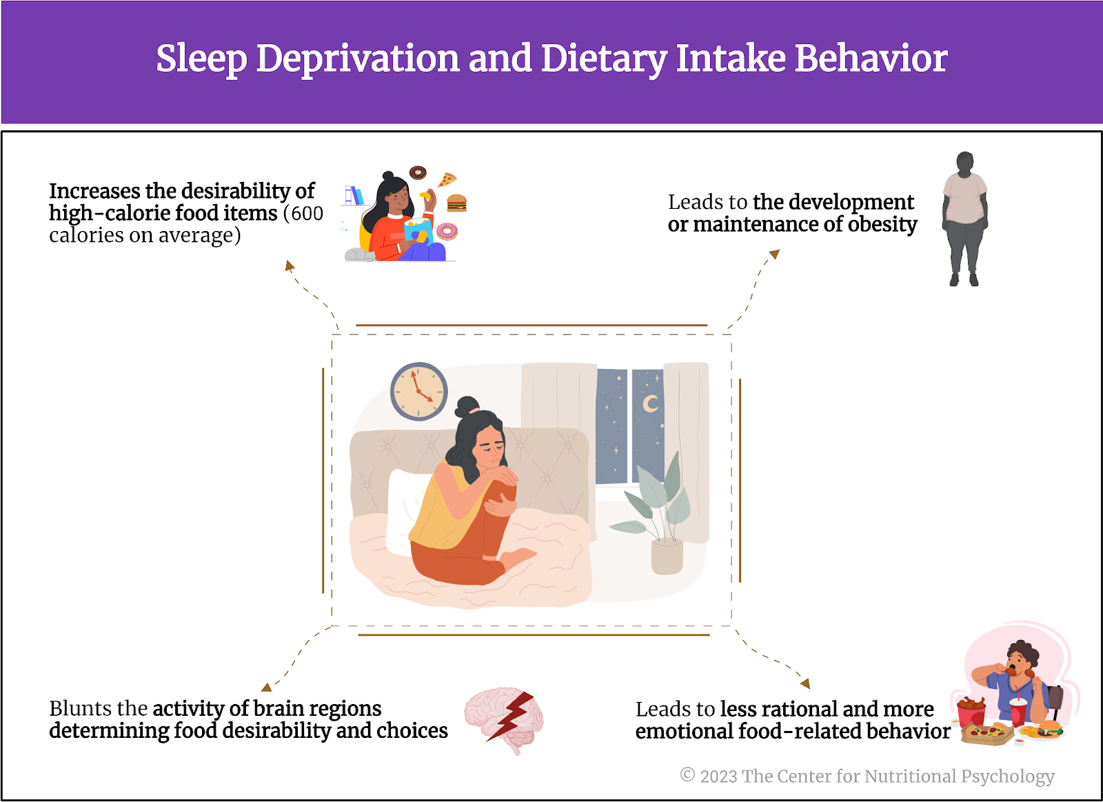
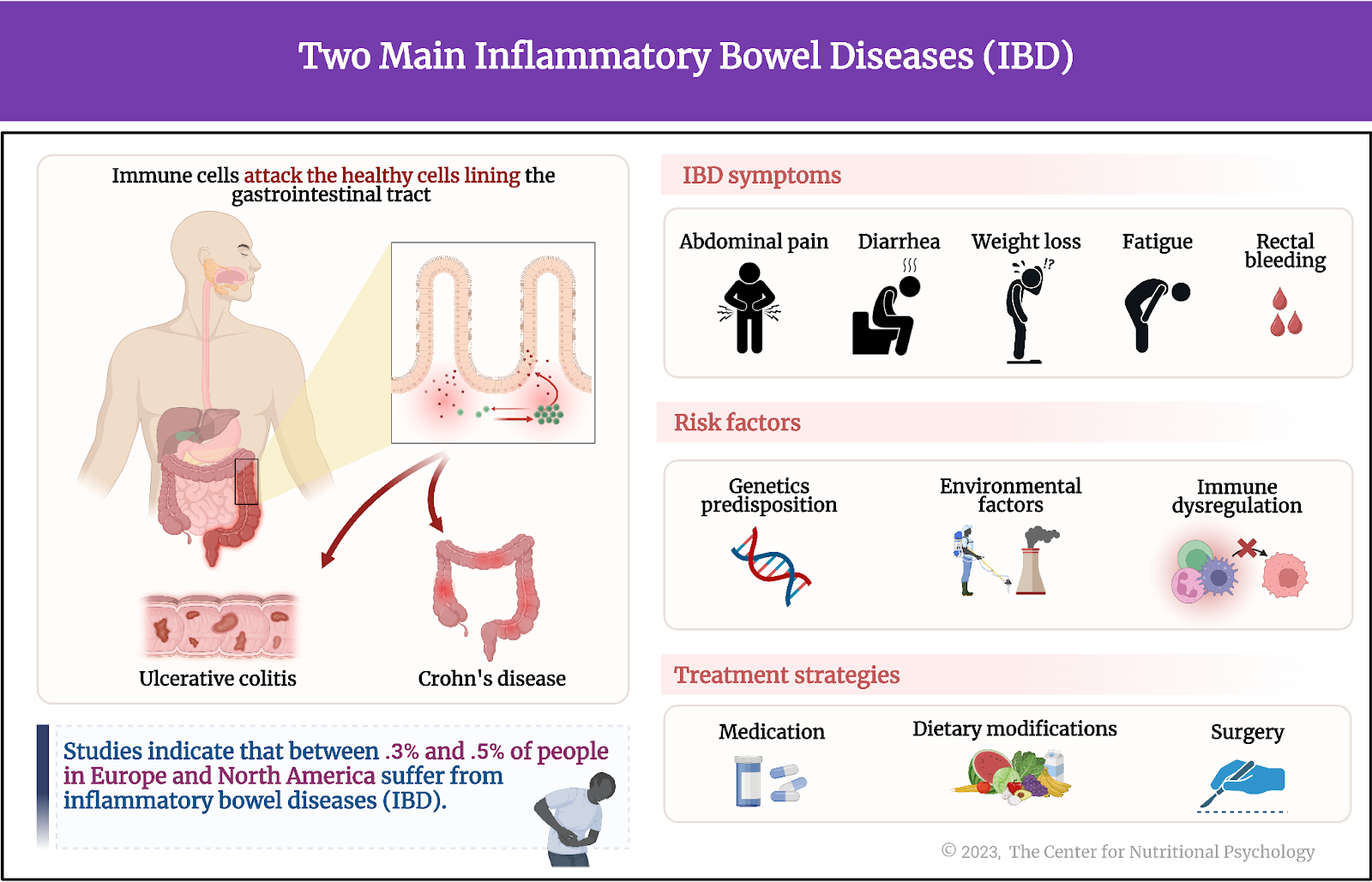
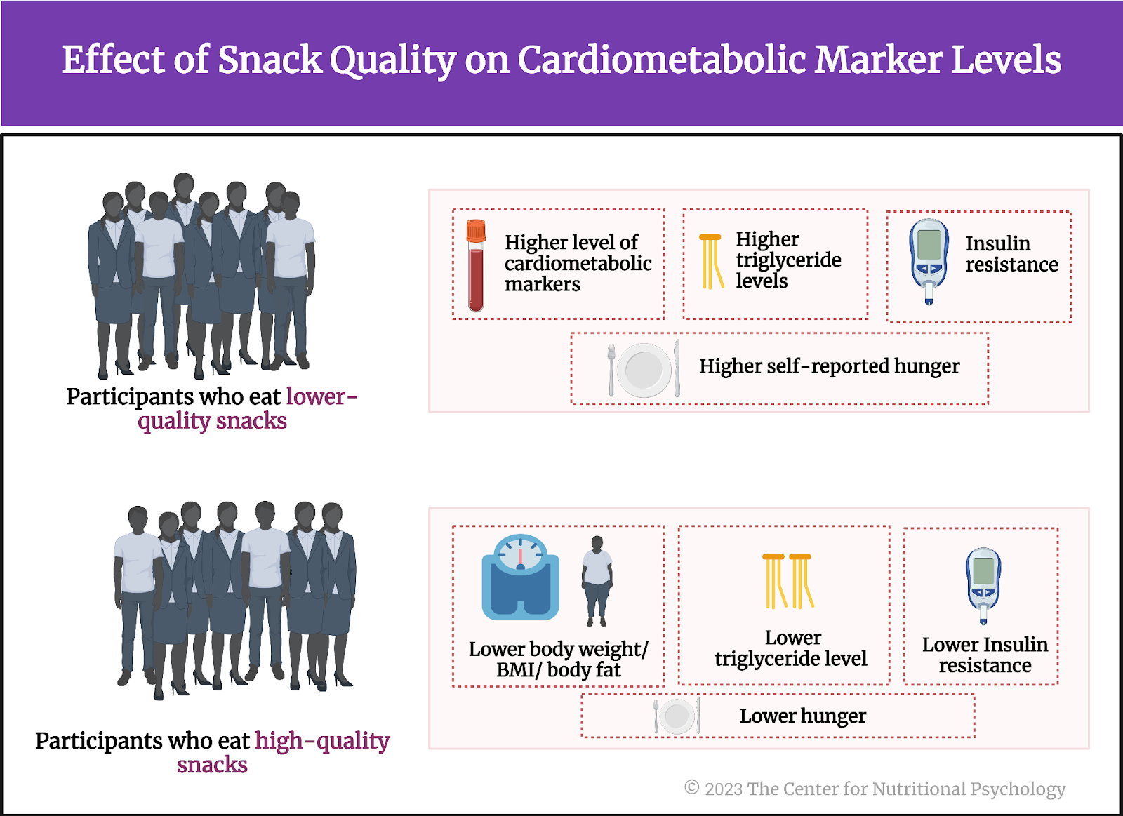
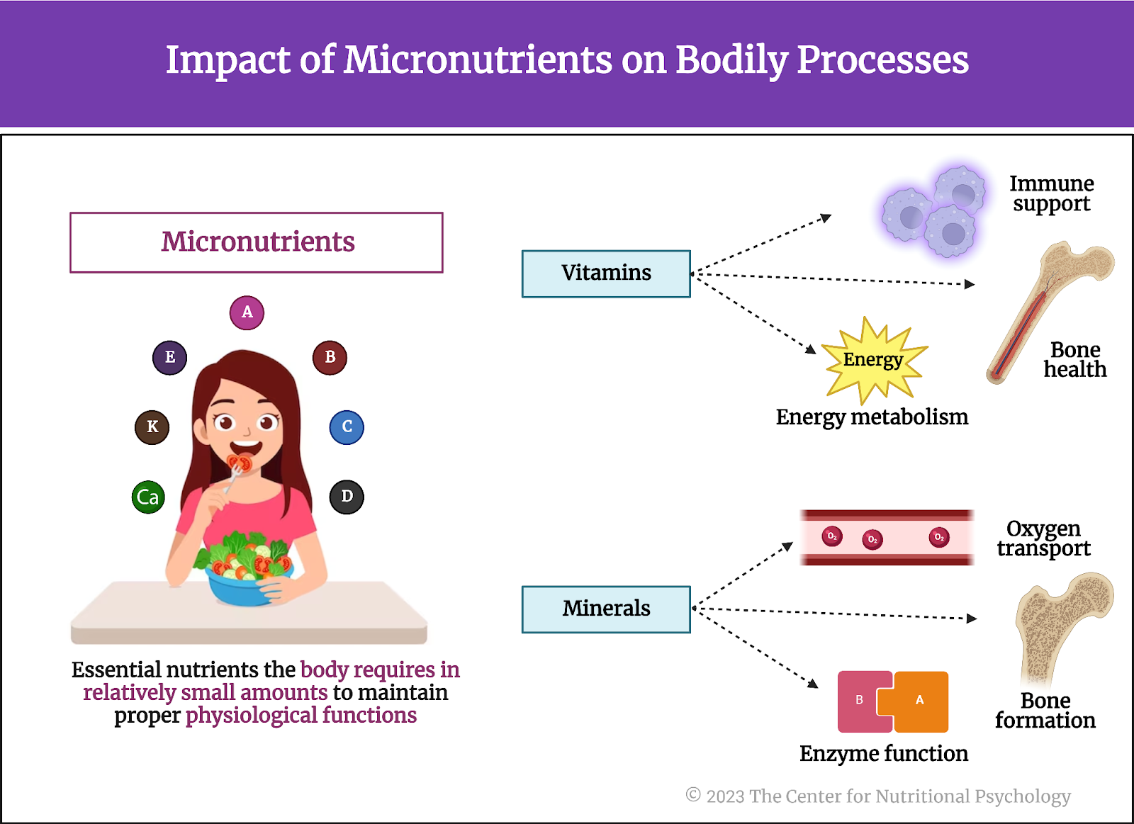
  • A series of experiments on mice published in Cell Reports found that hunger reduces inflammatory responses after an injury.
  • The anti-inflammatory effects of hunger were more robust than those of non-steroidal anti-inflammatory drugs.
  • AgRP neurons projecting to the paraventricular nucleus of the brain’s hypothalamus region mediate the majority of hunger’s anti-inflammatory effects.

People need food to survive. Severe malnutrition causes the body to break down its own tissues to meet energy needs. The body first utilizes the stored fat, but if starvation continues, it eventually uses muscle mass to fulfill its nutritional needs. Prolonged starvation impairs vital functions and leads to organ failure, weakened immune response, and severe hormonal imbalances (Sidiropoulos, 2007). Cognitive functions also deteriorate. But what happens when people restrict their food intake only for limited periods?

Restricted food intake
While the consequences of a chronic lack of food are dire, cultures worldwide have long believed that restricted food intake for limited periods of time can produce beneficial psychological and physiological consequences.

The practice of fasting, or abstaining from all or some kinds of food or drink for a specific period of time, is an important part of almost all major religions. Religions practice fasting as a means of spiritual discipline, self-reflection, and expressing devotion or penitence. It is often seen as a way to cleanse the body and mind, allowing for deeper religious or spiritual contemplation and connection with the divine or higher power.


Cultures worldwide have long believed that restricted food intake for limited periods of time can produce beneficial psychological and physiological consequences


Research studies show that restricted food intake for a limited period leads to complex physiological changes in the central and peripheral nervous systems. These changes can inhibit inflammation by preventing the activation of inflammasomes, multi-protein complexes that activate an inflammatory response. Additionally, restricted food intake reduces the production and release of pro-inflammatory cytokines, a group of signaling proteins secreted by immune cells that promote inflammation (Klima et al., 2023). On the psychological side, however, restrained eating can increase the feeling of hunger and food craving (Dicker-Oren et al., 2022).

Restricted food intake for a limited period can inhibit inflammation by preventing the activation of inflammasomes, multi-protein complexes that activate an inflammatory response, and reduce the production and release of pro-inflammatory cytokines 

While inflammation is an adaptive response, helping the body fight off infection or facilitating the repair of damaged tissue, long-term inflammation can become maladaptive and impair basic actions necessary for survival. It is also the cause or a contributing factor to many diseases and adverse medical conditions.


On the psychological side, however, restrained eating can increase the feeling of hunger and food craving (Dicker-Oren et al., 2022)


Agouti-related peptide (AgRP) neurons – hunger neurons
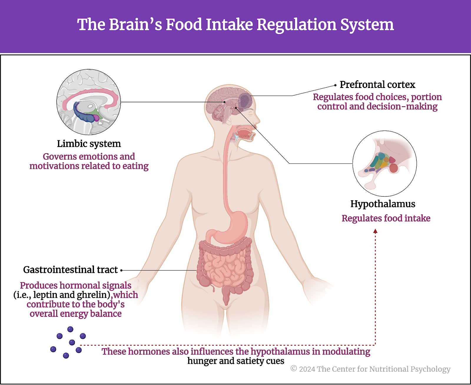
An important part of the neural circuits that react to restricted food intake are agouti-related peptide neurons, or AgRP neurons for short. Often referred to as “hunger neurons,” they are a specific type of neuron located in the brain’s arcuate nucleus of the hypothalamus region. These neurons play a critical role in regulating feeding behavior and energy homeostasis. AgRP neurons produce neuropeptides, including agouti-related peptide (AgRP) and neuropeptide Y (NPY), which are potent stimulators of appetite (Sternson & Atasoy, 2014) (see Figure 1).


%learn about nutrition mental health %The Center for Nutritional Psychology

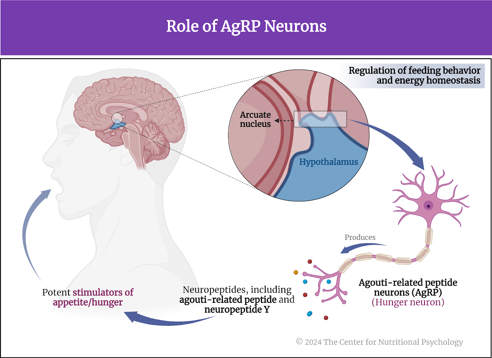
Figure 1. Role of AgRP neurons


The activity of AgRP neurons increases food intake and reduces energy expenditure, leading to weight gain. They are part of a complex neural network that responds to various signals related to energy status, such as hormones indicating hunger (ghrelin) or satiety (leptin and insulin). In conditions of energy deficit, such as fasting, AgRP neurons are stimulated, promoting hunger and encouraging food-seeking behavior (Atasoy et al., 2012; Chen et al., 2016).


“Hunger neurons” (AgRP) are a specific type of neuron located in the brain’s arcuate nucleus of the hypothalamus region and play a critical role in regulating feeding behavior and energy homeostasis


This system is crucial for survival, ensuring that energy intake is increased when energy stores are low. However, dysregulation of AgRP neurons and the pathways they influence can contribute to eating disorders and obesity (see Figure 2).


%learn about nutrition mental health %The Center for Nutritional Psychology

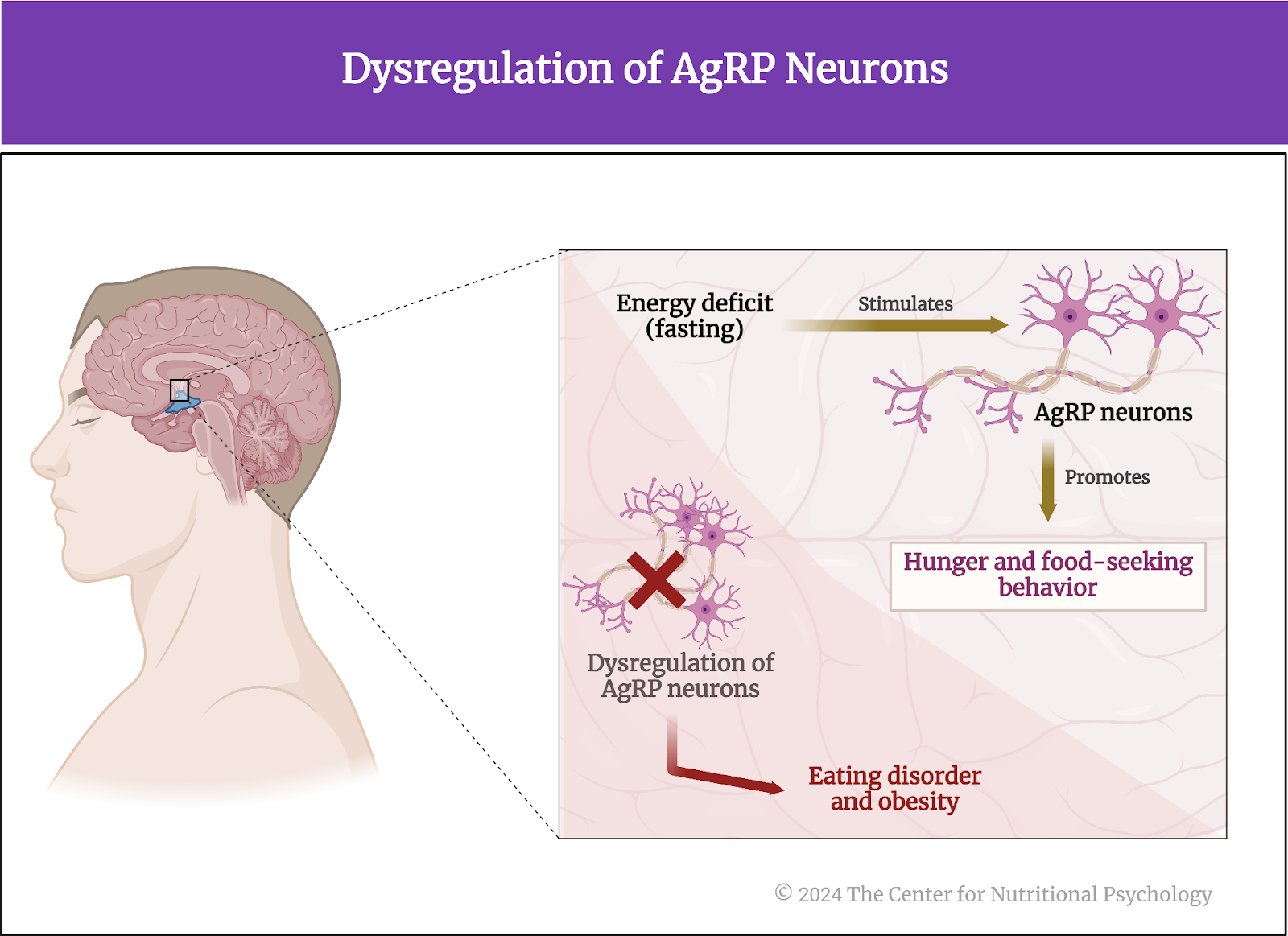
Figure 2. Dysregulation of AgRP neurons


The current study
Study author Michelle L. Klima and her colleagues wanted to map the neural pathways from the brain to the periphery that convey the anti-inflammatory effects of food deprivation. They note that it is likely that hunger circuits (AgRP neurons) in the central nervous system play a role in this because previous studies have shown that manipulations of gene expressions in these neurons can influence immune responses (Klima et al., 2023).

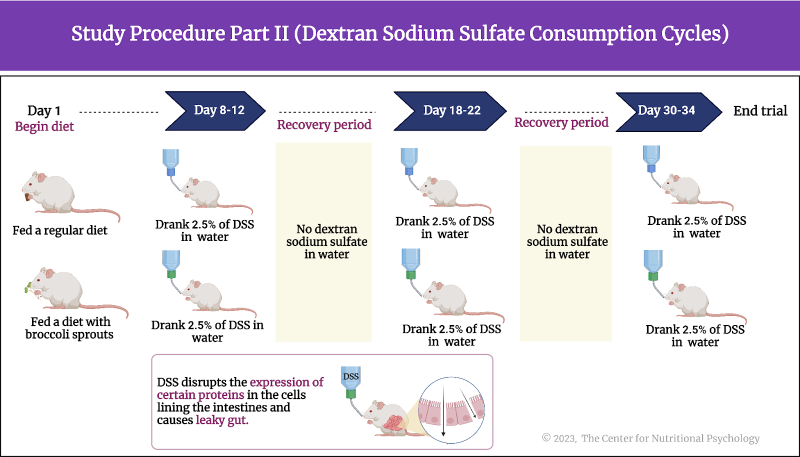
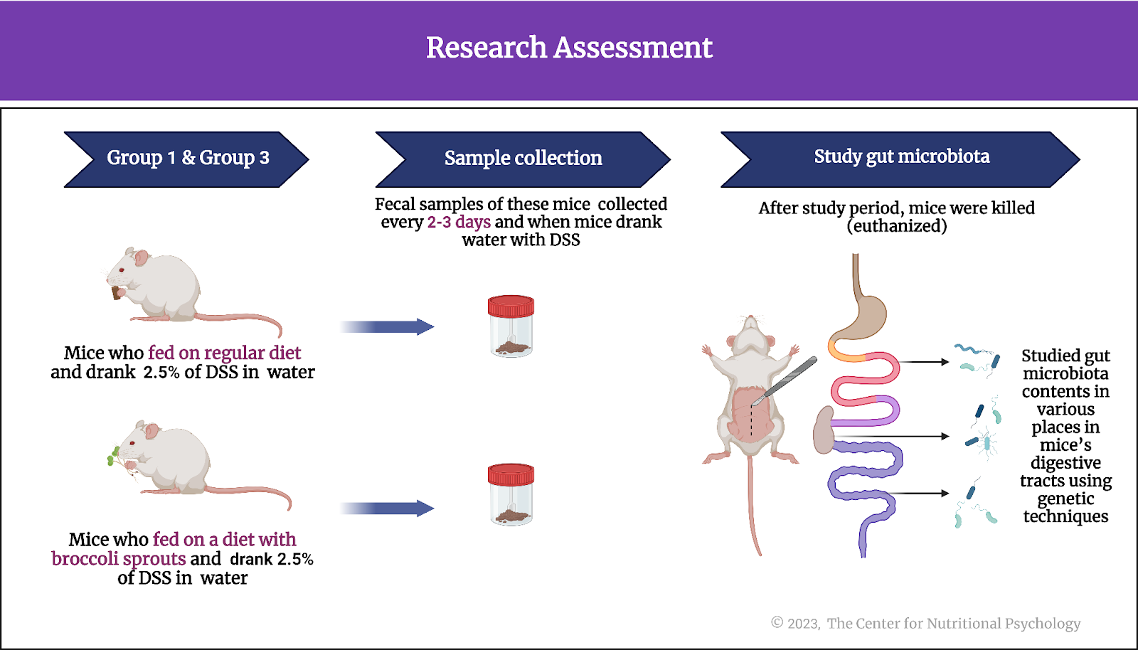
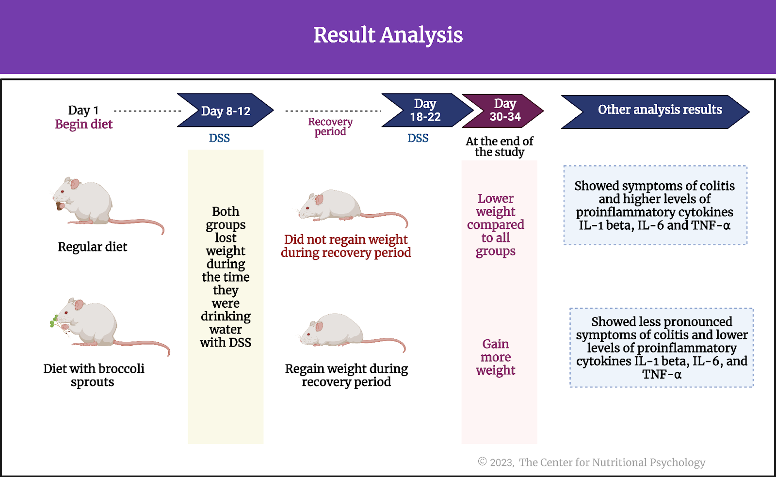
They conducted a series of experiments on mice. The mice used in this study were male and female, kept in group housing, and at least eight weeks old.

Hunger reduces inflammation in response to irritants
In the first experiment, study authors injected either formalin, complete Freund’s adjuvant (CFA), or a saline solution into the paws of these mice. Formalin and CFA are strong irritants that cause an inflammatory reaction and swelling of the paw. The saline solution injection was a control procedure. It is not expected to produce an inflammatory response, so the researchers use it as a reference for comparison with the reactions caused by the injections of the other two substances.

In the experiment, the study authors had two groups of mice – 8 mice that were kept without food for 24 hours before the experiment, i.e., hungry, and another eight that had free access to food. Some mice were injected with irritants and others with a saline solution within the two groups.

Results showed that the paws of mice injected with irritants developed strong inflammation, visibly increasing in size. However, this inflammatory reaction was attenuated by approximately 50% in hungry mice compared to mice fed normally. Female mice had stronger paw inflammation after the CFA injection than male mice, but the inflammation reduction effects of hunger were similar in both sexes.

Concentrations of inflammatory cytokine tumor necrosis factor-alpha, an indicator of inflammation, were much lower in hungry mice, confirming that hunger had a strong anti-inflammatory effect. Researchers also compared the anti-inflammatory effects of hunger with the effects of non-steroidal anti-inflammatory drugs ketoprofen and ketorolac. They found that hunger reduced paw inflammation 20% more than these two drugs (see Figure 3).


%learn about nutrition mental health %The Center for Nutritional Psychology

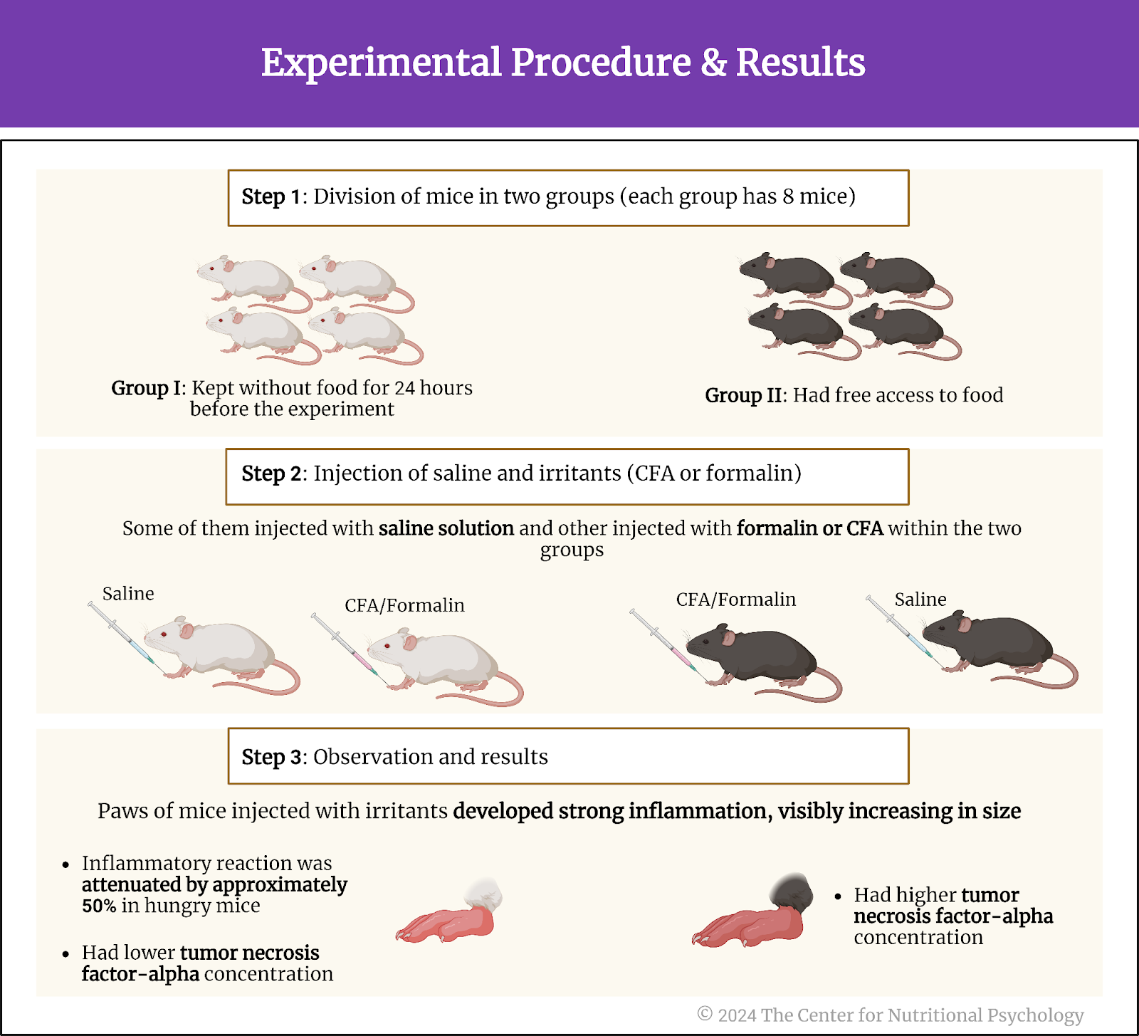
Figure 3. Experimental procedure and results 


Anti-inflammatory effects of hunger go from the brain through the vagus nerve
In their next experiment, study authors disabled the vagus nerve of the mice either by cutting it below the diaphragm or by injecting chemicals to disable parts of this nerve.

The vagus nerve extends from the brainstem to the abdomen, innervating various organs. It is crucial in the parasympathetic nervous system, responsible for the body’s rest and digest functions. The vagus nerve helps regulate heart rate, digestion, and respiratory rate but is also involved in inflammatory responses. It also conveys sensory information from the internal organs to the brain.

Results showed that the anti-inflammatory effects of hunger were suppressed when the vagus nerve was disabled in this way. This happened both when the nerve was physically cut and when chemicals were used.

Further experimentation showed that the anti-inflammatory effect is suppressed only when neurons going from the brain to the periphery (efferent neurons) are destroyed or disabled. Destroying or disabling neurons that carry information from the periphery to the brain (afferent neurons) did not affect the anti-inflammatory effects of hunger. This told researchers that the anti-inflammatory effects of hunger go from the brain through the vagus nerve to other areas of the body (see Figure 4).


%learn about nutrition mental health %The Center for Nutritional Psychology

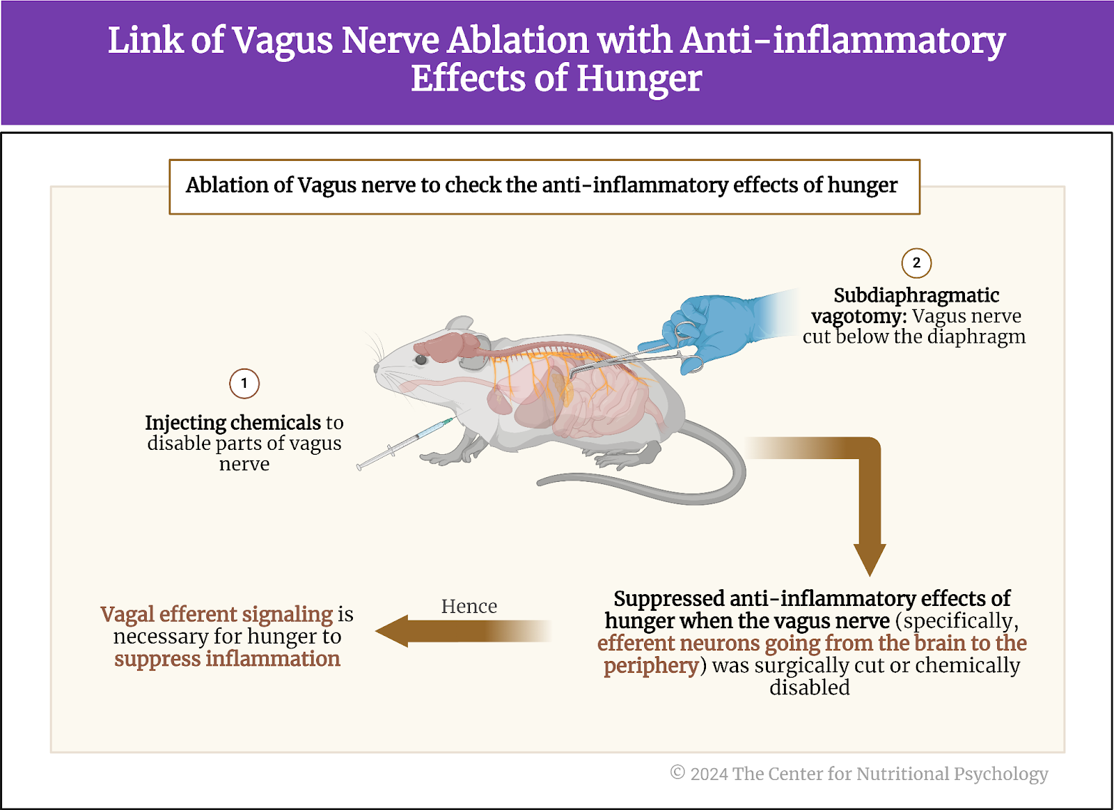
Figure 4. Link of vagus nerve ablation with anti-inflammatory effects of hunger 


Activation of AgRP neurons reduces inflammation
Next, the study authors looked for neural circuits in the brain that trigger the anti-inflammatory response to hunger. Knowing that AgRP neurons are activated by hunger, they first looked at them. These researchers genetically modified the AgRP neurons in a group of mice to be activated by specific chemicals or light.

Results showed an anti-inflammatory response followed when they artificially activated these neurons without restricting food to mice. Paws of mice in which study authors artificially activated AgRP neurons showed a much less intense inflammatory reaction after an irritant (CFA) injection than mice from the control group that ate normally and whose AgRp neurons were not artificially activated. The anti-inflammatory reaction caused by the activation of AgRP neurons was rapid, taking less than 1 hour for the reduction in multiple indicators of inflammation in the paw to become visible (see Figure 5).


%learn about nutrition mental health %The Center for Nutritional Psychology

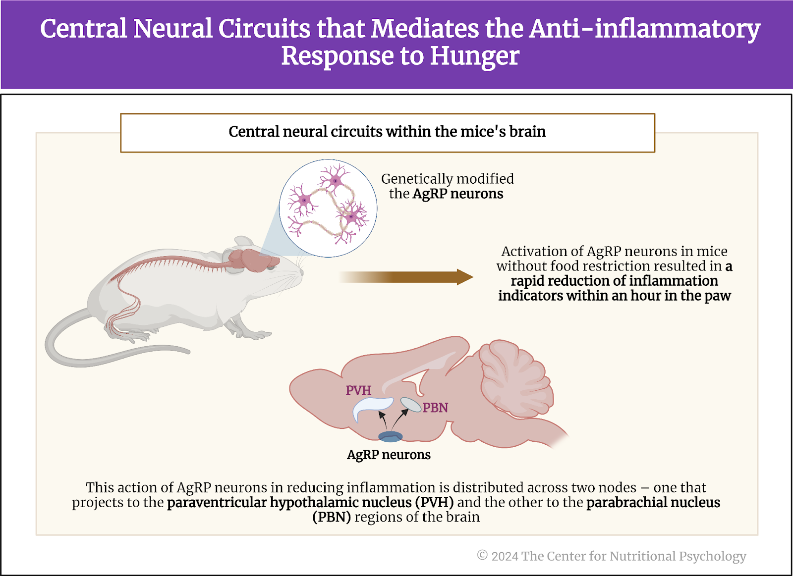
Figure 5. Central neural circuits that mediate the anti-inflammatory response to hunger


The study authors further sought to identify which AgRP neurons specifically activated the anti-inflammatory response. To do this, they activated specific groups of AgRP neurons one by one artificially in normally fed mice. They found that the anti-inflammatory action of AgRP neurons is distributed over two nodes – one projecting to the paraventricular hypothalamic nucleus and the other to the parabrachial nucleus regions of the brain. The magnitude of the anti-inflammatory effect of the first one was larger, mediating the bulk of the anti-inflammatory effects of these neurons.

Conclusions
Through a series of experiments on mice, this study showed that hunger produces an anti-inflammatory effect stronger than the effects of non-steroid anti-inflammatory medications such as ketoprofen and ketorolac. It showed that the bulk of the anti-inflammatory effect is mediated by hunger neurons, i.e., Agouti-related peptide (AgRP) neurons in the arcuate nucleus in the brain’s hypothalamus region. Even artificially triggering the activity of these neurons when a mouse is not hungry causes a rapid anti-inflammatory response.

These findings improve scientific understanding of the neural pathways of inflammation and can help healthcare researchers develop better ways to treat inflammation. Many diseases, from autoimmune diseases to skin disorders, are caused or aggravated by maladaptive immune responses of the body. Finding ways to better modulate or control the body’s immune response may be key to developing more effective treatments for them.

The paper “Anti-inflammatory effects of hunger are transmitted to the periphery via projection-specific AgRP circuits” was authored by Michelle L. Klima, Kayla A. Kruger, Nitsan Goldstein, Santiago Pulido, Aloysius Y.T. Low, Charles-Antoine Assenmacher, Amber L. Alhadeff, and J. Nicholas Betle.


References

Atasoy, D., Betley, J. N., Su, H. H., & Sternson, S. M. (2012). Deconstruction of a neural circuit for hunger. Nature, 488(7410), 172–177. https://doi.org/10.1038/nature11270

Chen, Y., Lin, Y.-C., Zimmerman, C. A., Essner, R. A., & Knight, Z. A. (2016). Hunger neurons drive feeding through a sustained, positive reinforcement signal. ELife, 5, e18640. https://doi.org/10.7554/eLife.18640.001

Dicker-Oren, S. D., Gelkopf, M., & Greene, T. (2022). The dynamic network associations of food craving, restrained eating, hunger and negative emotions. Appetite, 175(March), 106019. https://doi.org/10.1016/j.appet.2022.106019

Klima, M. L., Kruger, K. A., Goldstein, N., Pulido, S., Low, A. Y. T., Assenmacher, C. A., Alhadeff, A. L., & Betley, J. N. (2023). Anti-inflammatory effects of hunger are transmitted to the periphery via projection-specific AgRP circuits. Cell Reports, 42(11). https://doi.org/10.1016/j.celrep.2023.113338

Sidiropoulos, M. (2007). Anorexia Nervosa: The physiological consequences of starvation and the need for primary prevention efforts. CASE PRESENTATION. McGill Journal of Medicine, 10(1), 20–25.

Sternson, S. M., & Atasoy, D. (2014). Agouti-related protein neuron circuits that regulate appetite. Neuroendocrinology, 100, 95–102. https://doi.org/10.1159/000369072


Frequent Consumption of Fried Foods is Associated With a Slightly Higher Risk of Depression and Anxiety

  • A study of UK Biobank data published in PNAS reported that frequent consumption of fried food increased the risk of anxiety by 12% and the risk of depression by 7%.
  • Fried potato consumption was specifically associated with a somewhat increased risk of depression and anxiety symptoms.
  • An experiment on zebrafish confirmed that chronic exposure to acrylamide, a substance created when food is fried, creates anxiety-like symptoms in these fish.

The consolidation of research in the diet-mental health relationship within nutritional psychology has shown that our food choices and patterns can influence our moods and state of mind, and our moods and state of mind can influence our food choices and patterns (NPRL). People tend to eat when stressed or bored (Dicker-Oren et al., 2022; Stevenson et al., 2023). They can also feel angry when hungry (called hanger), in which one experiences anger due to hunger (Hedrih, 2023a).


Food choices and patterns can influence our moods and state of mind, which can influence our food choices and patterns


But could our dietary choices be associated with serious mental health disorders? Disorders like anxiety or depression?

Depression and anxiety
Depression and anxiety are two of the most frequent mental health disorders. Depression is characterized by persistently low mood and a lack of interest or pleasure in activities. A depressed individual will often feel sad, hopeless, and with little energy. This can lead to adverse changes in appetite and sleep patterns, but sometimes also thoughts of suicide.

Anxiety, on the other hand, is a condition marked by persistent and excessive worry that interferes with daily activities. It is typically accompanied by physical symptoms like restlessness, fatigue, difficulty concentrating, irritability, muscle tension, and sleep disturbances. While both conditions involve emotional distress, depression primarily affects mood and motivation, whereas anxiety is centered around fear and nervousness (see Figure 1).


%learn about nutrition mental health %The Center for Nutritional Psychology

Figure 1. Characteristics of and differences between depression and anxiety


Studies indicate that the share of individuals suffering from depression has been increasing both in the U.S. and globally in the past decades (Steffen et al., 2020; Weinberger et al., 2018). Anxiety and depression often occur together. Estimates state that the number of people suffering from anxiety increased after the global COVID-19 pandemic by 26% (Wang et al., 2023).


The number of people suffering from anxiety increased after the global COVID-19 pandemic by 26% (Wang et al., 2023)


Diet and mental health
While the precise causes of anxiety and depression are unknown, a great number of genetic, biological, environmental, and psychological factors have been found to play a role in their development. Recent studies have also linked certain dietary habits, such as the consumption of ultra-processed foods or sweetened beverages, to increased depression risks (Hedrih, 2023b; Samuthpongtorn et al., 2023).

Other researchers bring into focus the so-called Western diet, a diet based on fried and processed foods, refined grains, sugary products, and beer, as a dietary pattern associated with an increased risk of depression and anxiety (Wang et al., 2023). Within this diet, researchers single out fried food as particularly contributing to these adverse links with mental health.


The Western diet is a dietary pattern associated with an increased risk of depression and anxiety


Fried food and health
Frying is a method of food preparation that people widely use at home and in restaurants. However, the process of frying changes the nutrient composition of the food and can produce various hazardous compounds. One such substance is acrylamide. Acrylamide is created during the process of frying foods that are rich in carbohydrates, such as potatoes. Acrylamide is known to be toxic for neurons in higher concentrations. Studies have linked its prolonged intake to an increased risk of cardiovascular disease, neurological disorders, obesity, metabolic syndrome, and depression.


The process of frying changes the food’s nutrient composition and can produce various hazardous compounds 


The current study
Study author Anli Wang and her colleagues wanted to examine the association between the intake of a substance found in fried food called acrylamide and depression and anxiety in a large sample from the general population. Acrylamide is a substance found in fried food.

These authors expected that high consumption of fried food and, consequently, higher intake of acrylamide would be associated with more severe symptoms of anxiety and depression. Ingested acrylamide would lead to abnormal levels of lipids, such as cholesterol and triglycerides, in the blood and inflammation. This would, in turn, impact the likelihood that depression or anxiety would develop (see Figure 2).


%learn about nutrition mental health %The Center for Nutritional Psychology

Figure 2. Effects of acrylamide ingested from fried foods


In this study, the authors analyzed human data from the UK Biobank and conducted an experiment on zebrafish.

The UK Biobank
The UK Biobank is a large-scale biomedical database and research resource containing in-depth genetic and health information from half a million UK participants. The project, which began in 2006 and is ongoing, aims to improve the prevention, diagnosis, and treatment of a wide range of serious and life-threatening illnesses.

UK Biobank participants provided blood, urine, and saliva samples and detailed health and lifestyle information, which researchers worldwide can access to conduct health-related research. Many valuable scientific discoveries and insights have come from this database, and new studies using UK Biobank data are continuously being published.

For this study, researchers analyzed data from 140,728 individuals from the UK Biobank. Of these, during the 11.3 years of follow-up, 8,294 had symptoms of anxiety, and 12,735 had depression symptoms (see Figure 3).


%learn about nutrition mental health %The Center for Nutritional Psychology

Figure 3. Study procedure


The Zebrafish experimentI
n addition to analyzing the UK Biobank data, the researchers conducted an experiment on zebrafish. This experiment exposed zebrafish groups to different acrylamide concentrations for 180 days. After the exposure period, they conducted a series of tests to evaluate anxiety-like symptoms in these fish. At the end of the experiment, they killed the fish and harvested their brains to analyze tissue changes.

Zebrafish are a small, freshwater fish species extensively used in biomedical research due to their genetic similarity to humans, transparent embryos that allow easy observation of developmental processes, and rapid reproduction.

These researchers looked for specific symptoms in zebrafish exposed to acrylamide –specifically thigmotaxis and scototaxis. Scototaxis is a tendency of fish to prefer darker areas over lighter ones in their environment. Thigmotaxis is a preference for staying close to physical objects or walls in their aquarium. In research, both of these tendencies are indications that the fish is experiencing anxiety-like conditions and that it is under stress. The more pronounced they are, the higher the anxiety-like conditions in the fish are.


UK Biobank People consuming fried food more often had somewhat higher risks of depression and anxiety


Results from the UK Biobank showed that individuals consuming at least one fried meal per day were more likely to be younger, male, and active smokers than those consuming fried food less often. After taking into account the age and sex of participants, statistical analyses showed that individuals consuming fried food more often were somewhat more likely to have anxiety and depression symptoms.

Notably, individuals consuming fried food were 12% more likely to have anxiety symptoms and 7% more likely to have symptoms of depression. Results of looking at specific food types highlighted fried potatoes and fried white meat. Consumption of each of these foods was associated with a 4% higher risk of anxiety and a 7% higher risk of depression. However, after adjusting for various other factors, the link with fried white meat consumption disappeared, but the one with fried potato consumption remained (see Figure 4).


%learn about nutrition mental health %The Center for Nutritional Psychology

Figure 4. Fried food consumption and depression and anxiety symptoms 


Individuals consuming fried food were 12% more likely to have anxiety symptoms and 7% more likely to have symptoms of depression


Zebrafish exposed to acrylamide developed behaviors indicative of stress and anxiety
The experiment on zebrafish showed that fish chronically exposed to acrylamide in a concentration of 0.5 mM decreased in body mass and length. Fish exposed to even lower concentrations of acrylamide (0.125-0.5 mM) showed signs of scototaxis. They spent more time in the dark zones than zebrafish without this exposure. The time the fish spent in the dark zones was associated with the concentration of acrylamide used.

Other behavioral comparisons between zebrafish exposed to acrylamide and those in the control group also confirmed the finding about anxiety-like symptoms in fish exposed to this substance. Analysis of tissues of zebrafish showed that exposure to acrylamide disturbed their lipid metabolism and initiated an inflammatory response, which, in turn, likely led to the observed behavioral changes (see Figure 5).


%learn about nutrition mental health %The Center for Nutritional Psychology

Figure 5. Behavioral changes and physical changes in Zebrafish exposed to acrylamide


Conclusion
An analysis of UK Biobank data showed that individuals frequently consuming fried food have a slightly higher risk of having depression or anxiety symptoms. An experiment on zebrafish confirmed that chronic exposure to acrylamide, a substance created when food rich in carbohydrates is fried, might cause this link. Exposure to acrylamide can disturb the metabolism of lipids and initiate an inflammatory response that may, in turn, contribute to the development of anxiety and depression symptoms. 


Chronic exposure to acrylamide, a substance created when food rich in carbohydrates is fried, might cause this link


These findings contribute to a better scientific understanding of the diet-mental health relationship (DMHR). They may help individuals and mental health practitioners prevent or better treat anxiety and depression conditions by modifying their diet. The insights brought by this study may also allow for the creation of better menus and meal plans in the hospitality industry and various institutional food services.

The paper “High fried food consumption impacts anxiety and depression due to lipid metabolism disturbance and neuroinflammation” was authored by Anli Wang, Xuzhi Wan, Pan Zhuang, Wei Jia, Yang Ao, Xiaohui Liu, Yimei Tian, Li Zhu, Yingyu Huang, Jianxin Yao, Binjie Wang, Yuanzhao Wu , Zhongshi Xu, Jiye Wang, Weixuan Yao, Jingjing Jiao, and Yu Zhang.


References

Dicker-Oren, S. D., Gelkopf, M., & Greene, T. (2022). The dynamic network associations of food craving, restrained eating, hunger and negative emotions. Appetite, 175(March), 106019. https://doi.org/10.1016/j.appet.2022.106019

Hedrih, V. (2023a). Food and Mood: Is the Concept of ‘Hangry’ Real? CNP Articles in Nutritional Psychology.
https://www.nutritional-psychology.org/food-and-mood-is-the-concept-of-hangry-real/

Hedrih, V. (2023b). Women Consuming Lots of Artificially Sweetened Beverages Might Have a Higher Risk of Depression, Study Finds. CNP Articles in Nutritional Psychology. https://www.nutritional-psychology.org/women-consuming-lots-of-artificially-sweetened-beverages-might-have-a-higher-risk-of-depression-study-finds/

Samuthpongtorn, C., Nguyen, L. H., Okereke, O. I., Wang, D. D., Song, M., Chan, A. T., & Mehta, R. S. (2023). Consumption of Ultraprocessed Food and Risk of Depression. JAMA Network Open, 6(9), e2334770. https://doi.org/10.1001/jamanetworkopen.2023.34770

Steffen, A., Thom, J., Jacobi, F., Holstiege, J., & Bätzing, J. (2020). Trends in prevalence of depression in Germany between 2009 and 2017 based on nationwide ambulatory claims data. Journal of Affective Disorders, 271, 239–247. https://doi.org/10.1016/J.JAD.2020.03.082

Stevenson, R. J., Bartlett, J., Wright, M., Hughes, A., Hill, B. J., Saluja, S., & Francis, H. M. (2023). The development of interoceptive hunger signals. Developmental Psychobiology, 65(2), 1–11. https://doi.org/10.1002/dev.22374

Wang, A., Wan, X., Zhuang, P., Jia, W., Ao, Y., Liu, X., Tian, Y., Zhu, L., Huang, Y., Yao, J., Wang, B., Wu, Y., Xu, Z., Wang, J., Yao, W., Jiao, J., & Zhang, Y. (2023). High-fried food consumption impacts anxiety and depression due to lipid metabolism disturbance and neuroinflammation. Proceedings of the National Academy of Sciences of the United States of America, 120(118). https://doi.org/10.1073/pnas.2221097120

Weinberger, A. H., Gbedemah, M., Martinez, A. M., Nash, D., Galea, S., & Goodwin, R. D. (2018). Trends in depression prevalence in the USA from 2005 to 2015: widening disparities in vulnerable groups. Psychological Medicine, 48(8), 1308–1315. https://doi.org/10.1017/S0033291717002781


Consuming Fat and Sugar (At The Same Time) Promotes Overeating, Study Finds

  • A study on mice published in Cell Metabolism found that gut-brain neural circuits conveying and processing information about the presence of fat and sugar in the gut are separate
  • Foods that contain both sugar and fats activate both circuits, releasing more dopamine compared to foods of equal caloric value containing only fat
  • The large amounts of dopamine released in this way create feelings of pleasure that promote overeating on the types of food that caused the release

When we want to find out what a piece of food is like, we can taste or smell it. When we smell something, sensory cells located in a small patch of tissue (olfactory epithelium) at the top of our nasal cavity react to the odorant molecules coming from that piece of food, and the olfactory nerve to carries the information to our brain, allowing us to experience smell. Similarly, when we taste a piece of food, taste buds in our mouth react to it, and specific nerves carry the information to our brain. While we eat, this evaluation of the qualities of food using our sensory organs happens continuously. However, not all of our food sensations come from our external senses. Our body also has sensory cells in the gut that inform the brain about what we eat.

The vagus nerve
After we ingest food through our mouth, it enters the esophagus. It continues towards the stomach and, after that, into the intestine. Food is further digested and broken down into components absorbed into the body as nutrients in these parts of the gastrointestinal system.

There are sensory cells located throughout the gastrointestinal system. The vagus nerve, one of the longest nerves in the body, connects these cells to the brain through a communication pathway called the gut-brain axis. In this way, it conveys sensory information about the state of the intestinal environment, including nutrient levels, gut microbiota activity, and intestinal wall integrity from the gut to the brain. These signals are then integrated into the brainstem, allowing the brain to monitor and respond to gut activity, influencing digestive processes, immune responses, and emotional states. (Bonaz et al., 2018) (see Figure 1).


%learn about nutrition mental health %The Center for Nutritional Psychology

Figure 1. Role of sensory cells and vagus nerve in the Gut-Brain Axis.


Among other things, the vagus nerve carries information to the brain about the nutritional value of our food. However, scientists so far have not fully understood the intricate details of how this system functions (McDougle et al., 2024).

The obesity pandemic
Recent decades have seen a continued increase in the share of obese individuals throughout the developed world. Many call this the obesity pandemic (Wong et al., 2022). This has motivated many studies into the causes of this pandemic. Results point to an intricate interplay between specific types of nutrients and other substances in the food and the properties of the human central nervous system as important contributing causes of obesity (Wilding, 2001).

Notably, while human brains (and brains of many other species) have a food intake regulation mechanism that determines when we will feel a desire to eat or when we will feel satiated, studies indicate that parts of this mechanism might be malfunctioning in obese individuals (Pujol et al., 2021; Seabrook et al., 2023). Studies indicate that feeding rodents high-fat diets instead of their regular chow can, in time, dysregulate their food intake regulation mechanisms, leading them to eat more calories than they need, resulting in obesity (Ikemoto et al., 1996). In humans, studies have linked the dysregulation of this mechanism with the consumption of foods that are both fatty and sweet (a property rarely found in natural foods) but also to various additives that create addiction-like reactions to foods that contain them (Hedrih, 2023).

The current study
Study author Molly McDougle and her colleagues wanted to explore the neural pathways through which the brain detects the intake of fats and sugars (i.e., fatty and sweet foods). They wanted to know whether the intake of these two types of nutrients triggers the same sensory neurons and whether they engage overlapping or separate neural circuits.

What was known before this study was that a direct infusion of fats or sugars into the gut (of mice) activates the vagus nerve. This, in turn, leads to the release of the neurotransmitter dopamine in the dorsal striatum region of the brain. The dorsal striatum is involved in reward and motivation processing, and dopamine is a key neurotransmitter that signals pleasure and reward (see Figure 2).


%learn about nutrition mental health %The Center for Nutritional Psychology

Figure 2. Vagal nerve activation from the direct infusion of fats or sugars into the gut


Study procedure
The study was conducted on 70 mice. The mice used in this study were between 6 and 20 weeks of age. Thirty-five mice were male, and 35 were female. They were housed at 22 degrees C and had free access to mouse food.

The study authors performed a series of experiments on these mice. The experiments involved surgical procedures that exposed or cut the vagus nerve and/or exposed the nodose ganglion (a part of the vagus nerve) for further activities. These surgeries also involved the implantation of catheters that allowed researchers to inject fluids directly into the gut of these mice, bypassing the mouth and, thus, taste receptors.

These researchers used 2-photon imaging to study the activity of the nodose ganglion in the mice (while the mice were alive) and of brain areas involved in sensing and processing information about nutrient intake. Two-photon imaging is a fluorescence microscopy technique that allows for deep-tissue, high-resolution imaging of live cells and tissues. It uses two photons of lower energy to excite a fluorophore simultaneously. Fluorophores are special molecules that emit a visible glow when excited by light of a specific wavelength, thus allowing researchers to track and observe different biological processes or structures.

Other experimental procedures included behavioral tests, techniques using light to control neurons that have been genetically modified to express ion channels sensitive to light (optogenetics), sampling extracellular fluid from the dorsal striatum region of the brain to measure the quantities of dopamine (microdialysis) and others. In the end, the study authors harvested and analyzed the brain tissues of these mice (see Figure 3).


%learn about nutrition mental health %The Center for Nutritional Psychology

Figure 3. Study procedure summary (McDougle et al., 2024).


Fats and sugars are sensed by separate neurons of the vagal nerve
Results showed that fats and sugars increased the activity of neurons in the vagus nerve, but mostly not of the same ones. Neurons comprising the nodose ganglion mostly reacted to the presence of fat or sugars. Very few neurons reacted to both. These neurons reacting to nutrients in the intestine comprised around 17% of the neurons in the nodose ganglion. The nodose ganglion is a cluster of sensory neurons located on the vagus nerve, transmitting visceral sensory information from the body to the brain.

When researchers infused sugar or fat into the guts of mice, the neurons of the nodose ganglion they previously marked increased their frequencies of nerve impulses. Analysis of the neural circuits activated by fats and sugars showed that they differ. They were largely separate. Information about sugar and fats in the gut is carried to the brain through two distinct pathways. These pathways convey information about the type of nutrient in the gut, but they also likely convey information about its concentration (see Figure 4).


%learn about nutrition mental health %The Center for Nutritional Psychology

Figure 4. Sensing fats and sugars in the gut


The duodenum is the key sensing site for both sugar and fat
Tracing the locations of sensory cells, these neurons led these researchers to discover an extensive network of sensory cells in the duodenum. Almost all sensory cells detecting fats and many of those detecting sugars were located there. The duodenum is the first section of the small intestine, connecting the stomach to the jejunum. It plays a crucial role in the initial phase of digestion by receiving partially digested food from the stomach and digestive enzymes from the pancreas and liver. On the other hand, study authors found that sugar-sensing also happens in the hepatic portal vein, a blood vessel that carries nutrient-rich blood from the gastrointestinal tract and spleen to the liver (see Figure 5).


%learn about nutrition mental health %The Center for Nutritional Psychology

Figure 5. Detection of fats and sugars by a network of sensory cells in the duodenum 


Reinforcement learning based on sugar and fat happens through separate circuits
Experiments where researchers destroyed fat-sensing neurons but left the sugar-sensing ones intact or vice versa indicated that reinforcement learning, a type of learning where a mouse can associate a specific object or sensation with a specific nutrient, can be conducted using only the neurons sensing that specific nutrient. Mice without fat-sensing neurons but with intact sugar-sensing neurons were able to form a preference for a flavor associated with sugar intake.


Mice without fat-sensing neurons but with intact sugar-sensing neurons were able to form a preference for a flavor associated with sugar intake


In the nigrostriatal region of the brain, both information about fat and sugar in the gut led to the release of dopamine (thought to produce the feeling of pleasure and a rewarding experience). However, these nutrients activated parallel and largely separate neuronal populations at each node of the reward circuit in this part of the brain. This means that distinguishable neural circuits are responsible for reinforcement learning for fat and sugar, i.e., for learning to associate stimuli with pleasure caused by the ingestion of sugar and pleasure caused by the ingestion of fat (see Figure 6).


%learn about nutrition mental health %The Center for Nutritional Psychology

Figure 6. Release of dopamine in response to fat and sugar


Consuming fat and sugar at the same time promotes overeating for pleasure
In another group of experiments, study authors found that when they gave mice a choice between taking solutions of fat, sugar (sucrose), or a mixture of the two nutrients, mice consumed equal amounts of fat or sugar solutions. However, the number of licks of the solution containing both sugar and fats nearly doubled compared to just sugar or just fat alone.


Consuming fat and sugar at the same time promotes overeating for pleasure


Researchers then offered mice a non-nutrient-flavored solution to lick, injecting sugar, fat, or a combination of the two directly into their guts (using the catheters they installed). After training to pair a non-nutritive flavor with a solution, mice again showed a preference for the mixed solution (both sugar and fat), indicating that the preference for this solution is independent of taste. This means that the preference for the sugar and fat combination has to do with sensing those nutrients in the gut and not with their taste in the mouth.


The preference for the sugar and fat combination has to do with sensing those nutrients in the gut and not with their taste in the mouth


Study authors conclude that this is because this mixed solution activates both the brain’s rewarding circuits for fat and sugar, increasing the overall feeling of pleasure. Analysis of the quantity of released dopamine in the dorsal striatum region of the brain found that it is much higher after the injection of the mixed sugar-fat solution than after the injection of a solution with equal caloric value but containing only fat.

This suggests that foods rich in fat and sugar produce higher activation of reward circuits for the same amount of calories than foods containing just one of these two nutrients. This would translate to higher feelings of pleasure after consumption, motivating the individual to consume more food of that type, which could lead to obesity (see Figure 7).


%learn about nutrition mental health %The Center for Nutritional Psychology

Figure 7. Activation of the brain’s rewarding circuits with combined sugar and fat


Conclusion
The study showed that sensing the presence of sugars and fats in the intestines of mice is achieved through different sets of neurons. The study also found that these two types of nutrients activate parallel but largely separate reward circuits in the brain. Due to this, consuming foods that contain both sugars and fats activates reward circuits in the brain for both of these substances, resulting in a higher release of dopamine and, thus, higher feelings of pleasure, compared to consuming foods of equal caloric value but containing only fats or only sugars. These peculiarities of neural pathways make mice, and possibly humans, more likely to overeat foods rich in fats and sugars driven by feelings of pleasure the simultaneous activation of these two neural pathways creates.

The discovery of these separate mechanisms and understanding of the ways they work can help scientists. Still, people working in nutrition better understand how the properties of certain food items lead to obesity. This can, in sequence, help better plan individual diets and develop policies to help stop the ongoing obesity epidemic.

The paper “Separate gut-brain circuits for fat and sugar reinforcement combine to promote overeating” was authored by Molly McDougle, Alan de Araujo, Arashdeep Singh, Mingxin Yang, Isadora Braga, Vincent Paille, Rebeca Mendez-Hernandez, Macarena Vergara, Lauren N. Woodie, Abhishek Gour, Abhisheak Sharma, Nikhil Urs, Brandon Warren, and Guillaume de Lartigue.


References

Bonaz, B., Bazin, T., & Pellissier, S. (2018). The vagus nerve at the interface of the microbiota-gut-brain axis. In Frontiers in Neuroscience (Vol. 12, Issue FEB). Frontiers Media S.A. https://doi.org/10.3389/fnins.2018.00049

Hedrih, V. (2023). Scientists Propose that Ultra-Processed Foods be Classified as Addictive Substances. CNP Articles in Nutritional Psychology. https://www.nutritional-psychology.org/scientists-propose-that-ultra-processed-foods-be-classified-as-addictive-substances/

Ikemoto, S., Takahashi, M., Tsunoda, N., Maruyama, K., Itakura, H., & Ezaki, O. (1996). High-fat diet-induced hyperglycemia and obesity in mice: Differential effects of dietary oils. Metabolism, 45(12), 1539–1546. https://doi.org/10.1016/S0026-0495(96)90185-7

McDougle, M., de Araujo, A., Singh, A., Yang, M., Braga, I., Paille, V., Mendez-Hernandez, R., Vergara, M., Woodie, L. N., Gour, A., Sharma, A., Urs, N., Warren, B., & de Lartigue, G. (2024). Separate gut-brain circuits for fat and sugar reinforcement combine to promote overeating. Cell Metabolism. https://doi.org/10.1016/j.cmet.2023.12.014

Pujol, J., Blanco-Hinojo, L., Martínez-Vilavella, G., Deus, J., Pérez-Sola, V., & Sunyer, J. (2021). Dysfunctional Brain Reward System in Child Obesity. Cerebral Cortex, 31, 4376–4385. https://doi.org/10.1093/cercor/bhab092

Seabrook, L. T., Naef, L., Baimel, C., Judge, A. K., Kenney, T., Ellis, M., Tayyab, T., Armstrong, M., Qiao, M., Floresco, S. B., & Borgland, S. L. (2023). Disinhibition of the orbitofrontal cortex biases decision-making in obesity. Nature Neuroscience, 26(1), 92–106. https://doi.org/10.1038/s41593-022-01210-6

Wilding, J. P. H. (2001). Causes of obesity. Practical Diabetes International, 18(8), 288–292. https://doi.org/10.1002/PDI.277

Wong, M. C., Mccarthy, C., Fearnbach, N., Yang, S., Shepherd, J., & Heymsfield, S. B. (2022). Emergence of the obesity epidemic: 6-decade visualization with humanoid avatars. The American Journal of Clinical Nutrition, 115(4), 1189–1193. https://doi.org/10.1093/AJCN/NQAC005


Does Sleep Deprivation Increase Desire for High-Calorie foods?

  • A study published in Nature Communications found that sleep deprivation decreases the activity of frontal and insular cortices, parts of the human brain responsible for higher-order processes when making food choices.
  • At the same time, the activity of the amygdala region, responsible for processing emotions, increases.
  • When we lack sleep, food choices become less rational and more emotional, increasing the desire for high-calorie foods and potentially leading to weight gain.

Our minds don’t function as well when we continually get less sleep than we need. After a prolonged period of time with less sleep, concentrating becomes progressively more difficult, our reactions become slower, and our minds can often wander off. We may even fall asleep unintentionally. Our mind starts “drifting.“ Sometimes, consuming substances like coffee, similar caffeinated beverages, or even medications like modafinil (Wingelaar-Jagt et al., 2023) can help us remain vigilant for some additional time. However, we must eventually have a sufficiently long sleep period to recuperate and remain healthy.

Why is sleep important?


Sleep is a natural and recurring state of reduced consciousness and responsiveness, characterized by altered sensory perception and inactivity of voluntary muscles. Sleep consists of two main types: non-REM sleep, which includes four stages characterized by progressively deeper sleep, and REM (rapid eye movement) sleep, a stage associated with vivid dreams and heightened brain activity. During sleep, the body undergoes tissue repair, immune system strengthening, and the consolidation of memories. Sleep is crucial for maintaining physical and mental health. It plays a vital role in regulating mood, cognitive function, and overall well-being.

Insufficient or poor-quality sleep has been linked to various health issues, including impaired immune function, increased risk of chronic conditions, and negative effects on mood and cognitive performance (Hillman & Lack, 2013) (see Figure 1).


%learn about nutrition mental health %The Center for Nutritional Psychology

Figure 1. Types of sleep


Lack of sleep is associated with obesity


Studies have reported that sleep loss is one of the risk factors for obesity. This is the case both in children and in adults worldwide. There is mounting evidence that consuming food at night, at the time when we should normally be sleeping, is linked with poor health outcomes, such as worse cardiometabolic health (Bermingham et al., 2023), but also with an increased risk of obesity (Lent et al., 2022). Studies also indicate that food consumed at night tends to be less healthy. Individuals eating at night tend to consume less fruits and vegetables and more sugar-sweetened beverages and fast foods.


There is mounting evidence that consuming food at night, when we should be sleeping, is linked with poor health outcomes


There is an eating disorder called night eating syndrome. Night eating syndrome is a disordered eating pattern characterized by recurrent eating episodes during the night. A person suffering from this disorder typically awakes during the night and starts eating. Individuals with night eating syndrome often consume a significant portion of their daily food during these nocturnal eating episodes. In the morning, they may experience a lack of appetite. Because these eating episodes happen at night, interrupting the normal sleep cycle, the night eating syndrome is also considered a sleep disorder. It affects approximately 1.5% of adults in the U.S., but 9% of patients seeking weight-loss treatments, and 16% of individuals with binge eating disorder (Tzischinsky et al., 2021) (see Figure 2).


%learn about nutrition mental health %The Center for Nutritional Psychology

Figure 2. Characteristics of Night Eating Syndrome (NES)


Studies show that in parallel with the rising obesity rates, there is a continued decline in sleep duration in industrialized countries (Greer et al., 2013). What happens to our brain that makes us change our food-related behavior when we lack sleep?


In parallel with the rising obesity rates, there is a continued decline in sleep duration in industrialized countries


The current study


Study author Stephanie M. Greer and her colleagues wanted to find out. There is an abundance of research findings showing that lack of sleep changes our food-related behavior in a way that can lead to weight gain and obesity. Yet, study authors note in spite of this, the neural mechanisms through which this is achieved remain unknown (Greer et al., 2013).

Discovering these mechanisms would allow researchers to understand the link between sleep loss and obesity and potentially devise ways in which individuals could appropriately regulate dietary intake, thus preventing obesity. They conducted a study using functional magnetic resonance imaging (MRI) and focusing on cortical and subcortical regions of the brain, which are instrumental in food desire and evaluating food-related stimuli (see Figure 3). 


%learn about nutrition mental health %The Center for Nutritional Psychology

Figure 3. Cortical and subcortical regions of the brain involved in food desire and evaluation


These areas were the anterior insular cortex, lateral orbital frontal cortex, anterior cingulate cortex, amygdala, and the ventral striatum. The three cortex regions all have established roles in determining the value of various food cues, determining our food choices, and integrating various food features (e.g., odor or flavor) to create food preferences. The amygdala and the ventral striatum are strongly implicated in governing our motivation to eat. Activity in the ventral striatum can accurately predict immediate food intake, binge eating, and weight gain. 

The study procedure


Study participants were 23 healthy adults who agreed to abstain from drugs, alcohol, and caffeine for three days before the start of the study. They also kept a regular sleep schedule (7-9 hours per night) during this period. Thirteen participants were female. The average age of participants was 21 years, and they were of normal weight.

Each participant completed two experimental sessions – a night of normal sleep in the study authors’ lab monitored by polysomnography equipment and a night of total sleep deprivation (a night during which he/she did not sleep) monitored by lab personnel and by wrist actinography (a device detecting movements of the wrist that can be used to infer whether a person is sleeping). During the sleep deprivation night, participants had a snack around 2:30-3:00 am. The two nights were at least seven days apart.

Functional magnetic resonance imaging and the food-desire task


Participants completed functional magnetic resonance imaging (fMRI) sessions the morning after each experimental night. During the scan, participants completed a food-desire task. The task consisted of 80 pictures of food (without packaging) the researchers collected online. The food items were evenly distributed into five categories – salty, sweet, starchy, fruit, or dairy, and varied in calorie content. The two imaging sessions used the same 80 items with different pictures.

Participants’ task was to rate each food item on a scale ranging from 1 to 4 on how much they wanted that particular food item right now. Researchers did not tell them about the study hypotheses nor the calorie contents of the food items. They told participants that they have two of the shown food items in the lab and that each participant will receive the food item he/she rated higher. Researchers did this to increase the likelihood that participants would rate the food items according to their preferences (see Figure 4).


%learn about nutrition mental health %The Center for Nutritional Psychology
Study Procedure Nutrition psychology course (Greer, 2013)

Figure 4. Study procedure (Greer, 2013)


Sleep deprivation diminished activity in the three cortical regions


The activity in the three studied cortex regions was much lower when participants did not sleep (the sleep deprivation night) compared to the night when they slept well. Participants’ overall food desire also increased. The decrease in activity was the most pronounced in the anterior cingulate region. 

Activity in the amygdala region increased after the sleepless night


In contrast to the decrease in the cortex activity, the activity in the amygdala brain region in response to desirable food items increased after the sleepless nights when a participant saw a picture of a food item they desired (in the food-desire task taken during brain imaging).

Knowing the function of the amygdala, this increased activity likely indicates increased salience of the food items. After the sleepless night, desirable food items more easily capture participants’ attention. Interestingly, self-reported hunger levels were not different after the two experimental nights (Figure 5).


%learn about nutrition mental health %The Center for Nutritional Psychology

Figure 5. Research Findings


Sleep deprivation increases the desirability of high-calorie food items


After the sleepless night, participants found high-calorie food items much more desirable than the ratings they gave them after the night when they slept normally. However, there were no changes in the desirability of low-calorie items. As a consequence of these changes, the total calorie content of all wanted food items taken together was much higher after the sleepless night. The difference between the food item selections from the two nights was 600 calories on average.


Conclusion


These findings indicate that sleep deprivation blunts the activity of brain regions determining food desirability and choices.  While areas responsible for higher-order processes become less reactive to food, those governing our emotional reactions to food increase activity. Consequently, our food-related behaviors become less rational and more emotional when sleep-deprived.

We become more prone to eating tasty food. Since tasty and emotionally pleasing foods also tend to be rich in calories, the calorie intake also increases. The described mechanism explains how sleep loss can lead to the development or maintenance of obesity. Because of this, it is important that treatments for obesity or plans for maintaining a healthy weight also consider this mechanism and include sufficient and undisturbed sleep as one of the factors necessary for maintaining a healthy weight and overall health (see Figure 6).


%learn about nutrition mental health %The Center for Nutritional Psychology

Figure 6. Sleep deprivation and dietary intake behavior


The paper ”The impact of sleep deprivation on food desire in the human brain” was authored by Stephanie M. Greer, Andrea N. Goldstein, and Matthew P. Walker.

Find more studies on the Diet-Sleep Relationship in CNP’s Nutritional Psychology Research Library “Diet, Sleep and Fatigue” Research Category. 

References

Bermingham, K. M., May, A., Asnicar, F., Capdevila, J., Leeming, E. R., Franks, P. W., Valdes, A. M., Wolf, J., Hadjigeorgiou, G., Delahanty, L. M., Segata, N., Spector, T. D., & Berry, S. E. (2023). Snack quality and snack timing are associated with cardiometabolic blood markers: the ZOE PREDICT study. European Journal of Nutrition. https://doi.org/10.1007/s00394-023-03241-6

Greer, S. M., Goldstein, A. N., & Walker, M. P. (2013). The impact of sleep deprivation on food desire in the human brain. Nature Communications, 4. https://doi.org/10.1038/ncomms3259

Hillman, D. R., & Lack, L. C. (2013). Public health implications of sleep loss: The community burden. Medical Journal of Australia, 199(8), S7–S10. https://doi.org/10.5694/mja13.10620

Lent, M. R., Atwood, M., Bennett, W. L., Woolf, T. B., Martin, L., Zhao, D., Goheer, A. A., Song, S., McTigue, K. M., Lehmann, H. P., Holzhauer, K., & Coughlin, J. W. (2022). Night eating, weight, and health behaviors in adults participating in the Daily24 study. Eating Behaviors, 45. https://doi.org/10.1016/j.eatbeh.2022.101605

Tzischinsky, O., Latzer, I. T., Alon, S., & Latzer, Y. (2021). Sleep quality and eating disorder-related psychopathologies in patients with night eating syndrome and binge eating disorders. Journal of Clinical Medicine, 10(19). https://doi.org/10.3390/jcm10194613

Wingelaar-Jagt, Y. Q., Bottenheft, C., Riedel, W. J., & Ramaekers, J. G. (2023). Effects of modafinil and caffeine on night-time vigilance of air force crewmembers: A randomized controlled trial. Journal of Psychopharmacology, 37(2), 172–180. https://doi.org/10.1177/02698811221142568


Study Identifies Link Between Post-Traumatic Stress Disorder and Gut Microbiome Composition in a Cohort of Women

  • A study published in Nature Mental Health examined the links between post-traumatic stress disorder (PTSD), dietary patterns, and gut microbiome in U.S. nurses
  • Results showed that nurses with higher PTSD symptom levels tended to eat less plant-based food and more red/processed meat
  • Microbial processes related to the production of pantothenate and coenzyme A can potentially be protective against PTSD

When we are exposed to distressing events or conditions that overwhelm our ability to cope, our body can produce a severe emotional response, and we experience psychological trauma. We feel that we have lost control of events around us. Our ability to integrate these emotional experiences into the story of our lives is reduced. After experiencing psychological trauma, people often start dividing the subjective timeline of their lives into the time before and the time after the traumatic event. Long-lasting psychological and health consequences may often follow (Hamburger et al., 2021). Among other things, the experience of psychological trauma can lead to the development of a serious mental health disorder called post-traumatic stress disorder or PTSD.


After experiencing psychological trauma, people often start dividing the subjective timeline of their lives into the time before and after the traumatic event 


What is post-traumatic stress disorder?
Post-traumatic stress disorder is a mental health condition that can develop in individuals who have experienced or witnessed a traumatic event. Symptoms of PTSD include intrusive thoughts, nightmares, and flashbacks related to the traumatic experience, causing significant distress. Individuals with PTSD may actively avoid reminders of the trauma, experience heightened arousal, and have negative changes in mood and cognition.

Mainstream treatments for PTSD include psychotherapy, particularly cognitive-behavioral psychotherapy, and medications. Studies indicate that these treatments can be effective in reducing PTSD symptoms (see Figure 1). In some individuals, they are very effective (Watts et al., 2013). However, around 20% of patients suffering from PTSD drop out of treatment before symptoms have withdrawn, while up to 60% of individuals undergoing treatment do not respond to it, i.e., experience no reduction of symptoms as a result (Wittmann et al., 2021).


%learn about nutrition mental health %The Center for Nutritional Psychology

Figure 1. PTSD symptoms and mainstream treatments 


For these reasons, researchers are intensely working on new treatment options. They are trying alternative ways to achieve a reduction of symptoms, such as acupuncture (Watts et al., 2013) or psychedelic drugs (Barone et al., 2019). Researchers are also looking at chemicals that could potentially prevent the disorder from forming in the first place if taken immediately after the traumatic event, such as hydrocortisone (Hennessy et al., 2022).

Health impact of PTSD
Individuals suffering from PTSD often suffer from other psychiatric disorders as well. Epidemiological surveys indicate that the vast majority of individuals with PTSD meet the criteria for at least one other disorder, while a substantial percentage have three or more other psychiatric disorders. These most commonly include major depressive disorder, substance use disorder, and anxiety disorders (Brady et al., 2000).


The majority of individuals with PTSD meet the criteria for at least one other disorder, while a substantial percentage have three or more other psychiatric disorders


Individuals with PTSD also have a higher likelihood of various somatic diseases, particularly chronic ones. These include asthma, cardiovascular disease, chronic pain and inflammation, obesity, type 2 diabetes, gastrointestinal disorders, and cognitive decline (Ke et al., 2023).

However, researchers are still looking for mechanisms through which this association between PTSD and somatic disorders is achieved. One promising avenue of research is the study of the gut microbiome, the community of organisms living in the human digestive system. The recent discovery of the microbiota-gut-brain axis, a bidirectional communication pathway through which gut microorganisms affect processes in the brain and vice versa (Carbia et al., 2023; García-Cabrerizo et al., 2021), has made it even more likely that at least a part of the link between PTSD and somatic disorder includes the gut microbiome.


Researchers are looking for mechanisms connecting PTSD and somatic disorders -with one promising avenue of research being the gut microbiome


The current study
Study author Shanlin Ke and his colleagues hypothesized that gut microbiome might play a role in PTSD. They note that previous studies have already linked the gut microbiome with various other mental health disorders through the microbiota-gut-brain axis (Hedrih, 2023; Leclercq et al., 2020; Valles-Colomer et al., 2019). Additionally, studies show that the gut microbiome influences the brain region involved in learned fear. Fear is a key feature of PTSD. Gut microbiota depends on food intake, while individuals with PTSD tend to be more prone to eating unhealthy foods (Ke et al., 2023).


Previous studies have already linked the gut microbiome with various other mental health disorders through the microbiota-gut-brain axis (MGBA)


With this in mind, these researchers analyzed data from two studies of female registered nurses in the U.S. in order to systematically examine the associations of trauma exposure and PTSD status with dietary and microbiome data.

The study procedure
Data came from 191 nurses who were part of the NHS-II study. NHS-II is a large longitudinal study of U.S. women with over 100,000 registered nurses that started in 1989. Nurses whose data were analyzed here participated in two substudies – the 2008 PTSD substudy that collected data about trauma exposure and PTSD symptoms and the 2013 mind-body study that, among other things, collected up to four stool samples. Stool samples were collected 48-72 hours apart. This was followed by a second set of collections six months later. Stool samples were analyzed to make inferences about the composition of the gut microbiome.

Of the nurses included in this study, 44 had probable PTSD, 119 were exposed to trauma but did not develop PTSD, and 28 participants had no trauma exposure (see Figure 2).


%learn about nutrition mental health %The Center for Nutritional Psychology

Figure 2. Study Procedure


No association between PTSD and overall microbiome structure
Statistical analyses showed no associations between overall gut microbiota composition and PTSD status. Gut microbiota diversity was also not associated with PTSD.

However, there were associations between various other factors and the overall microbiome structure. Body mass index, depression, and the use of antidepressants were all associated with the overall composition of gut microbiome species.

Childhood trauma experiences and antidepressant use were associated with the relative abundance of species forming different metabolic pathways. The gut microbiome metabolic pathways refer to types of activities within the gastrointestinal tract that specific species of microorganisms perform.

Individuals with PTSD eat less plant-based food and more red/processed meat
Examination of associations between dietary habits and PTSD revealed that individuals with more pronounced PTSD symptoms tend to eat less plant-based foods and more red/processed meats compared to individuals with lower PTSD symptom levels. The diets of these individuals were less healthy overall. 


Individuals with more pronounced PTSD symptoms tend to eat less plant-based foods and more red/processed meats compared to individuals with less PTSD symptoms 


Individuals with PTSD symptoms had lower abundances of Eubacterium eligens
After statistical analyses showed no associations between the overall gut microbiome composition and PTSD, these authors examined associations between PTSD symptoms and the abundance of individual species of bacteria. They looked for species of microorganisms that differed in abundance in individuals with different levels of PTSD symptoms. Three species were clearly more abundant in individuals with more PTSD symptoms – Parabacteroides goldsteinii, Barnesiella intestinihominis, and Paraprevotella unclassified. Seven species with the highest negative association with PTSD symptoms, i.e., species less abundant in individuals with more PTSD symptoms, were also identified (Eubacterium eligens, Parabacteroides distasonis, Akkermansia muciniphila, Bacteroides massiliensis, Bifidobacterium longum, Dialister invisus, and Roseburia inulinivorans). These species play diverse roles in the human digestive system, contributing to functions ranging from fermentation of dietary fibers to protection of the gut lining.

Eubacterium eligens (a good bacteria) was less abundant in individuals eating more pies, carbonated beverages with sugars, candy without chocolate, hot dogs, bacon, and processed meats. It tended to be more abundant in individuals eating more vegetables (for example, raw carrot, spinach/collard greens cooked, and yellow squash), fruits (for example, orange and banana), and fish. The abundance of this bacteria was the most strongly associated with PTSD symptoms of all analyzed microorganisms. Eubacterium eligens in the gut synthesize pantothenate and Coenzyme A (see Figure 3).


%learn about nutrition mental health %The Center for Nutritional Psychology

Figure 3. Microorganisms associated with PTSD


Researchers tested a number of statistical models proposing that specific dietary habits mediate the impact of PTSD symptoms on the abundance of specific species of gut bacteria. These analyses revealed that it is possible that the impact of PTSD symptoms on the abundance of Eubacterium eligens is mediated by the consumption of raw carrots. Similarly, it is possible that Adlercreutzia equolifaciens mediates the link between PTSD symptoms and the intake of dairy-cottage/ricotta cheese.

Conclusion
In summary, the study showed that PTSD symptoms in U.S. nurses are not associated with the overall structure of the gut microbiome, but the abundance of several bacterial species was associated with the severity of PTSD symptoms in spite of this. Additionally, it turned out that participants with PTSD tend to eat less plant-based food and more red/processed meat. Their overall dietary habits tended to be less healthy (see Figure 4). 


%learn about nutrition mental health %The Center for Nutritional Psychology

Figure 4. Research findings (Ke et al., 2023)


The study also established statistical links between PTSD symptoms, dietary habits, and bacteria. Future studies could explore the nature of these links in more detail. Potentially, these could lead to the discovery of ways to affect PTSD symptoms through dietary interventions and modifying the abundance of certain species of gut bacteria. However, the feasibility of such an approach is yet to be established.

The paper “Association of probable post-traumatic stress disorder with dietary pattern and gut microbiome in a cohort of women” was authored by Shanlin Ke, Xu-Wen Wang, Andrew Ratanatharathorn, Tianyi Huang, Andrea L. Roberts, Francine Grodstein, Laura D. Kubzansky, Karestan C. Koenen, and Yang-Yu Liu.

Visit the research category within the Nutritional Psychology Research Library on “Diet, Trauma and PTSD” to learn more about the connection between diet and trauma. To access evidence-based continuing education on the connection between the microbiome and mental health, see the CNP Education page.


References

Barone, W., Beck, J., Mitsunaga-Whitten, M., & Perl, P. (2019). Perceived Benefits of MDMA-Assisted Psychotherapy beyond Symptom Reduction: Qualitative Follow-Up Study of a Clinical Trial for Individuals with Treatment-Resistant PTSD. Journal of Psychoactive Drugs, 51(2), 199–208. https://doi.org/10.1080/02791072.2019.1580805

Brady, K., Killeen, T., Brewerton, T., & Lucerini, S. (2000). Comorbidity of Psychiatric Disorders and Posttraumatic Stress Disorder. Journal of Clinical Psychiatry, 61(suppl 7), 22–32.

Carbia, C., Bastiaanssen, T. F. S., Iannone, F., García-cabrerizo, R., Boscaini, S., Berding, K., Strain, C. R., Clarke, G., Stanton, C., Dinan, T. G., & Cryan, J. F. (2023). The Microbiome-Gut-Brain axis regulates social cognition & craving in young binge drinkers. EBioMedicine, 89, 104442. https://doi.org/10.1016/j.ebiom.2023.104442

García-Cabrerizo, R., Carbia, C., O´Riordan, K. J., Schellekens, H., & Cryan, J. F. (2021). Microbiota-gut-brain axis as a regulator of reward processes. Journal of Neurochemistry, 157(5), 1495–1524. https://doi.org/10.1111/JNC.15284

Hamburger, A., Hancheva, C., & Volkan, V. (Eds.). (2021). Social Trauma – An Interdisciplinary Textbook. Springer Nature Switzerland AG. https://doi.org/10.1007/978-3-030-47817-9

Hedrih, V. (2023). Women Consuming Lots of Artificially Sweetened Beverages Might Have a Higher Risk of Depression, Study Finds. CNP Articles in Nutritional Psychology. https://www.nutritional-psychology.org/women-consuming-lots-of-artificially-sweetened-beverages-might-have-a-higher-risk-of-depression-study-finds/

Hennessy, V. E., Troebinger, L., Iskandar, G., Das, R. K., & Kamboj, S. K. (2022). Accelerated forgetting of a trauma-like event in healthy men and women after a single dose of hydrocortisone. Translational Psychiatry, 12(1). https://doi.org/10.1038/s41398-022-02126-2

Ke, S., Wang, X.-W., Ratanatharathorn, A., Huang, T., Roberts, A. L., Grodstein, F., Kubzansky, L. D., Koenen, K. C., & Liu, Y.-Y. (2023). Association of probable post-traumatic stress disorder with dietary pattern and gut microbiome in a cohort of women. Nature Mental Health, 1(11), 900–913. https://doi.org/10.1038/s44220-023-00145-6

Leclercq, S., Le Roy, T., Furgiuele, S., Coste, V., Bindels, L. B., Leyrolle, Q., Neyrinck, A. M., Quoilin, C., Amadieu, C., Petit, G., Dricot, L., Tagliatti, V., Cani, P. D., Verbeke, K., Colet, J. M., Stärkel, P., de Timary, P., & Delzenne, N. M. (2020). Gut Microbiota-Induced Changes in β-Hydroxybutyrate Metabolism Are Linked to Altered Sociability and Depression in Alcohol Use Disorder. Cell Reports, 33(2). https://doi.org/10.1016/J.CELREP.2020.108238

Valles-Colomer, M., Falony, G., Darzi, Y., Tigchelaar, E. F., Wang, J., Tito, R. Y., Schiweck, C., Kurilshikov, A., Joossens, M., Wijmenga, C., Claes, S., Van Oudenhove, L., Zhernakova, A., Vieira-Silva, S., & Raes, J. (2019). The neuroactive potential of the human gut microbiota in quality of life and depression. Nature Microbiology, 4(4), 623–632. https://doi.org/10.1038/s41564-018-0337-x

Watts, B. V., Schnurr, P. P., Mayo, L., Young-Xu, Y., Weeks, W. B., & Friedman, M. J. (2013). Meta-analysis of the efficacy of treatments for posttraumatic stress disorder. Journal of Clinical Psychiatry, 74(6). https://doi.org/10.4088/JCP.12r08225

Wittmann, L., Muller, J., Morina, N., Maercker, A., & Schnyder, U. (2021). Predicting Treatment Response in Psychotherapy for Postraumatic Stress Disorder: a Pilot Study. Psihologija, 54(1), 1–14. https://doi.org/10.2298/PSI190905007W


Can Symptoms of Alzheimer’s be Transferred to Rats via the Gut Microbiota of Alzheimer’s Patients?

  • A study published in Brain showed that symptoms of Alzheimer’s disease can be transferred to young rats via the gut microbiota of Alzheimer’s patients
  • Transplantation of these microorganisms into the guts of healthy rats induced cognitive deficits
  • The deficits resulted from the disruption of adult hippocampal neurogenesis, the capacity of the hippocampus to produce new neurons

Our gut is home to trillions of different microorganisms. These microorganisms sustain themselves using the food we eat and help our digestion process. For example, many food items contain substances called resistant starches. Our digestive system cannot digest these resistant starches, but some bacteria in our gut can. They ferment those types of starches, creating substances called short-chain fatty acids (SCFA) that our bodies can use (Li et al., 2023) (see Figure 1). 


%learn about nutrition mental health %The Center for Nutritional Psychology

Figure 1. Digestion of resistant starch by gut bacteria


Similarly, these bacteria help digest substances like dietary fiber, other complex polysaccharides, lignans, and many others, converting them into substances our body can use as nutrients or that convey various health benefits. However, when pathogenic, unhealthy microorganisms infect our digestive tract, we experience an upset stomach. This includes symptoms such as nausea, vomiting, diarrhea, abdominal pain, and others.

The microbiota-gut-brain axis (MGBA)
This community of microorganisms living in our gut is called the gut microbiota. However, its effects on us go far beyond helping digestion. Gut microbiota can also influence our central nervous system –and be influenced by it. The mechanism through which this bidirectional communication link between gut microbiota and the brain is achieved is called the microbiota-gut-brain axis (MGBA). It is crucial in regulating various physiological and psychological processes (Carbia et al., 2023; García-Cabrerizo et al., 2021).


The Microbiota-Gut-Brain Axis is crucial in regulating various physiological and psychological processes


This bidirectional communication mechanism occurs via several biomolecules, including the hormone cortisol, short-chain fatty acids (SCFAs), and tryptophan. Emerging studies reveal that the gut microbiota produces substances (called ‘signaling molecules’) that can influence the brain’s activity and responses to stress and emotions (more about this can be found in NP 120 Parts I & II). The MGBA is closely tied to the immune system, influencing the body’s inflammatory responses and potentially contributing to neuroinflammation (Zhu et al., 2023) (see Figure 2).


%learn about nutrition mental health %The Center for Nutritional Psychology

Figure 2. MGBA, bidirectional communication, and signaling molecules


What is Alzheimer’s disease?
Alzheimer’s disease is a progressive neurodegenerative disorder characterized by a decline in cognitive function, memory loss, and changes in behavior. It is the most common form of dementia, affecting primarily older adults. The exact cause of Alzheimer’s disease is not fully understood. Still, it is associated with the accumulation of abnormal protein deposits in the brain, called beta-amyloid plaques and tau tangles. These deposits disrupt communication between nerve cells and eventually lead to their degeneration and death (Grabrucker et al., 2023) (see Figure 3).


%learn about nutrition mental health %The Center for Nutritional Psychology

Figure 3. Alzheimer’s Disease (AD)


Alzheimer’s disease and adult hippocampal neurogenesis
The hippocampus is a brain area that is particularly vulnerable to Alzheimer’s disease. It plays a crucial role in learning, memory, and emotional regulation. The hippocampus hosts a population of neural stem cells, special undifferentiated cells that can produce new neurons throughout their lifespan. This process of creating new neural cells is called adult hippocampal neurogenesis. It is crucial for cognitive processes like spatial learning, distinguishing between similar events and environments, and emotion regulation (see Figure 4).


%learn about nutrition mental health %The Center for Nutritional Psychology

Figure 4. Adult hippocampal neurogenesis


Interestingly, hippocampal neurogenesis is impaired in Alzheimer’s even before abnormal protein deposits can be detected in the brain, including in the hippocampus. This indicates that dysfunction of this system is an early indicator that Alzheimer’s disease is developing (Grabrucker et al., 2023).


The hippocampus is a brain area that is particularly vulnerable to Alzheimer’s disease. It plays a crucial role in learning, memory, and emotional regulation


Causes of Alzheimer’s
Genetic factors, such as specific gene mutations, are linked to early-onset Alzheimer’s, while other risk factors include age, family history, and certain lifestyle factors. Despite ongoing research, there is currently no cure for Alzheimer’s disease, and treatment focuses on managing symptoms and improving the quality of life for affected individuals.

However, the recent discovery of the MGBA has opened a new avenue of research demonstrating that this communication pathway is a significant mediator of behavior throughout the lifespan. Studies indicate clear links between gut microbiota composition and behavior (e.g., Leclercq et al., 2020; Valles-Colomer et al., 2019).


Studies indicate clear links between gut microbiota composition and behavior


Recently, links between the MGBA and Alzheimer’s have begun to appear. Studies on mice indicate that transplanting gut microbiota from Alzheimer’s patients into mice can cause adverse cognitive changes in these mice (Kim et al., 2021; Wang et al., 2022) (see Figure 5). But could it also disrupt the adult hippocampal neurogenesis? And would these changes correlate with the level of cognitive impairment of the person the transplanted microbiota came from?


%learn about nutrition mental health %The Center for Nutritional Psychology

Figure 5. Gut microbiota transplanted from Alzheimer’s patients 


The current study
Current studies clearly implicate the gut microbiota in the pathological features of Alzheimer’s disease, but until this particular study, it has remained unclear whether cognitive symptoms in human Alzheimer’s patients and underlying cellular changes (such as the disruption of adult hippocampal neurogenesis) could be transmitted to a healthy organism via the gut microbiota. Study author Stefanie Grabrucker and her colleagues wanted to find out. They also wanted to uncover the mechanism through which this happens.

These authors conducted a study in which they took fecal samples containing gut microbiota from humans suffering from Alzheimer’s disease and transplanted them into the guts of young adult male rats.


Current studies implicate gut microbiota in the pathological features of AD, but until now, it has remained unclear whether cognitive symptoms and underlying cellular changes in human Alzheimer’s patients could be transmitted to a healthy organism via the gut microbiota


The study procedure
Study participants were 69 patients with Alzheimer’s disease, and 64 healthy individuals were included as controls. They were recruited at the IRCCS Centro San Giovanni di Dio Fatebenefratelli, Brescia, Italy. They all underwent clinical assessment for cognitive function and a physical examination. All participants gave blood samples for further analysis. Fifty-four participants with Alzheimer’s disease and 41 healthy participants gave stool samples in a sterile cup at their homes. Researchers processed these samples for transplanting gut microbiota found in feces into rats.

Animals used in the study were male Sprague-Dawley rats aged 11 weeks. They were kept in environmentally controlled conditions at a temperature of 21oC and under a 12-hour light-dark cycle (12 hours in light and 12 hours in the dark).

Transplanting gut microbiota from stool samples into rats
Researchers kept rats for two weeks after arrival at the laboratory without any treatment to let them acclimatize. After this period, they added a powerful cocktail of antibiotics to their water for seven consecutive days. This combination of antibiotics consisted of ampicillin (1 g/l), vancomycin (500 mg/l), ciprofloxacin HCL (200 mg/l), and imipenem (250 mg/l). This treatment aimed to deplete rats’ own microbiota so that their digestive system could readily accept the microorganisms to be transplanted from human participants.

After the antibiotic treatment, the study authors randomly allocated the rats into two groups. one was to receive microbiota transplants from Alzheimer’s patients, while the other would receive it from healthy control participants.

They then applied oral gavage of homogenized fecal slurry of human participants (from stool samples) on them for three days. Gavage is a force-feeding method involving inserting a tube into the animal’s esophagus and delivering a measured amount of food directly into the stomach. In this way, study authors transplanted the gut microbiota of human study participants into the guts of these rats (see Figure 6).

Behavior testing and other analyses
Ten days after transplanting the gut microbiota to rats, these researchers conducted a series of behavioral tests on the rats. They conducted the tests during the day (i.e., light cycle), between 9:00 and 19:00. There was a minimum interval of 36 hours between behavioral tests. The tests used were Open Field, Elevated Plus Maze, Modified Spontaneous Location Recognition Test, Novel Object Recognition, Novel Location Recognition, Morris Water Maze, and Forced Swim Test. Their goal was to examine the rats’ cognitive capacity.

Study authors also collected and analyzed fecal samples of the rats during the study. They took blood from their tails ten days after microbiota transplantation for analysis. At the end of the study, the rats were killed, and additional analyses were done on their brain tissue and blood from the trunk (see Figure 6).


%learn about nutrition mental health %The Center for Nutritional Psychology

Figure 6. Research procedure


Microbiota composition differed between Alzheimer’s patients and controls
Researchers used metagenomics (bacterial 16S rRNA gene sequencing) on participants’ stool samples to estimate the composition of participants’ gut microbiota. Results revealed no differences in microbiota diversity between participants with and without Alzheimer’s.

However, there were differences in the abundances of specific groups of bacterial species. Alzheimer’s patients had a higher abundance of Bacteroides (particularly of various species associated with inflammatory processes) and Desulfovibrio genera of bacteria. They had lower abundances of Clostridium sensu stricto 1 and the short-chain fatty acid butyrate-producing genera Coprococcus.

Further analyses revealed that participants with Alzheimer’s who had better cognitive function and higher mental clarity (assessed using the Mini-Mental State Examination) tended to have higher abundances of Coproccocus bacteria. These individuals tended to have lower abundances of Desulfovibrio and Dialister species of bacteria.

Gut microbiota from Alzheimer’s patients induced cognitive deficits in rats
Analyses of rat stool samples indicated that gut microbiota transplantation was successful. Comparing rats with microbiota from healthy human participants and those from Alzheimer’s patients, study authors noted that the fecal matter of rats with microbiota from Alzheimer’s patients had higher water content. These rats also increased their water intake, and their colon length decreased. There were other structural changes in the colons of these rats as well.

Behavioral tests showed no changes in rats that received microbiota transplants from healthy human participants. On the other hand, rats with microbiota from Alzheimer’s patients showed impaired ability to recognize familiar locations. They also showed impairment in tasks that relied on different types of memory. Study authors note that all of these cognitive functions depend on the normal functioning of hippocampal neurogenesis.


Rats with microbiota from Alzheimer’s patients showed an impaired ability to recognize familiar locations


Hippocampal neurogenesis was reduced in rats with microbiota from Alzheimer’s patients
Direct assessment of hippocampal neurogenesis in rats that received gut microbiota from Alzheimer’s patients showed that hippocampal neurogenesis was indeed disrupted, confirming the study authors’ suspicions. These rats had substantially fewer new neurons than those that received microbiota from healthy human participants.

Serum of patients with Alzheimer’s reduced the capacity of human brain cells to multiply
The study authors then conducted an in vitro experiment on embryonic human hippocampal progenitor cells (undifferentiated cells found in the hippocampus of the brain, obtained from female human fetuses that were medically terminated). After treating these cells with serum (the liquid component of blood that remains after blood coagulation) taken from the two groups of human study participants, researchers noted that the serum from Alzheimer’s patients decreased the capacity of these cells to multiply.

The capacity of these cells to multiply after treatment depended on the human participant serum from which they came. More specifically, the capacity of these cells to multiply tended to be better after treatment with serum from Alzheimer’s patients with better cognitive functions. The prevalence of indicators of neuron development was higher if serum from Alzheimer’s patients with better cognitive function assessments was used.

Conclusion
The study showed that it is possible to transfer symptoms of Alzheimer’s disease to young, healthy rats by using the gut microbiota of humans suffering from Alzheimer’s disease. This transfer induced a number of changes in the digestive tract and disrupted cognitive functions that depend on preserved hippocampal neurogenesis -–the capacity of stem cells in the hippocampus to generate new neurons (see Figure 7).


%learn about nutrition mental health %The Center for Nutritional Psychology

Figure 7. Transfer of AD symptoms via gut microbiota


These results demonstrate that gut microbiota has a causal role in Alzheimer’s disease and that adult hippocampal neurogenesis is central for cognitive impairments in its course. But most importantly, they indicate that Alzheimer’s disease can potentially be transmitted through the fecal-oral route. These findings will likely lead to new ways to prevent and possibly even treat Alzheimer’s disease very soon.

The paper “Microbiota from Alzheimer’s patients induce deficits in cognition and hippocampal neurogenesis” was authored by Stefanie Grabrucker, Moira Marizzoni, Edina Silajdžić, Nicola Lopizzo, Elisa Mombelli, Sarah Nicolas, Sebastian Dohm-Hansen, Catia Scassellati, Davide Vito Moretti, Melissa Rosa, Karina Hoffmann, John F. Cryan, Olivia F. O’Leary, Jane A. English, Aonghus Lavelle, Cora O’Neill, Sandrine Thuret, Annamaria Cattaneo, and Yvonne M. Nolan.


References

Carbia, C., Bastiaanssen, T. F. S., Iannone, F., García-cabrerizo, R., Boscaini, S., Berding, K., Strain, C. R., Clarke, G., Stanton, C., Dinan, T. G., & Cryan, J. F. (2023). The Microbiome-Gut-Brain axis regulates social cognition & craving in young binge drinkers. EBioMedicine, 89, 104442. https://doi.org/10.1016/j.ebiom.2023.104442

García-Cabrerizo, R., Carbia, C., O´Riordan, K. J., Schellekens, H., & Cryan, J. F. (2021). Microbiota-gut-brain axis as a regulator of reward processes. Journal of Neurochemistry, 157(5), 1495–1524. https://doi.org/10.1111/JNC.15284

Grabrucker, S., Marizzoni, M., Silajdžić, E., Lopizzo, N., Mombelli, E., Nicolas, S., Dohm-Hansen, S., Scassellati, C., Moretti, D. V., Rosa, M., Hoffmann, K., Cryan, J. F., O’Leary, O. F., English, J. A., Lavelle, A., O’Neill, C., Thuret, S., Cattaneo, A., & Nolan, Y. M. (2023). Microbiota from Alzheimer’s patients induce deficits in cognition and hippocampal neurogenesis. Brain. https://doi.org/10.1093/brain/awad303

Kim, N., Jeon, S. H., Ju, I. G., Gee, M. S., Do, J., Oh, M. S., & Lee, J. K. (2021). Transplantation of gut microbiota derived from Alzheimer’s disease mouse model impairs memory function and neurogenesis in C57BL/6 mice. Brain, Behavior, and Immunity, 98, 357–365. https://doi.org/10.1016/J.BBI.2021.09.002

Leclercq, S., Le Roy, T., Furgiuele, S., Coste, V., Bindels, L. B., Leyrolle, Q., Neyrinck, A. M., Quoilin, C., Amadieu, C., Petit, G., Dricot, L., Tagliatti, V., Cani, P. D., Verbeke, K., Colet, J. M., Stärkel, P., de Timary, P., & Delzenne, N. M. (2020). Gut Microbiota-Induced Changes in β-Hydroxybutyrate Metabolism Are Linked to Altered Sociability and Depression in Alcohol Use Disorder. Cell Reports, 33(2). https://doi.org/10.1016/J.CELREP.2020.108238

Li, C., Hu, Y., Li, S., Yi, X., Shao, S., Yu, W., & Li, E. (2023). Biological factors controlling starch digestibility in human digestive system. In Food Science and Human Wellness (Vol. 12, Issue 2, pp. 351–358). KeAi Communications Co. https://doi.org/10.1016/j.fshw.2022.07.037

Valles-Colomer, M., Falony, G., Darzi, Y., Tigchelaar, E. F., Wang, J., Tito, R. Y., Schiweck, C., Kurilshikov, A., Joossens, M., Wijmenga, C., Claes, S., Van Oudenhove, L., Zhernakova, A., Vieira-Silva, S., & Raes, J. (2019). The neuroactive potential of the human gut microbiota in quality of life and depression. Nature Microbiology, 4(4), 623–632. https://doi.org/10.1038/s41564-018-0337-x

Wang, F., Gu, Y., Xu, C., Du, K., Zhao, C., Zhao, Y., & Liu, X. (2022). Transplantation of fecal microbiota from APP/PS1 mice and Alzheimer’s disease patients enhanced endoplasmic reticulum stress in the cerebral cortex of wild-type mice. Frontiers in Aging Neuroscience, 14. https://doi.org/10.3389/fnagi.2022.858130

Zhu, X., Sakamoto, S., Ishii, C., Smith, M. D., Ito, K., Obayashi, M., Unger, L., Hasegawa, Y., Kurokawa, S., Kishimoto, T., Li, H., Hatano, S., Wang, T. H., Yoshikai, Y., Kano, S. ichi, Fukuda, S., Sanada, K., Calabresi, P. A., & Kamiya, A. (2023). Dectin-1 signaling on colonic γδ T cells promotes psychosocial stress responses. Nature Immunology. https://doi.org/10.1038/s41590-023-01447-8


What is “Food Noise” and How Does it Influence the DMHR?

  • Patients struggling with food intake regulation often report obsessively thinking about food for prolonged periods and spending lots of time doing things related to food.
  • A study published in Nutrients proposes that this phenomenon of heightened reactivity to food cues be termed “food noise.”
  • It proposes a conceptual model describing factors linking food cues and consequences of heightened food cue reactivity, including ways to regulate it.

Traditionally, people widely believed that individuals gain weight simply because they are not careful and eat too much. Religious teachings, for example, speak about gluttony, one of the deadly sins symbolizing primarily excessive or overindulgent eating. In this view, people become overweight more or less because their willpower is not strong enough to avoid the temptation to overeat. Similarly, to lose weight, they need to “tough it out” and show sufficient willpower to resist the urge to overeat.


In this view, people become overweight more or less because their willpower is not strong enough to avoid the temptation to overeat. They need to “tough it out” and show sufficient willpower to resist the urge to overeat


However, we are all aware of people who fail to lose weight or maintain healthy body weight in spite of significant efforts. Others maintain a healthy physique without paying much attention to their diets.

Given these observations, can being overweight or maintaining a healthy weight really be just a matter of willpower? Scientific discoveries made in recent decades say otherwise.


Can being overweight or maintaining a healthy weight really just be just a matter of willpower?


What causes obesity?
The obvious answer is that obesity results from consuming more calories than one expends. However, things are far from being so simple. For instance, our food intake is guided by processes in our brain that tell us when we need to eat and when to stop eating. This is the case in humans and most other complex species (Wilding, 2001). 

The mechanism of hunger creates a sensation of hunger when our body needs nutrients and a sensation of satiety when we eat enough. These sensations make us start or stop eating. However, studies show that this hunger-satiety regulation system is dysregulated in many individuals. This dysregulation can give rise to dysfunctional eating behaviors. When this happens, individuals may either consume less nutrients than they need, as observed in the case of anorexia, or more than their body needs, contributing to overweight and obesity (Pujol et al., 2021) (see Figure 1). 


%learn about nutrition mental health %The Center for Nutritional Psychology

Figure 1. The Hunger-Satiety Regulation System


The food intake regulation system
The brain’s hypothalamus region regulates food intake through a complex system of neural circuits. However, this neural network in the hypothalamus interacts with many other systems of the body, such as the limbic system, which governs emotions and motivations related to eating. Higher cognitive processes, mediated by regions like the prefrontal cortex, play a crucial role in food choices and portion control decision-making. Furthermore, hormonal signals from the gastrointestinal tract, such as leptin and ghrelin, contribute to the body’s overall energy balance and influence the hypothalamus in modulating hunger and satiety cues. This intricate interplay among neural circuits, emotional centers, cognitive functions, and hormonal systems collectively orchestrates the complex regulation of food intake (see Figure 2).


%learn about nutrition mental health %The Center for Nutritional Psychology

Figure 2. The brain’s food intake regulation system


As with any highly complex system, many things can lead to the dysregulation of the food intake control system. Studies show that genetic factors, such as those leading to the deficit of leptin, the hormone responsible for inhibiting feelings of hunger, can dysregulate this system and lead to obesity. Similar effects were observed in individuals with damage to the hypothalamus regions of the brain (Wilding, 2001). 

The strong increase in the share of obese individuals throughout the world in recent decades, the obesity pandemic, has pointed to additional factors leading to the dysregulation of the food intake control system. Studies find that diets based on certain types of food, such as highly processed foods or foods high in fat and sugars, dysregulate food intake, leading to obesity (Hedrih, 2023). Experiments show that feeding mice high-fat diets disrupts their food intake regulation and makes them develop obesity (Ikemoto et al., 1996), but also changes certain structures in the brains of their offspring (Lippert et al., 2020).


Diets based on highly processed foods or foods high in fat and sugars disrupt the regulation of food intake, consequently contributing to the development of obesity


Finally, research suggests that human food consumption is influenced not solely by a deficiency of nutrients in the body but frequently by learned food cues from childhood and throughout life  (Hedrih, 2023a; Schulte et al., 2019). These are the associations between our perceptions of food items and our experiences (taste, smell, etc.) of them. New studies indicate that people might differ in how reactive they are to these food queues, with some people being very much overwhelmed by them. This led researchers to coin the term “food noise” to describe this situation (Hayashi et al., 2023).


Humans consume food often in response to food cues learned in childhood and throughout life


What is ‘food noise’?
Our brains excel at triggering motivational responses when exposed to food cues (a concept involved with the term “availability” within nutritional psychology) (Morphew-Lu et al., 2021). Simply put, our brains are very good at making us desire the foods and beverages we see, smell, hear, or sense in another way (Hayashi et al., 2023). For example, when we smell the aroma of freshly baked pastries, hear the sizzle of bacon in a skillet, or see desserts at a party or in a grocery store, we often develop a desire to consume that food. This responsiveness to food cues constitutes our reactivity to them.


New studies indicate that people might differ in how reactive they are to food cues, with some people being very much overwhelmed by them


From an evolutionary perspective, being reactive to food cues has contributed to the survival of humans in times of food scarcity. It made them use opportunities to meet their nutritional needs whenever they arose, regardless of whether their body needed those nutrients at that very moment or not. However, in modern industrial societies, highly palatable and energy-dense foods are widely available, and the environment tends to be full of food cues. These include foods exposed for sale in grocery stores, food supplies kept at home, and many food advertisements found across various media channels.

People vary in their responsiveness to food cues. While some individuals can easily overlook the numerous food cues they encounter, others exhibit heightened reactivity. The latter group can be described as experiencing ‘food noise.’


People vary in their responsiveness to food cues


The authors of this paper, Daisuke Hayashi and his colleagues, define food noise as “heightened and/or persistent manifestations of food cue reactivity, often leading to food-related intrusive thoughts and maladaptive eating behaviors.” Individuals experiencing food noise find themselves constantly thinking about food, checking food ordering websites, and being obsessively preoccupied with food. This then easily leads them to act on these thoughts, resulting in overeating, binge eating, and weight gain as a consequence.


Food noise is defined as “heightened and/or persistent manifestations of food cue reactivity, often leading to food-related intrusive thoughts and maladaptive eating behaviors”


How was food noise discovered?
In recent decades, the global population has witnessed a significant increase in the prevalence of overweight and obese individuals (Wong et al., 2022).  This obesity epidemic has coincided with a surge in the number of people affected by type 2 diabetes, a chronic condition characterized by ineffective cell responses to insulin (the hormone that facilitates glucose uptake into cells of the body). This inefficiency leads to impaired glucose absorption and elevated blood sugar levels. Health professionals widely prescribe a type of medicine called GLP-1Ras or glucagon-like peptide-1 receptor agonists to treat type 2 diabetes. GLP-1RAs mimic the action of the glucagon-like peptide-1 hormone, helping to lower blood sugar levels. They do this by increasing insulin production and reducing glucagon secretion, a hormone that raises blood sugar levels, through several other mechanisms (see Figure 3).


%learn about nutrition mental health %The Center for Nutritional Psychology

Figure 3. GLP-1Ras mechanism


Very soon after the use of GLP-1Ras became widespread medical practitioners noted that these medicines often also promote weight loss. Scientists identified multiple physiological mechanisms through which this effect can be achieved. However, many practitioners noted that patients using GLP-1Ras sometimes report that the “food noise” in their heads has decreased after using them. They reported that they stopped constantly thinking about foods or the next meal they planned to consume. Generally, the amount of thinking about food or food cue reactivity has been reduced (Hayashi et al., 2023).


The Cue–Influencer–Reactivity–Outcome (CIRO) model of food cue reactivity
Based on these and various other findings, Daisuke Hayashi and his colleagues proposed a conceptual model of factors influencing food cue reactivity. They called this model CIRO, which is short for the Cue–Influencer–Reactivity–Outcome. This model proposes that food cues can be internal, like hunger signals coming from the body or thoughts of food and eating, or external, like sensory cues (e.g., sight or smell of food), environmental (e.g., being in a place associated with eating like a restaurant or a cafeteria), or social (e.g., other people talking about food) (more about the Diet Sensory-Perceptual Relationship and the Diet-Interoceptive Relationship can be found in NP 110: Introduction to Nutritional Psychology Methods) (see Figure 4).


%learn about nutrition mental health %The Center for Nutritional Psychology

Figure 4. Conceptual model of factors influencing food cue reactivity (Adapted from: https://doi.org/10.3390/nu15224809)


The presence of these food cues elicits different degrees of food cue reactivity. These degrees depend on various factors that modify food cue reactivity. Some of these factors are constant. These include the genetic makeup of the individual, weight status, appetitive traits, food preferences, and emotion regulation and coping skills. Others are transient. These include the time of day (e.g., a person will likely be more reactive to food cues at a time he/she usually eats), the environment, physical activity, sleep (lack of sleep tends to make one more prone to eat), stress, emotional state, or appetite-regulating hormones (e.g., level of leptin, ghrelin and other hormones in circulation in the body).


The presence of these food cues elicits different degrees of food cue reactivity 


Depending on the combination of present food cues and these modifying factors, the body will react more or less strongly (or not at all) to these cues. The manifestations of this reactivity can be biological or psychological. Biological manifestations include changes in heart rate, blood pressure, skin conductance, gastric activity, salivation, or region-specific brain activity. Psychological manifestations include increased attention to food (attention bias), food craving, anticipation of relief (if food is eaten), anticipation of positive reinforcement, preoccupation with food, and awareness of physiological hunger (the feeling of hunger).

Food cue reactivity consequently leads to a series of outcomes, some of which are short-term, while others are long-term. Short-term outcomes of heightened food reactivity include increased food intake and food-seeking behaviors. Long-term outcomes represent the results of repeated instances of exposure to food cues accompanied by heightened food cue reactivity (see Figure 5).


%learn about nutrition mental health %The Center for Nutritional Psychology

Figure 5. Food cue reactivity outcomes (Adapted from: https://doi.org/10.3390/nu15224809)


This involves long-term behavioral outcomes that appear over longer periods. For example, it includes making food cues more powerful in encouraging overeating, a phenomenon known as ‘incentive sensitization’ (being extra sensitive to rewards). It also involves the direct connection between food cues and food intake, referred to as ‘Pavlovian conditioning’ (similar to how we associate a bell ringing with mealtime).

Moreover, food-seeking behaviors may intensify due to the rewarding nature of highly palatable foods, a process called ‘operant conditioning’ (like training ourselves to want certain things). Over time, heightened reactivity to food cues in environments with abundant food can lead to weight gain or regain, disordered eating, and a decline in overall quality of life, as illustrated in Figure 5 above.


Food-seeking behaviors can also become more pronounced due to the rewarding nature of highly palatable foods (operant conditioning)


Conclusion
This conceptual paper proposes the concept of “food noise,” defined as heightened and/or persistent manifestations of reactivity to food cues, often leading to food-related intrusive thoughts and maladaptive eating behaviors. In modern societies, where food abundance is prevalent , heightened food reactivity, i.e., food noise, may  induce  both biological and psychological changes that contribute to weight gain, disordered eating, and obesity.


Food noise may  induce  both biological and psychological changes that contribute to weight gain, disordered eating, and obesity


The paper’s authors also proposed a theoretical CIRO model of food cue reactivity that identifies factors that modify food cue reactivity. Controlling these factors can reduce food noise and thus help individuals maintain a healthy and balanced diet and a healthy weight.  Most notably, the model proposes that food noise can be reduced by modifying the environment to reduce people’s exposure to food cues and influencing the transient factors that modify food cue reactivity.

The review paper “What Is Food Noise? A Conceptual Model of Food Cue Reactivity” was authored by Daisuke Hayashi, Caitlyn Edwards, Jennifer A. Emond, Diane Gilbert-Diamond, Melissa Butt, Andrea Rigby, and Travis D. Masterson.

More about dietary intake behaviors and neural mechanisms can be found in online courses through CNP entitled NP 110: Introduction to Nutritional Psychology Methods, NP 120 Part I: Microbes in our Gut: An Evolutionary Journey into the World of the Microbiota Gut-Brain Axis and the DMHR, and NP 120 Part II: Gut-Brain Diet-Mental Health Connection: Exploring the Role of Microbiota from Neurodevelopment to Neurodegeneration. 


References
Hayashi, D., Edwards, C., Emond, J. A., Gilbert-Diamond, D., Butt, M., Rigby, A., & Masterson, T. D. (2023). What Is Food Noise? A Conceptual Model of Food Cue Reactivity. In Nutrients (Vol. 15, Issue 22). Multidisciplinary Digital Publishing Institute (MDPI). https://doi.org/10.3390/nu15224809

Hedrih, V. (2023a). Are Hunger Cues Learned in Childhood? CNP Articles. https://www.nutritional-psychology.org/are-hunger-cues-learned-in-childhood/

Hedrih, V. (2023b). Scientists Propose that Ultra-Processed Foods be Classified as Addictive Substances. CNP Articles in Nutritional Psychology. https://www.nutritional-psychology.org/scientists-propose-that-ultra-processed-foods-be-classified-as-addictive-substances/

Ikemoto, S., Takahashi, M., Tsunoda, N., Maruyama, K., Itakura, H., & Ezaki, O. (1996). High-fat diet-induced hyperglycemia and obesity in mice: Differential effects of dietary oils. Metabolism, 45(12), 1539–1546. https://doi.org/10.1016/S0026-0495(96)90185-7

Lippert, R. N., Hess, S., Klemm, P., Burgeno, L. M., Jahans-Price T, Walton, M. E., Kloppenburg, P., & Brüning, J. C. (2020). Maternal high-fat diet during lactation reprograms the dopaminergic circuitry in mice. Journal of Clinical Investigation, 130(7), 3761–3776.

Morphew-Lu, E., Lokken, K., Doswell, C., Protogerous, C., Greunke, S. (2021). Module 3: The Diet-Behavior Relationship. In E. Lu (Ed.), NP 110: Introduction to Nutritional Psychology. The Center for Nutritional Psychology. https://www.nutritional-psychology.org/np110/

Pujol, J., Blanco-Hinojo, L., Martínez-Vilavella, G., Deus, J., Pérez-Sola, V., & Sunyer, J. (2021). Dysfunctional Brain Reward System in Child Obesity. Cerebral Cortex, 31, 4376–4385. https://doi.org/10.1093/cercor/bhab092

Schulte, E. M., Yokum, S., Jahn, A., & Gearhardt, A. N. (2019). Food Cue Reactivity in Food Addiction: a Functional Magnetic Resonance Imaging Study HHS Public Access. Physiol Behav, 208, 112574. https://doi.org/10.1016/j.physbeh.2019.112574

Wilding, J. P. H. (2001). Causes of obesity. Practical Diabetes International, 18(8), 288–292. https://doi.org/10.1002/PDI.277

Wong, M. C., Mccarthy, C., Fearnbach, N., Yang, S., Shepherd, J., & Heymsfield, S. B. (2022). Emergence of the obesity epidemic: 6-decade visualization with humanoid avatars. The American Journal of Clinical Nutrition, 115(4), 1189–1193. https://doi.org/10.1093/AJCN/NQAC005

Can Broccoli Sprouts Alleviate Symptoms of Bowel Inflammation?

  • A study published in mSystems found that feeding mice a diet consisting of 10% steamed broccoli sprouts alleviated the symptoms of experimentally induced bowel inflammation
  • These mice gained more weight during the study period
  • The mice showed lower inflammation indicators and richer bacterial communities in all parts of the gut compared to mice with induced bowel inflammation but on a regular diet

Inflammatory bowel diseases

Inflammatory bowel diseases constitute a group of chronic, inflammatory disorders primarily affecting the gastrointestinal tract. The two main types of these diseases are Crohn’s and ulcerative colitis. Both conditions arise from an abnormal immune response in which the immune system mistakenly attacks healthy cells lining the digestive tract, leading to chronic inflammation (see Figure 1).


%learn about nutrition mental health %The Center for Nutritional Psychology

Figure 1. Two main inflammatory bowel diseases


Symptoms of inflammatory bowel diseases can include abdominal pain, diarrhea, weight loss, fatigue, and sometimes rectal bleeding. These diseases are complex conditions usually caused by multiple factors. Genetic predisposition, environmental factors, and immune system dysregulation play a role in their development (Ramos & Papadakis, 2019). While there is currently no cure for inflammatory bowel diseases, various treatments such as medications, dietary modifications, and, in some cases, surgery can help manage symptoms and improve the quality of life for individuals suffering from them (Holman et al., 2023).

Could inflammatory bowel disease symptoms be improved through diet?

Studies indicate that between 0.3% and 0.5% of people in Europe and North America suffer from inflammatory bowel diseases (Ng et al., 2017). While these percentages might seem small, they represent millions of individuals worldwide. Considering that these diseases’ symptoms are often devastating, it is quite understandable that finding ways to treat inflammatory bowel diseases has attracted much research attention.

A significant portion of this research focused on identifying dietary elements that could alleviate the symptoms of these diseases because changing a diet is considered an easy way to address symptoms if a viable dietary approach existed. In this regard, cruciferous vegetables are seen as a promising avenue of research, as their consumption is associated with reduced inflammation and a lower risk of cancer (Holman et al., 2023; Tilg, 2015). Some common cruciferous vegetables include broccoli, cauliflower, cabbage, Brussels sprouts, kale, and collard greens.


%learn about nutrition mental health %The Center for Nutritional Psychology


Broccoli sprouts

Broccoli sprouts are young, edible shoots that grow from germinated broccoli seeds. They are known for their high nutritional value, particularly for being rich in glucosinolates –compounds that can be converted into bioactive substances with potential health benefits, including antioxidant and anti-inflammatory properties. The presence of glucosinolates is particularly high in immature broccoli sprouts.

One of these bioactive substances is sulforaphane. Studies have shown that sulforaphane inhibits the action of certain immune factors responsible for the upregulation of proinflammatory proteins in the body, known as cytokines.

However, when broccoli is eaten raw, enzymes in it will convert most glucosinolates into an inactive substance. Steaming or cooking fresh broccoli sprouts alters these plant enzymes’ activity and leaves glucosinolates intact. This allows a specific type of gut bacteria to convert glucosinolates into sulforaphane (Holman et al., 2023) (see Figure 2).


%learn about nutrition mental health %The Center for Nutritional Psychology

Figure 2. Broccoli sprout properties


The current study

Study author Johanna M. Holman and her colleagues aimed to investigate whether adding steamed broccoli sprouts to mice’s diets could improve chronic, relapsing colitis symptoms. Simultaneously, they wanted to see how these sprouts affect gut microbiota composition under the same conditions.

Gut microbiota, also known as gut flora or gut microbiome, is the diverse community of microorganisms, including bacteria, viruses, fungi, and archaea, that inhabit the gastrointestinal tract of humans and other animals. They play a crucial role in digestion and metabolism, being also involved in several other bodily functions (Carbia et al., 2023; Leclercq et al., 2020).

The procedure

The study was conducted on 40 mice divided into 2×2 groups. Researchers fed one group of mice a regular diet while adding steamed broccoli sprouts to the other group’s diet. Researchers chemically induced bowel inflammation (colitis) in one-half of the mice from each group, while the other half did not undergo this induction. In that way, there were four groups, with ten mice in each – the group fed a regular diet with induced colitis, the group fed a regular diet without induced colitis, the group fed a diet with broccoli sprouts without induced colitis, and the group fed a diet with broccoli sprouts with induced colitis. The study started when the mice were seven weeks old and continued for 34 days.

Researchers prepared the broccoli sprouts for 10 minutes in a double boiler. They then cooled the steamed broccoli sprouts down and stored them in a -80oC freezer until they freeze-dried. Researchers then ground the freeze-dried broccoli sprouts into a fine powder and added the powder to the regular mice food, constituting 10% of the food by weight (see Figure 3.1).


%learn about nutrition mental health %The Center for Nutritional Psychology

Figure 3.1. Study Procedure (Part I)


Dextran sodium sulfate (DSS) in water

The study authors added dextran sodium sulfate (DSS) to the water two groups (group I & group 3) of mice drank to induce colitis. When consumed, dextran sodium sulfate disrupts the expression of certain proteins in the cells lining the intestines. This disruption causes the cells lining the intestine walls to become permeable, leading to a leaky barrier. The leakiness of this barrier triggers a host of events that result in inflammation and symptoms similar to colitis in humans.

Researchers started putting dextran sodium sulfate in the water the mice drank starting from the 7th day of the study. Its concentration in water was 2.5%. They gave mice water with dextran sodium sulfate for five days. This was followed by a recovery period of 5-7 days (no dextran sodium sulfate in water), after which they repeated the procedure. There were three cycles during which water with dextran sodium sulfate was given to mice (Figure 3.2).


%learn about nutrition mental health %The Center for Nutritional Psychology

Figure 3.2. Study procedure Part II (Dextran sodium sulfate consumption cycles)


Assessments

The study authors assessed the severity of symptoms caused by the treatment through a combination of factors, including the mice’s body weight, fecal consistency, and the presence of blood in feces. They collected fecal samples of these mice every 2-3 days and every day during periods when mice drank water with dextran sodium sulfate.

After the study period, mice were euthanized, and researchers studied gut microbiota contents in various places in mice’s digestive tracts using genetic techniques. They also determined various inflammation indicators (cytokines) levels from their blood (see Figure 4).


%learn about nutrition mental health %The Center for Nutritional Psychology

Figure 4. Research Assessment


Broccoli sprouts alleviated the symptoms of colitis

Results showed that, during the study period, mice with induced colitis who ate a diet with broccoli sprouts gained more weight than mice with induced colitis on the regular diet. Although mice were still in their growth phase and expected to gain weight naturally, the first two cycles of dextran sodium sulfate treatments led to weight loss. However, mice on the broccoli sprout diet regained the lost weight by the beginning of the next cycle, while mice on the regular diet did not. At the end of the study, mice fed a regular diet with induced colitis had the lowest weight of all groups.

Mice with induced colitis who were fed a diet with broccoli sprouts had less pronounced symptoms of colitis and lower levels of some of the inflammation markers (proinflammatory cytokines IL-1 beta, IL-6, and tumor necrosis factor-alpha (TNF-α) compared to mice with induced colitis on the regular diet (see Figure 5).


%learn about nutrition mental health %The Center for Nutritional Psychology

Figure 5. Result analysis (Holman et al., 2023)


Broccoli sprouts protected against changed to bacterial community induced by colitis

Mice on a broccoli sprout diet with induced colitis had significantly more bacterial richness in their colon contents and on the colon walls (materials scraped from the colon) compared to mice with induced colitis on the regular diet. Comparing all four groups, results showed that the two groups that consumed diets with broccoli sprouts had higher bacterial richness in all gut locations, with the most notable difference observed in the colon.
Further analysis showed that the gut bacterial community of mice with induced colitis that consumed a diet with broccoli sprouts differed in which bacterial species were present from those of mice with induced colitis that consumed the regular diet. This indicated that broccoli sprouts strongly protected against changes to bacterial communities induced by dextran sodium sulfate.

Conclusion

Overall, the study results show that adding steamed broccoli sprouts to the diet of mice alleviates the effects of chemically induced bowel inflammation – colitis, but also protects microbial communities in their guts from changes induced by the treatment designed to induce colitis. Bacterial richness in various locations in the gut was similar between mice with induced colitis consuming broccoli sprouts and mice without colitis on a regular diet.

Although these findings were obtained on mice using chemically induced bowel inflammation, similarities with human biochemistry and inflammatory bowel diseases might be sufficient for similar effects. Since broccoli sprouts are widely available and affordable food items, adding them to a diet might be an easily implementable way to reduce symptoms of inflammatory bowel diseases and protect the gut microbiome. Of course, further study is needed to confirm that the effects observed in this study would also be present in humans with inflammatory bowel diseases. However, adding broccoli sprouts to the diet is a promising strategy.


Since broccoli sprouts are widely available and affordable food items, adding them to a diet might be an easily implementable way to reduce symptoms of inflammatory bowel diseases and protect the gut microbiome


The paper “Steamed broccoli sprouts alleviate DSS-induced inflammation and retain gut microbial biogeography in mice” was authored by Johanna M. Holman, Louisa Colucci, Dorien Baudewyns, Joe Balkan, Timothy Hunt, Benjamin Hunt, Marissa Kinney, Lola Holcomb, Allesandra Stratigakis, Grace Chen, Peter L. Moses, Gary M. Mawe, Tao Zhang, Yanyan Li, and Suzanne L. Ishaq.


References
Carbia, C., Bastiaanssen, T. F. S., Iannone, F., García-cabrerizo, R., Boscaini, S., Berding, K., Strain, C. R., Clarke, G., Stanton, C., Dinan, T. G., & Cryan, J. F. (2023). The Microbiome-Gut-Brain axis regulates social cognition & craving in young binge drinkers. EBioMedicine, (In press), 104442. https://doi.org/10.1016/j.ebiom.2023.104442

Holman, J. M., Colucci, L., Baudewyns, D., Balkan, J., Hunt, T., Hunt, B., Kinney, M., Holcomb, L., Stratigakis, A., Chen, G., Moses, P. L., Mawe, G. M., Zhang, T., Li, Y., & Ishaq, S. L. (2023). Steamed broccoli sprouts alleviate DSS-induced inflammation and retain gut microbial biogeography in mice. MSystems. https://doi.org/10.1128/msystems.00532-23

Leclercq, S., Le Roy, T., Furgiuele, S., Coste, V., Bindels, L. B., Leyrolle, Q., Neyrinck, A. M., Quoilin, C., Amadieu, C., Petit, G., Dricot, L., Tagliatti, V., Cani, P. D., Verbeke, K., Colet, J. M., Stärkel, P., de Timary, P., & Delzenne, N. M. (2020). Gut Microbiota-Induced Changes in β-Hydroxybutyrate Metabolism Are Linked to Altered Sociability and Depression in Alcohol Use Disorder. Cell Reports, 33(2). https://doi.org/10.1016/J.CELREP.2020.108238

Ng, S. C., Shi, H. Y., Hamidi, N., Underwood, F. E., Tang, W., Benchimol, E. I., Pannacione, R., Ghosh, S., Wu, J. C. Y., Chan, F. K. L., Sung, J. J. Y., & Kaplan, G. G. (2017). Worldwide incidence and prevalence of inflammatory bowel disease in the 21st century: a systematic review of population-based studies. The Lancet, 390(10114), 2769–2778.

Ramos, G. P., & Papadakis, K. A. (2019). Mechanisms of Disease: Inflammatory Bowel Diseases. Mayo Clinic Proceedings, 94(1), 155–165. https://doi.org/10.1016/j.mayocp.2018.09.013

Tilg, H. (2015). Cruciferous vegetables: prototypic anti-inflammatory food components. Tilg Clinical Phytoscience, 1(10). https://doi.org/10.1186/s40816-015-0011-2

Do the Quality and Timing of Your Snacks Affect Your Cardiometabolic Health?

  • A study published in the European Journal of Nutrition found that individuals consuming poor-quality snacks tend to have poorer cardiometabolic health indicators
  • Individuals snacking late in the evening, after 9 pm, tended to have poorer cardiometabolic health indicators than those not snacking late
  • The number of consumed snacks per day was not associated with cardiometabolic health indicator levels

Snacking
Snacking is consuming small, often casual, food portions between regular meals. Individuals typically do this to curb hunger, satisfy cravings, ease stress/boredom/nerves, or provide a quick energy boost. Snacks can vary widely in terms of their nutritional content and may range from healthy options like fruits and nuts to less nutritious choices like chips and candy.


%learn about nutrition mental health %The Center for Nutritional Psychology


A study in the U.S. indicated that 97% of people practiced snacking in 2006. The same study reported that the share of snack calories in the total daily energy intake stood at 24%. This was a substantial increase compared to findings in previous years. Not only did snacking become more widespread, but the energy density of snacks consumed increased (Piernas & Popkin, 2010). This increase in snacking coincided with the worldwide obesity pandemic (Wong et al., 2022).


A study in the U.S. in 2006 indicated that 97% of people practiced snacking and the share of snack calories in their total daily energy was 24%


Cardiometabolic blood markers
When studying the effects of various dietary patterns or differences between groups of individuals practicing different dietary patterns, researchers rely on various indicators of the functioning of study participants’ metabolisms and various physiological parameters to estimate possible links between dietary patterns and health. One very frequently used group of indicators is cardiometabolic blood markers, a group of indicators that can be derived from a blood sample.

Cardiometabolic blood markers are a group of specific substances found in the bloodstream that provide information about an individual’s cardiovascular and metabolic health. These markers include cholesterol levels, particularly low-density lipoprotein (LDL) cholesterol (“bad” cholesterol), which is associated with an increased risk of heart disease when elevated. Additionally, triglycerides (TGs), a type of fat in the blood, and blood glucose levels are important indicators of metabolic health. Elevated markers can signify an increased risk of diabetes, obesity, and heart disease, making them crucial for assessing and managing overall health and wellness (see Figure 1).


%learn about nutrition mental health %The Center for Nutritional Psychology

Figure 1. Cardiometabolic blood markers and associated health risks


Snacking and health
On the general level, snacking can be beneficial for health. It distributes energy and nutrient intake across multiple occasions in a day. Frequent snacks also present more opportunity for an individual to consume specific nutrients required by the body, thereby completing main meals (Marangoni et al., 2019)


On the general level, snacking can be beneficial for health 


However, this largely depends on what the snacks are, i.e., their quality. For example, a recent study showed that snacking on a high-quality snack, i.e., whole almonds for six weeks, improved endothelial function, i.e., the ability of a thin layer of cells lining the inner surface of blood vessels to regulate various physiological processes in the cardiovascular system. These snacks also reduced concentrations of LDL cholesterol (also known as “bad“ cholesterol) in the blood (Dikariyanto et al., 2020).

On the other hand, consuming low-quality snacks, snacks consisting of ultra-processed foods, and foods with poor nutritional values can have opposite effects. Studies have linked frequent consumption of ultra-processed foods to adverse health outcomes (Monteiro et al., 2019; Samuthpongtorn et al., 2023), and it makes little difference whether these foods are perceived as snacks or as the main meals.


Frequent consumption of ultra-processed foods leads to adverse health outcomes 


The current study
Study author Kate M. Bermingham and her colleagues wanted to explore the relationship between snacking habits – frequency of snacks, their quality, and timing with cardiometabolic blood markers, body measures, and the gut microbiome. They analyzed data from the ZOE PREDICT 1 study.

ZOE PREDICT 1 was a diet intervention study conducted between June 2018 and May 2019 that examined interactions between diet and cardiometabolic markers. The study participants were 967 healthy individuals from the UK. They were between 18 and 65 years of age. 73% of the participants were females. The study lasted for two weeks. Participants visited the clinic on the first day to take measurements and logged their dietary behavior for the next 13 days.

Participants logged food intake through an app
Researchers running the ZOE PREDICT 1 study trained study participants to accurately record their food intake using photos, product barcodes, portion sizes, and weighing food items on digital scales. Participants used a specially developed app called ZOE to record their food intake data throughout the study. Researchers collected data on the nutrient compositions of food from a nutrient database, while data on the contents of branded food items came from supermarket websites.

Participants reported the meal type (i.e., snack, breakfast, lunch, dinner, or drink), when they had it, and the food items consumed. A meal was when a participant consumed food or drinks separated by at least 30 minutes from a previous occasion. All food and drinks taken within 30 minutes of each other were considered a single meal. Participants consumed standardized meals on multiple study days to allow researchers to test their effects, but data from those days were not included in these analyses (see Figure 2).


%learn about nutrition mental health %The Center for Nutritional Psychology

Figure 2. Recording data on food consumption


Snacking habits and other data
Study authors considered snacks to be food or drinks consumed between main meals. They could consist of a single or multiple types of foods. However, the study authors did not count drinks of up to 50 kcal (e.g., drinking a glass of water) as snacks (if nothing else was consumed along with those drinks).

From the snacking data, researchers assessed the quality of snacks and inferred typical consumption times. The quality of snacks was related to the level of processing. Poor-quality snacks were ultra-processed, while unprocessed or minimally processed foods were considered high-quality.

Participants reported their hunger levels daily through the ZOE app. They did this at the time of the first logging into the app of the day and at regular intervals later. There were up to 7 hunger ratings per day. Participants also self-reported their general activity levels over the past year (“In the past year, how frequently have you typically engaged in physical exercises that raise your heart rate and last for 20 min at a time?”). They provided stool samples to allow researchers to examine their gut microbiome composition and gave blood samples at the start of the study for measuring cardiometabolic markers.


People who snack have 2.28 snacks per day on average


Results showed that 95% of participants snacked. On average, participants who snacked had 2.28 snacks per day: 19% had one snack per day, 47% had two snacks/day, and 29% had more than 2. The more snacks an individual had, the higher the share of snacks in their total daily energy intake was. Participants with larger shares of sugar and fats in their diets tended to consume more daily snacks, and their snacks tended to be higher in energy. Compared to main meals, snacks had higher shares of fats and sugars but lower protein contents (see Figure 3). 


%learn about nutrition mental health %The Center for Nutritional Psychology

Figure 3. Snacking and snack vs. main meals


Cakes, pies, cereals, and ice cream were snacks with the highest contribution to energy intake
The most popular foods consumed as snacks were drinks (milk, tea, coffee, fruit drinks), candy, cookies and brownies, nuts, seeds and fruits (apples, bananas, citrus fruits), crisps, bread, cheese and butter, cakes and pies, and granola or cereal bars.

However, snacks with the highest contributions to total daily energy intakes were cakes and pies (14% of energy intake), cereals (13%), ice cream and frozen dairy products (12%), donuts and pastries (11%), candies, cookies and brownies (11%), nuts and seeds (11%) and corn snacks, chips and puffs (11%). There were no differences between genders on the average share of energy derived from snacks. The same was true with different age groups and people with different overall physical activity levels (see Figure 4).


%learn about nutrition mental health %The Center for Nutritional Psychology

Figure 4. Snacks with the highest contributions to total daily energy intake


People who eat better-quality snacks tend to have more favorable cardiometabolic blood marker levels


There were no differences in cardiometabolic blood marker levels between people who ate different numbers of snacks per day, nor between those who ate and those who did not eat snacks. There was also no association between the quantity of energy derived from snacks and cardiometabolic marker levels. Gut microbiota composition was not associated with snacking habits.


The number of snacks in a day and the quantity of energy derived from them were not associated with cardiometabolic blood markers (the quality of snacks was)


However, the quality of snacks was associated with cardiometabolic marker levels. Analysis showed that, on average, individuals who eat lower-quality snacks have higher levels of cardiometabolic markers than those who eat better-quality snacks. More specifically, these individuals had higher triglyceride levels and were more likely to show insulin resistance. They also had higher average levels of self-reported hunger. Participants eating high-quality snacks tended to have lower body weight, body mass index values, and body fat (see Figure 5).                                                                                    


%learn about nutrition mental health %The Center for Nutritional Psychology

Figure 5. Effect of snack quality on cardiometabolic marker levels


Late-evening snackers had poorer cardiometabolic blood marker levels
Analysis of the time of snack consumption showed that 13% of participants tended to mostly snack before noon (up to 50% of calories from snacks in that period), 39% were afternoon snackers (12 pm – 6 pm), and 31% were evening snackers (after 6 pm). 17% ate snacks equally throughout the day – there was no specific period when they ate more snacks. Additionally, researchers found that 32% of individuals tend to eat snacks (at least one) late in the evening – after 9 p.m. They referred to these individuals as late-evening snackers (see Figure 6). 


%learn about nutrition mental health %The Center for Nutritional Psychology

Figure 6. Snacking times


Statistical analyses showed that late-evening snackers tend to have poorer cardiometabolic marker levels than those who do not snack after 9 p.m. Notably, these individuals had heightened blood glucose and triglyceride levels after meals and higher glycated hemoglobin levels compared to those who ate their snacks during the day. These differences were even higher in late-evening snackers prone to consuming poor-quality snacks (see Figure 7).


%learn about nutrition mental health %The Center for Nutritional Psychology

Figure 7. Effect of late-evening snacking on cardiometabolic markers


Conclusion
The study showed that the number of snacks eaten throughout the day is not associated with cardiometabolic blood marker levels. The same was the case with the share of energy derived from snacks. However, these health indicators are related to the quality of snacks and the time of day consumed. Consuming poor-quality snacks, particularly late in the evening, was associated with poorer cardiometabolic health indicators.

These findings could be used to inform the general public, as well as metabolic and cardiovascular disease prevention programs, about the possible health effects of snacking habits. Although the study design does not allow any cause-and-effect conclusions to be drawn, i.e., it remains unknown whether changing snacking habits would affect cardiometabolic indicator levels, there is a possibility that simply switching to better-quality snacks and avoiding late-evening snacking might indeed improve cardiometabolic health indicators and reduce the risk of serious metabolic diseases by at least some extent.

The paper “Snack quality and snack timing are associated with cardiometabolic blood markers: the ZOE PREDICT study” was authored by Kate M. Bermingham, Anna May, Francesco Asnicar, Joan Capdevila, Emily R. Leeming, Paul W. Franks, Ana M. Valdes, Jonathan Wolf, George Hadjigeorgiou, Linda M. Delahanty, Nicola Segata, Tim D. Spector, and Sarah E. Berry.


References

Bermingham, K. M., May, A., Asnicar, F., Capdevila, J., Leeming, E. R., Franks, P. W., Valdes, A. M., Wolf, J., Hadjigeorgiou, G., Delahanty, L. M., Segata, N., Spector, T. D., & Berry, S. E. (2023). Snack quality and snack timing are associated with cardiometabolic blood markers: the ZOE PREDICT study. European Journal of Nutrition. https://doi.org/10.1007/s00394-023-03241-6

Dikariyanto, V., Smith, L., Francis, L., Robertson, M., Kusaslan, E., O’Callaghan-Latham, M., Palanche, C., D’Annibale, M., Christodoulou, D., Basty, N., Whitcher, B., Shuaib, H., Charles-Edwards, G., Chowienczyk, P. J., Ellis, P. R., Berry, S. E. E., & Hall, W. L. (2020). Snacking on whole almonds for 6 weeks improves endothelial function and lowers LDL cholesterol but does not affect liver fat and other cardiometabolic risk factors in healthy adults: the ATTIS study, a randomized controlled trial. The American Journal of Clinical Nutrition, 111(6), 1178–1189. https://doi.org/10.1093/AJCN/NQAA100

Marangoni, F., Martini, D., Scaglioni, S., Sculati, M., Donini, L. M., Leonardi, F., Agostoni, C., Castelnuovo, G., Ferrara, N., Ghiselli, A., Giampietro, M., Maffeis, C., Porrini, M., Barbi, B., & Poli, A. (2019). Snacking in nutrition and health. International Journal of Food Sciences and Nutrition, 70(8), 909–923. https://doi.org/10.1080/09637486.2019.1595543

Monteiro, C. A., Cannon, G., Levy, R. B., Moubarac, J. C., Louzada, M. L. C., Rauber, F., Khandpur, N., Cediel, G., Neri, D., Martinez-Steele, E., Baraldi, L. G., & Jaime, P. C. (2019). Ultra-processed foods: What they are and how to identify them. In Public Health Nutrition (Vol. 22, Issue 5, pp. 936–941). Cambridge University Press. https://doi.org/10.1017/S1368980018003762

Piernas, C., & Popkin, B. M. (2010). Snacking Increased among U.S. Adults between 1977 and 2006, ,. The Journal of Nutrition, 140(2), 325–332. https://doi.org/10.3945/JN.109.112763

Samuthpongtorn, C., Nguyen, L. H., Okereke, O. I., Wang, D. D., Song, M., Chan, A. T., & Mehta, R. S. (2023). Consumption of Ultraprocessed Food and Risk of Depression. JAMA Network Open, 6(9), e2334770. https://doi.org/10.1001/jamanetworkopen.2023.34770

Wong, M. C., Mccarthy, C., Fearnbach, N., Yang, S., Shepherd, J., & Heymsfield, S. B. (2022). Emergence of the obesity epidemic: 6-decade visualization with humanoid avatars. The American Journal of Clinical Nutrition, 115(4), 1189–1193. https://doi.org/10.1093/AJCN/NQAC005


Intake of Micronutrients May Quicken Recovery From Anxiety and Depressive Symptoms

  • An experimental study published in the Journal of Affective Disorders reports that intake of micronutrients might accelerate the improvement of depression and anxiety symptoms
  • Symptoms of the group taking micronutrients improved more quickly than those of the placebo group
  • The effect of the micronutrients was greater in younger participants, men, those from lower socioeconomic groups, and participants who had previously tried psychiatric medication

Everyone occasionally experiences situations in which they feel low, sad, or not interested in doing anything in particular, having difficulty gathering the motivation to perform daily activities. Similarly, we all feel anxious from time to time, particularly before important events, but the outcome of which is uncertain. However, in some individuals, these feelings become so persistent and frequent that they begin to impair their daily functioning. These are conditions that we refer to as depression (or major depressive disorder) and anxiety disorder.

What are depression and anxiety disorders?
Depression is a mental health disorder characterized by persistent feelings of sadness, hopelessness, and a lack of interest or pleasure in activities. Symptoms may include changes in appetite and sleep patterns, fatigue, difficulty concentrating, and thoughts of death or suicide. It significantly impacts a person’s emotional well-being and daily functioning.
Anxiety, on the other hand, is a condition marked by excessive worry, fear, or apprehension about future events. It can manifest in various forms, such as generalized anxiety disorder, panic disorder, social anxiety disorder, or various phobias. Physical symptoms like restlessness, muscle tension, and increased heart rate often accompany anxiety (see Figure 1).


%learn about nutrition mental health %The Center for Nutritional Psychology

Figure 1. Depression Vs. Anxiety


Epidemiological studies indicate that the number of people suffering from anxiety and depression has been increasing across many world countries in recent decades. Analyses indicate that these increases are likely not solely the consequence of better diagnostics and propose that changes to the way we live our lives might have adverse mental health consequences as well (Baxter et al., 2014; Steffen et al., 2020; Weinberger et al., 2018).


The number of people suffering from anxiety and depression has been increasing across many world countries in recent decades


What causes depression and anxiety disorders?
Researchers currently believe that neither depression nor anxiety have a single cause. Available data indicate that they can stem from various factors, including genetics, imbalances in brain chemistry, trauma, and environmental stressors. Recent studies also linked these disorders to certain dietary habits and changes in gut microbiota (Craiovan, 2015; Hedrih, 2023; Leclercq et al., 2020; Samuthpongtorn et al., 2023; Valles-Colomer et al., 2019).

How are these disorders treated?
Currently, psychiatric medications are an accessible treatment option for many people with depression and anxiety disorders (Blampied et al., 2023). Medications are often combined with psychotherapy. However, the effectiveness of these treatments is far from 100%. In many individuals, standard treatment protocols do not result in the withdrawal of symptoms. They sometimes fail to produce even a reduction of symptoms. This has given rise to concepts such as treatment-resistant depression (Fava, 2003). Also, it motivates researchers to seek alternative treatment options (e.g., Zavaliangos-Petropulu et al., 2023) or additions to the existing protocols that could improve their effectiveness.

Among other things, researchers proposed lifestyle changes and physical exercise as potential ways to improve symptoms of depression and anxiety. However, studies of the effectiveness of such treatments indicate mixed results (Kvam et al., 2016; Serrano Ripoll et al., 2015).

Studies conducted in recent decades identified associations between depression and mental health in general with dietary habits and properties of the gut microbiota (Hedrih, 2023a, 2023b; Leclercq et al., 2020; Valles-Colomer et al., 2019). This opened another possible venue for developing potential mental health disorder treatments – dietary intervention.

The current study
Study author Meredith Blampied and her colleagues wanted to explore the potential of dietary intervention in treating anxiety and depression. They note that poverty of diet is a well-established characteristic of people with these two types of disorders. These researchers considered adding micronutrients to patients’ diets as a promising option for a dietary intervention (Blampied et al., 2023).

Some previous studies already established that dietary interventions might effectively improve mental health symptoms. However, to be truly effective, dietary changes introduced through dietary interventions need to be maintained long-term. This is an issue in many patients (Blampied et al., 2023).


Poverty of diet is a well-established characteristic of people with anxiety and depression


What are micronutrients?
Micronutrients are essential nutrients the body requires in relatively small amounts to maintain proper physiological functions. These include vitamins and minerals, each playing unique roles in supporting various bodily processes. Vitamins such as A, B, C, D, E, and K are organic compounds that contribute to immune support, bone health, and energy metabolism. Minerals, including calcium, iron, zinc, and magnesium, are inorganic elements vital for bone formation, oxygen transport, and enzyme function (see Figure 2).


%learn about nutrition mental health %The Center for Nutritional Psychology

Figure 2. Impact of micronutrients on Bodily functions


There are several possible mechanisms for how micronutrients might benefit mental health. Some micronutrients are necessary components for the production of neurotransmitters. Other micronutrients help reduce inflammation and oxidative stress or maintain the balance of microbes in the digestive tract. Due to all this, the authors of this study believed that adding a broad spectrum of micronutrients to the diets of individuals suffering from depression or anxiety might lead to a greater reduction of symptoms during treatment (compared to placebo) (Blampied et al., 2023).


There are several possible mechanisms for how micronutrients might benefit mental health


Study participants
Study participants were 150 adults from Canterbury, New Zealand, reporting functionally impairing symptoms of anxiety and/or depression. Functionally impairing symptoms are symptoms that adversely affect their relationships, ability to work and/or engage in meaningful activity, and/or that prevent them from engaging in activities of daily living. Researchers recruited these participants between 2018 and 2020 via referrals from general practitioners and through self-referrals.

Study procedure
The study’s authors divided participants randomly into two groups of equal size. Both groups received pills that they were supposed to take over a ten-week study period. The pills and their packaging looked identical. Researchers sent participants the packages with pills for the study period via courier service. There were 12 pills participants had to consume each day, in three doses, four pills per dose.

However, pills delivered to one group (the micronutrient group) contained essential micronutrients, while those delivered to the other group contained maltodextrin (a carbohydrate derived from starch), fiber acacia gum (a natural thickening agent), and very small amounts of cocoa and riboflavin powders (for flavor) (see Figure 3).


%learn about nutrition mental health %The Center for Nutritional Psychology

Figure 3. Study Procedure (Blampied et al., 2023)


A full daily dose of micronutrient pills contained vitamins A, C, D, E, B6, B12, thiamin, riboflavin, niacin, biotin, pantothenic acid, calcium, iron, phosphorous, iodine, magnesium, zinc, selenium, copper, manganese, chromium, molybdenum, potassium, and several other ingredients.

Of all the individuals involved in the study procedure, only the pharmacist who prepared the pills had access to the group membership list, i.e., knew which participant was in which group. No one else knew this, including the study participants themselves. This was necessary to ensure that participants in both groups remained uncertain whether they were taking micronutrient capsules or a placebo.

Once per week, participants completed online assessments of depression (the Patient Health Questionnaire – 9 item scale, PHQ-9), anxiety (the Generalized Anxiety Disorder-7 question Scale, GAD-7), and a modified questionnaire used to assess side-effects of antidepressants (the Antidepressant Side-Effect Checklist, ASEC). Additionally, a clinical psychologist monitored the participants’ condition during the trial. This included assessment phone calls at the start and the end of the study and weekly text message reminders to complete the online assessments.

Symptoms improved faster in the micronutrient group
Results showed that anxiety and depression symptom severity decreased in both groups as the study progressed. However, the pace of decrease was faster in the group that consumed micronutrients. This was the case with both symptoms of depression and anxiety (See Figure 4).


%learn about nutrition mental health %The Center for Nutritional Psychology

Figure 4. Symptoms improved faster in the micronutrient group


To verify that these are the effects of micronutrients (and not, e.g., of participants’ expectations), study authors asked participants to report which group they think they are in. Results showed that 62% of participants in the placebo group and 55% from the micronutrient group believed they were in the placebo group. The small difference in percentages indicated to the researchers that their attempt to not let participants know which group they were in was successful. It also increased the likelihood that the observed differences between groups are the effects of micronutrient supplements (and not some other uncontrolled factor).

Results showed that anxiety and depression symptom severity decreased in both groups as the study progressed. However, the decrease pace was faster in the group that consumed micronutrients

Effects of micronutrients depend on age, previous psychiatric treatments, and socioeconomic status
Further analysis revealed that the effects of micronutrients on depressive symptoms depended on age – younger participants in the micronutrient group showed stronger improvements compared to the placebo group as time progressed.

The effects of micronutrients on depressive symptoms depended on age – younger participants in the micronutrient group showed greater improvements compared to the placebo group as time progressed.

Depression symptoms of participants in the placebo group also improved more quickly if they had not previously tried psychiatric medication. This effect was absent for anxiety symptoms. However, the effects of micronutrient intake on the pace of improvement of both depression and anxiety symptoms were greater in participants who previously used psychiatric medication. In a similar manner, depression symptoms of participants with better socioeconomic status in the placebo group improved more quickly. Still, the effects of micronutrients on the improvement of both types of symptoms were greater in participants with lower socioeconomic status.

Men’s symptoms improved slower than women’s, but micronutrients eliminated the difference
Men showed slower improvement in the placebo condition than women. However, in the micronutrient group, there was no difference in the pace of symptom improvement between men and women. This indicates that micronutrient intake accelerated the pace of symptom improvement in men specifically – men’s response to micronutrient intake was stronger.

By the end of the trial, both groups showed similar levels of improvement
Of participants who entered the study with depression symptom severity that indicated depression disorder, 61% from the micronutrient group and 49% from the placebo group achieved clinically significant symptom improvements by the end of the study.

Of participants who started the study with levels of anxiety symptoms indicating anxiety disorder, 62% from the micronutrient group and 56% from the placebo group achieved clinically significant reductions in symptoms.

In a similar fashion, males and females showed similar levels of improvement by the end of the study, and the same was the case with participants who had and those who had not used psychiatric medications earlier. Clinicians’ assessments of levels of improvement in the two groups indicated similar levels of improvement.

Conclusion
Overall, results showed that micronutrient intake might help existing treatments for anxiety and depression by accelerating the pace of recovery. The effects of this dietary intervention seem to be particularly visible in younger individuals, men, those of low socioeconomic status, and individuals with a previous history of psychiatric medication use.

Overall, results showed that micronutrient intake might help existing treatments for anxiety and depression by accelerating the pace of recovery

While it remains unclear why micronutrients showed greater effects in these categories, an important possibility is that they help alleviate dietary deficiencies in some members of these groups, producing greater overall effects in the group as a whole. This indicates that it might be useful for future depression and anxiety treatment programs, but also programs aimed at prevention, to look at the dietary habits of affected individuals along with their psychological status.

The paper “Efficacy and safety of a vitamin-mineral intervention for symptoms of anxiety and depression in adults: A randomized placebo-controlled trial “NoMAD” was authored by Meredith Blampied, Jason M. Tylianakis, Caroline Bell, Claire Gilbert, and Julia J. Rucklidge.


References
Baxter, A. J., Vos, T., Scott, K. M., Ferrari, A. J., & Whiteford, H. A. (2014). The global burden of anxiety disorders in 2010. Psychological Medicine, 44(11), 2363–2374. https://doi.org/10.1017/S0033291713003243

Blampied, M., Tylianakis, J. M., Bell, C., Gilbert, C., & Rucklidge, J. J. (2023). Efficacy and safety of a vitamin-mineral intervention for symptoms of anxiety and depression in adults: A randomised placebo-controlled trial “NoMAD.” Journal of Affective Disorders, 339, 954–964. https://doi.org/10.1016/j.jad.2023.05.077

Craiovan, P. M. (2015). Burnout, Depression and Quality of Life among the Romanian Employees Working in Non-governmental Organizations. Procedia – Social and Behavioral Sciences, 187, 234–238. https://doi.org/10.1016/j.sbspro.2015.03.044

Fava, M. (2003). Diagnosis and Definition of Treatment-Resistant Depression. Biol Psychiatry, 53, 649–659. https://doi.org/10.1016/S0006-3223(03)00231-2

Hedrih, V. (2023a). The Diet-Mental Health Relationship in Astronaut Performance. In CNP Articles. https://www.nutritional-psychology.org/the-diet-mental-health-relationship-in-astronaut-performance/

Hedrih, V. (2023b). Women Consuming Lots of Artificially Sweetened Beverages Might Have a Higher Risk of Depression, Study Finds. CNP Articles in Nutritional Psychology. https://www.nutritional-psychology.org/women-consuming-lots-of-artificially-sweetened-beverages-might-have-a-higher-risk-of-depression-study-finds/

Kvam, S., Lykkedrang Kleppe, C., Nordhus, I. H., & Hovland, A. (2016). Exercise as a treatment for depression: A meta-analysis. Journal of Affective Disorders, 202, 67–86. https://doi.org/10.1016/j.jad.2016.03.063

Leclercq, S., Le Roy, T., Furgiuele, S., Coste, V., Bindels, L. B., Leyrolle, Q., Neyrinck, A. M., Quoilin, C., Amadieu, C., Petit, G., Dricot, L., Tagliatti, V., Cani, P. D., Verbeke, K., Colet, J. M., Stärkel, P., de Timary, P., & Delzenne, N. M. (2020). Gut Microbiota-Induced Changes in β-Hydroxybutyrate Metabolism Are Linked to Altered Sociability and Depression in Alcohol Use Disorder. Cell Reports, 33(2). https://doi.org/10.1016/J.CELREP.2020.108238

Samuthpongtorn, C., Nguyen, L. H., Okereke, O. I., Wang, D. D., Song, M., Chan, A. T., & Mehta, R. S. (2023). Consumption of Ultraprocessed Food and Risk of Depression. JAMA Network Open, 6(9), e2334770. https://doi.org/10.1001/jamanetworkopen.2023.34770

Serrano Ripoll, M. J., Oliván-Blázquez, B., Vicens-Pons, E., Roca, M., Gili, M., Leiva, A., García-Campayo, J., Demarzo, M. P., & García-Toro, M. (2015). Lifestyle change recommendations in major depression: Do they work? Journal of Affective Disorders, 183, 221–228. https://doi.org/10.1016/j.jad.2015.04.059

Steffen, A., Thom, J., Jacobi, F., Holstiege, J., & Bätzing, J. (2020). Trends in prevalence of depression in Germany between 2009 and 2017 based on nationwide ambulatory claims data. Journal of Affective Disorders, 271, 239–247. https://doi.org/10.1016/J.JAD.2020.03.082

Valles-Colomer, M., Falony, G., Darzi, Y., Tigchelaar, E. F., Wang, J., Tito, R. Y., Schiweck, C., Kurilshikov, A., Joossens, M., Wijmenga, C., Claes, S., Van Oudenhove, L., Zhernakova, A., Vieira-Silva, S., & Raes, J. (2019). The neuroactive potential of the human gut microbiota in quality of life and depression. Nature Microbiology, 4(4), 623–632. https://doi.org/10.1038/s41564-018-0337-x

Weinberger, A. H., Gbedemah, M., Martinez, A. M., Nash, D., Galea, S., & Goodwin, R. D. (2018). Trends in depression prevalence in the USA from 2005 to 2015: widening disparities in vulnerable groups. Psychological Medicine, 48(8), 1308–1315. https://doi.org/10.1017/S0033291717002781

Zavaliangos-Petropulu, A., McClintock, S. M., Khalil, J., Joshi, S. H., Taraku, B., Al-Sharif, N. B., Espinoza, R. T., & Narr, K. L. (2023). Neurocognitive effects of subanesthetic serial ketamine infusions in treatment-resistant depression. Journal of Affective Disorders, 333, 161–171. https://doi.org/10.1016/j.jad.2023.04.015

Recent Articles

SUPPORT THE FIELD

CNP is a non-profit that relies on our small team of staff and our many dedicated volunteers.

If you find nutritional psychology meaningful, please consider supporting our mission in one of the following ways:

We would also love to connect with you on social media!

CONTINUING EDUCATION