Women Consuming Lots of Artificially Sweetened Beverages Might Have a Higher Risk of Depression, Study Finds

What is food processing?
Almost all foods are processed to some extent (Monteiro et al., 2019). Humans process food to preserve it so that it would not spoil, to make it edible, to destroy harmful microorganisms or chemical compounds, to improve its taste, and for many other reasons.

Many foods are not edible unless processed. For example, cassava, a root vegetable that is one of the staple foods in many parts of the world, contains large amounts of cyanogenic glycosides. If consumed raw, these compounds can release the poison cyanide. Thorough processing, which involves peeling, soaking, and cooking, is necessary to make cassava safe to eat. This thorough processing turns a poisonous plant into an important source of nutrients for humans.


Many foods are not edible unless processed


Other types of food would readily spoil without processing. For example, left on their own, meat, fish, and many fruits and vegetables would spoil very quickly in most environments. However, drying, curing, and canning allow us to preserve these foods for long periods, ensuring food is available throughout the year. Food processing such as this is crucial for creating a stable and dependable human food supply. Due to all this, it is not helpful to be critical of food processing in general. Food processing is necessary. 

It is worth noting, however, that the way humans process foods – even the same food – can vary significantly depending on various factors, for example, the setting. Processing food at home (e.g., cooking a meal) differs from processing food on an industrial level (e.g., preparing the same meal industrially for mass distribution). The latter involves specific manufacturing techniques and often uses additives (e.g., colorants, flavor enhancers, preservatives, pesticides, etc.) even in relatively unprocessed foods. 


With regard to food processing, researchers have developed classifications of various food items


The NOVA food classification system
With regard to processing, researchers have developed various classifications of food items. One very popular classification is the NOVA classification system (Monteiro et al., 2018), which groups foods into four categories according to the extent and purpose of processing they undergo.

The NOVA system categories are (see Figure 1):

  • Group 1. Unprocessed or minimally processed foods – natural foods altered only by processes that include removing inedible or unwanted parts and preparation for storage (e.g., vacuum-packaging, drying, pasteurization, refrigeration, freezing, roasting…)
  • Group 2. Processed culinary ingredients – derived from group 1 foods and processed to make durable products suitable for kitchens to prepare, season, and cook group 1 foods. These processes include milling, grinding, refining, pressing, and drying. Examples are oils, butter, sugar, and salt.
  • Group 3. Processed foods – created by adding salt, oil, sugar, or other substances from group 2 to group 1 foods. Examples include bottled vegetables, canned fish, fruits in syrup, cheeses, and freshly made bread.
  • Group 4. Ultra-processed foods (UPFs) are formulations made mostly or entirely from substances derived from foods and additives with little if any, intact group 1 food (Monteiro et al., 2018).


%learn about nutrition mental health %The Center for Nutritional Psychology

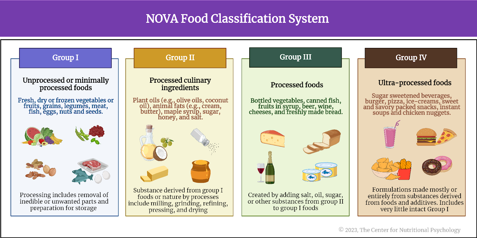
Figure 1. Nova food classification system and examples of foods in each class


Why are ultra-processed foods special?
Ultra-processed foods (UPFs) are heavily processed before consumption. They typically contain numerous artificial additives, preservatives, and flavor enhancers. These additives include dyes, color stabilizers, non-sugar sweeteners, de-foaming, anti-caking or glazing agents, emulsifiers, humectants, and many others. Some processes used in preparing these foods, such as hydrogenation, hydrolyzation, or extrusion, are exclusively industrial processes that cannot be performed in a regular kitchen (see Figure 2).


%learn about nutrition mental health %The Center for Nutritional Psychology

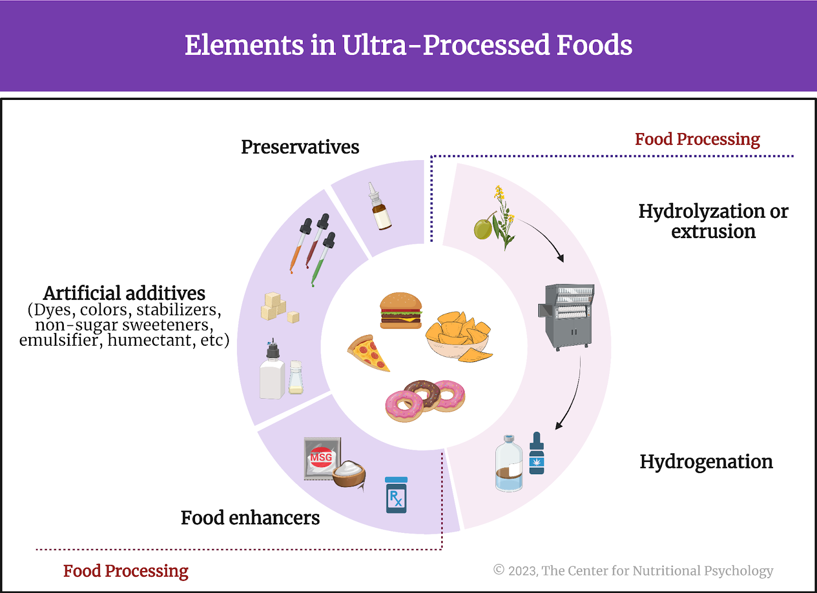
Figure 2. Elements in ultra-processed foods


Ultra-processed foods are often low in nutritional value, high in calories, and packed with unhealthy ingredients like refined sugars, trans fats, and excessive sodium. Examples of ultra-processed foods include instant noodles, artificial sweeteners, artificially sweetened beverages, sugary cereals, microwaveable meals, reconstituted meat products, sweet and savory packaged snacks, pre-prepared frozen dishes, and soft drinks. The overall purpose of ultra-processing is to create a durable product that is highly palatable but also highly profitable due to the low cost of ingredients. Ultra-processed foods usually have attractive packaging and are marketed intensely (see Figure 3).


%learn about nutrition mental health %The Center for Nutritional Psychology

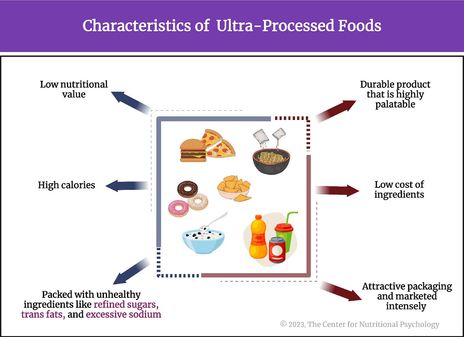
Figure 3. Characteristics of ultra-processed foods


Ultra-processed foods and health risks
One of the concerning aspects of ultra-processed foods is their link to various health problems. Studies have linked the regular consumption of these foods with a higher risk of obesity, type 2 diabetes, heart disease, and certain types of cancer. Ultra-processed foods often lack essential nutrients like fiber, vitamins, and minerals important for overall health.

Despite these health risks, over half of the dietary energy consumed by individuals from high-income countries such as the USA, Canada, or the UK comes from ultra-processed foods. In middle-income countries such as Brazil, Mexico, and Chile, the share of dietary calories from ultra-processed foods is estimated to be between 20% and 33%. Additionally, the sales of these foods are growing by about 1% per year in high-income countries and up to 10% per year in middle-income countries (Monteiro et al., 2019) (see Figure 4).


%learn about nutrition mental health %The Center for Nutritional Psychology

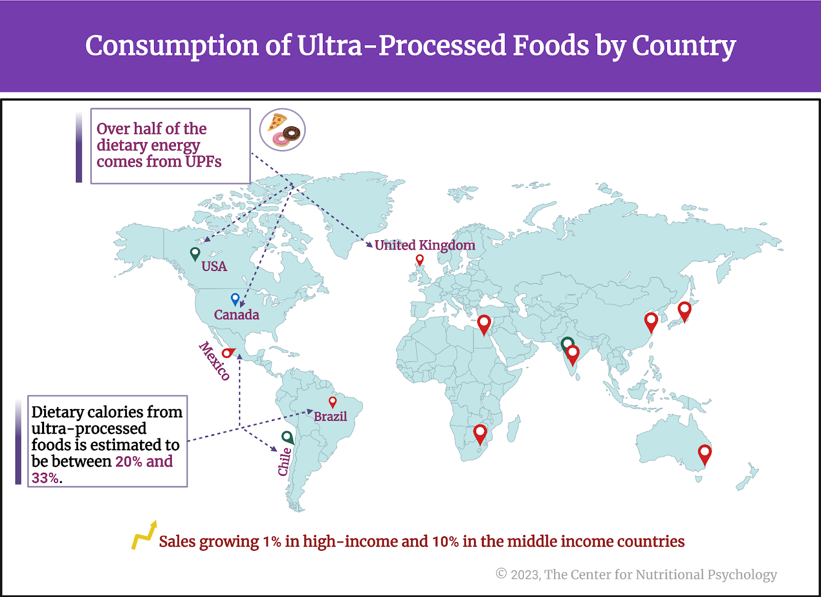
Figure 4. Consumption of UPFs by Country. 


Over half of the dietary energy consumed by individuals from high-income countries such as the USA, Canada, or the UK comes from ultra-processed foods


The current study
Study author Chatpol Samuthpongtorn and his colleagues wanted to examine whether consuming ultra-processed foods might be associated with the risk of depression. They were aware of findings linking the consumption of ultra-processed foods with many different diseases. Still, they noted that many studies that reported these findings relied only on short-term dietary data and could not account for various factors that could have influenced the results. They also wanted to know if any specific ultra-processed foods are linked to depression and whether the timing of ultra-processed food consumption might play a role.

The study participants were 31,712 females in the Nurses’ Health Study II conducted between 2003 and 2017.  Participants were between 42 and 62 years old at the start of the study.


Are any specific ultra-processed foods linked to depression, and does the timing of ultra-processed food consumption play a role?


The procedure
Participants completed a food frequency questionnaire based on the NOVA classification every four years. This questionnaire asked participants to report how often they consumed various foods from the four NOVA categories. The part about ultra-processed foods included questions about the frequencies of consuming ultra-processed grain foods, sweet snacks, ready-to-eat meals, fats and sauces, ultra-processed dairy products, savory snacks, processed meat, beverages, and artificial sweeteners. 

Participants reported whether they were diagnosed with depression and whether they consumed antidepressants regularly. Researchers also collected data on participants’ ages, total caloric intake, body mass index, physical activity, smoking status, menopausal hormone therapy, alcohol consumption, chronic diseases, marital status, family income, and other characteristics (see Figure 5).


%learn about nutrition mental health %The Center for Nutritional Psychology

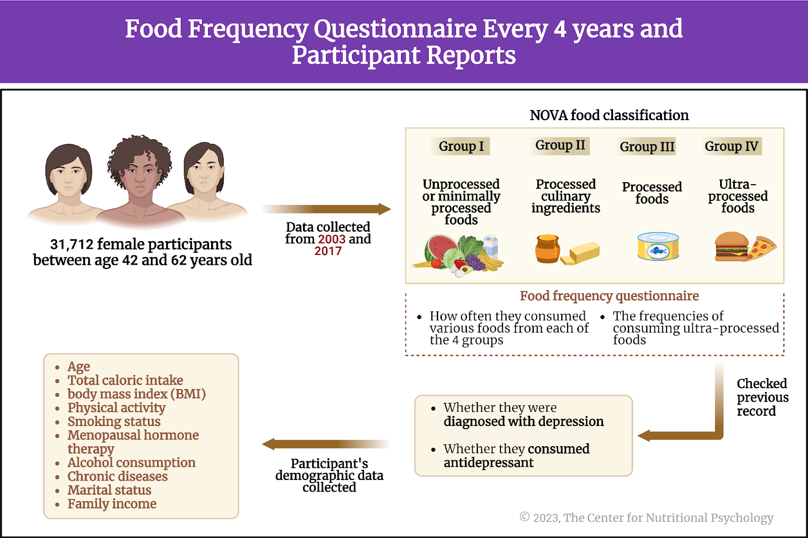
Figure 5. Food Frequency Questionnaire (FFQ) every four years, and participant reports 


Individuals eating more ultra-processed foods had higher body weight, smoked more, and were in poorer health


Participants who reported eating ultra-processed foods most frequently had greater body mass index, smoked more often, and suffered from diseases like diabetes, hypertension, and dyslipidemia compared to those who reported eating ultra-processed foods less often. These individuals were also less likely to exercise regularly. 

Dyslipidemia is a medical condition characterized by abnormal levels of lipids (fats) in the blood, typically involving elevated cholesterol and/or triglycerides, which can increase the risk of cardiovascular diseases.

Individuals frequently consuming ultra-processed foods were more often depressive
Depending on the definition of depression used, there were 2122 (using the stricter definition) or 4840 (using the broader definition) participants suffering from depression in the study. In both cases, participants who ate ultra-processed foods most frequently (the top 20% of the sample with the highest ultra-processed food consumption) were more likely to suffer from depression compared to those who ate the ultra-processed foods the least often (the bottom 20% on ultra-processed food consumption). 


Participants who ate ultra-processed foods most frequently were more likely to suffer from depression than those who ate ultra-processed foods least often


Depending on the definition of depression, the top consumers of ultra-processed foods had a 34% or 49% higher likelihood of depression compared to participants who consumed these types of food the least. This finding held when study authors considered various other factors and calculated the association between ultra-processed food consumption at one point and depression four years later (see Figure 6).


%learn about nutrition mental health %The Center for Nutritional Psychology

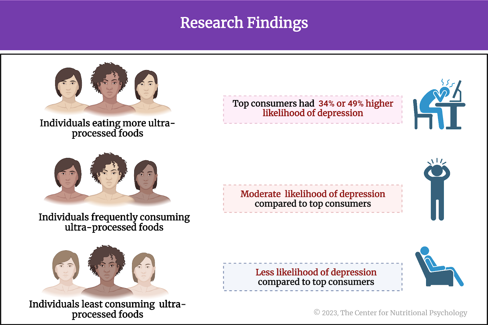
Figure 6. Research findings


The top consumers of ultra-processed foods had a higher likelihood of depression than participants who consumed these types of food the least


Sweetened beverages and artificial sweeteners are the ultra-processed foods most strongly linked with depression
Next, the study authors explored which ultra-processed foods are most linked with depression. Are all types of ultra-processed foods equally associated with depression, or is the high consumption of some specific types of ultra-processed foods linked with a higher likelihood of depression?

Analyses showed that only high consumption of artificially sweetened beverages and artificial sweeteners is associated with an increased likelihood of depression. These findings, however, don’t necessarily imply that all other UPFs consumed in the longitudinal study are exempt from having undesired effects on psychological well-being.


Analyses showed that only high consumption of artificially sweetened beverages and artificial sweeteners is associated with an increased likelihood of depression


Additional analyses indicated that individuals who reduced their intake of ultra-processed foods by at least three servings per day were at a lower risk of depression compared to individuals with a relatively stable intake of these foods in each four-year period.

Conclusion
Overall, the study results suggest that frequent intake of ultra-processed foods is associated with an increased risk of depression. This is particularly the case with high intake of artificially sweetened beverages and artificial sweeteners. While the nature of this association remains unknown, the large number of participants involved in this study and the fact that the association held across multiple years makes these findings particularly strong.

Given the current increase in the number of people suffering from depression in many world countries (e.g., Steffen et al., 2020; Weinberger et al., 2018) and also the increase in ultra-processed foods consumption (Monteiro et al., 2019; Samuthpongtorn et al., 2023), these findings could be used to inform policymakers and the general public about the importance of healthy food choices and how seemingly innocuous dietary decisions may have long-lasting health consequences.

The paper “Consumption of Ultraprocessed Food and Risk of Depression” was authored by Chatpol Samuthpongtorn, Long H. Nguyen, Olivia I. Okereke, Dong D. Wang, Mingyang Song, Andrew T. Chan, and Raaj S. Mehta.


References
Monteiro, C. A., Cannon, G., Levy, R. B., Moubarac, J. C., Louzada, M. L. C., Rauber, F., Khandpur, N., Cediel, G., Neri, D., Martinez-Steele, E., Baraldi, L. G., & Jaime, P. C. (2019). Ultra-processed foods: What they are and how to identify them. In Public Health Nutrition (Vol. 22, Issue 5, pp. 936–941). Cambridge University Press. https://doi.org/10.1017/S1368980018003762

Monteiro, C. A., Cannon, G., Moubarac, J. C., Levy, R. B., Louzada, M. L. C., & Jaime, P. C. (2018). The un Decade of Nutrition, the NOVA food classification and the trouble with ultra-processing. Public Health Nutrition, 21(1), 5–17. https://doi.org/10.1017/S1368980017000234

Samuthpongtorn, C., Nguyen, L. H., Okereke, O. I., Wang, D. D., Song, M., Chan, A. T., & Mehta, R. S. (2023). Consumption of Ultraprocessed Food and Risk of Depression. JAMA Network Open, 6(9), e2334770. https://doi.org/10.1001/jamanetworkopen.2023.34770

Steffen, A., Thom, J., Jacobi, F., Holstiege, J., & Bätzing, J. (2020). Trends in the prevalence of depression in Germany between 2009 and 2017 based on nationwide ambulatory claims data. Journal of Affective Disorders, 271, 239–247. https://doi.org/10.1016/J.JAD.2020.03.082

Weinberger, A. H., Gbedemah, M., Martinez, A. M., Nash, D., Galea, S., & Goodwin, R. D. (2018). Trends in depression prevalence in the USA from 2005 to 2015: widening disparities in vulnerable groups. Psychological Medicine, 48(8), 1308–1315. https://doi.org/10.1017/S0033291717002781


Gut Microbiota Play Crucial Role in Mediating Effects of Western Diet

Introduction

The past several decades have seen the rise of an obesity pandemic that is ongoing worldwide. While obese individuals were quite rare just a century ago, 2015-2018 estimates for the U.S. state that more than two-thirds of the adult population is overweight or obese (Wong et al., 2022). Determining the causes of this increase in obesity rates has attracted much research attention. Studies have revealed a complex interplay between diet components, environmental factors, and previously unknown psychological and physiological mechanisms resulting in overeating and obesity in the long term. These novel studies on the intersection of nutrition and psychology are part of a developing field of science called nutritional psychology (The Center for Nutritional Psychology, 2023) (see Figure 1).


%learn about nutrition mental health %The Center for Nutritional Psychology

Figure 1. Diet, environment, psychological, and physiological factors in nutritional psychology 


There is a complex interplay between diet, environmental factors, and psychological and physiological mechanisms resulting in overeating and obesity


Gut microbiota and the microbiota-gut-brain axis

The human gut microbiome consists of trillions of microorganisms that live in the human intestinal tract. These microorganisms play a key role in digesting the food we eat. However, their influence extends beyond the gut, encompassing crucial roles in metabolic regulation, body weight maintenance, and immune system modulation. 

This growing body of evidence suggests that these gut microorganisms also profoundly impact brain functions, mood, cognition, and emotional well-being  (Zhu et al., 2023). This topic is explored in continuing education curricula within nutritional psychology — particularly how the gut microbiota and the gut-brain axis interconnect with the diet-mental health relationship to influence psychological functioning and experience, shedding light on its potential therapeutic implications for mental health outcomes.


This growing body of evidence suggests that gut microorganisms profoundly impact brain functions, mood, cognition, and emotional well-being


Scientists have recently discovered a communication pathway connecting the gut microbiome and the brain. This pathway is called the microbiota-gut-brain axis. It is based on small proteins called cytokines and a number of other biomolecules, including the hormone cortisol, short-chain fatty acids (SCFAs), tryptophan, and others.

The Western diet

The Western diet is a modern dietary pattern prevalent in Western societies, characterized by a high intake of processed and hyperpalatable foods with increased contents of fat, sugary snacks, and refined grains. It typically includes low consumption of fruits, vegetables, unprocessed-high-quality proteins, nuts, and seeds. This diet’s excessive reliance on added sugars and unhealthy fats has been linked to an increased risk of obesity, metabolic syndrome, and various chronic diseases.

Studies have indicated that feeding mice a Western diet causes inflammation in the region of the brain called the hypothalamus (Heiss et al., 2021; Thaler et al., 2013). Inflammation of the hypothalamus damages the neurons and leads to the formation of scars made of glial cells. This is called gliosis. Inflammation of the hypothalamus often happens before a mouse starts gaining weight. Due to this, scientists believe it might cause weight gain by causing leptin resistance.


Studies have indicated that feeding mice a Western diet causes inflammation of the region of the brain called the hypothalamus


Leptin and leptin resistance

Leptin is a hormone produced by fat cells during eating. It regulates appetite and body weight and is produced in proportion to the amount of fat in the body. Leptin concentrations inform the brain of how much fat is stored. Increased leptin concentrations (normally caused by an abundance of body fat) “tell” the brain to decrease food intake and increase energy expenditure.


Leptin is a hormone produced by fat cells that regulate appetite and body weight


Factors such as chronic inflammation or eating high-fat diets (HFDs) may cause the body to be less receptive to leptin. This is called leptin resistance. Leptin resistance results in disrupted appetite and energy regulation, i.e., the brain does not reduce food intake in spite of the abundance of body fat. This can contribute to obesity and cause difficulty controlling body weight (Thaler et al., 2013) (see Figure 2).


%learn about nutrition mental health %The Center for Nutritional Psychology

Figure 2. Normal leptin cycle versus leptin resistance.


Gliosis, leptin resistance, and gut microbiota

Microglia are a type of immune cell in the central nervous system that helps protect and maintain the brain and spinal cord by detecting and responding to potential threats or damage. Studies have shown that activation of microglia cells that happens during inflammation of the hypothalamus might be causing leptin resistance. Removing these microglia cells from the hypothalamus has improved sensitivity to leptin. Improved sensitivity to leptin allows the brain to recognize when enough fat is stored in the body and reduce food intake. 


Studies have shown that the activation of microglia cells that happens during inflammation of the hypothalamus might be causing leptin resistance


Intriguingly, according to the scientific evidence presented in our recent NP 120 course, it has been discovered that the gut microbiota plays a significant role in regulating the development and maturation of microglia cells and influencing their function. Although the mechanism of this action remains unknown, it has led scientists to believe there might be a link between hypothalamus inflammation and gut microbiota (Heiss et al., 2021) (see Figure 3).


%learn about nutrition mental health %The Center for Nutritional Psychology

Figure 3. Link of Gut Microbiota in regulating development and maturation of microglia cells


There might be a link between hypothalamus inflammation and gut microbiota (Heiss et al., 2021)


The current study

Study author Christina N. Heiss and her colleagues wanted to know whether mice that lack gut microbiota are protected against diet-induced inflammation of the hypothalamus. They noted previous studies’ results showing mice with depleted gut microbiota and those treated with antibiotics to be protected from diet-induced obesity.

In other words, they noted that mice that consume a Western diet, a diet that normally leads to obesity in mice, do not become obese if microbiota are not present in their guts. This might be because the absence of microbiota prevents inflammation of the hypothalamus, which would, in turn, prevent leptin resistance from developing. If this is the case, the mechanism for preventing overeating based on leptin would remain intact, preventing mice from becoming obese. Alternatively, it could be that, without microbiota, the guts of mice could not digest complex nutrients from the food they eat, thus substantially reducing the amount of nutrition they can derive from food. In this case, obesity would be avoided because their bodies cannot use their food. But which of these is the case?

The procedure

The study authors used three groups of male mice, 10-13 weeks old – conventionally raised mice, germ-free mice, and antibiotic-fed mice. They used several genetic groups of mice, including a strain of genetically engineered mice that allow for controlled and regulated manipulation of specific genes in specific tissues (Tamoxifen-inducible Cre mice).

In the scope of the experiments, researchers fed mice either a chow diet or a Western diet. Western diet was given for either 2 days, 1 week, or 4 weeks, depending on the experiment conducted in the scope of the study.

The chow diet for mice is a nutritionally balanced and standardized diet formulated to provide essential nutrients required for the health and growth of laboratory mice. It typically consists of a combination of proteins, carbohydrates, fats, vitamins, and minerals in pellet or block form. The Western diet used in this study was high in fat and sucrose, with 40% of calories coming from fat.

All food for mice was sterilized, i.e., underwent procedures that killed all microorganisms in the food before mice ate it. The chow diet was sterilized in an autoclave, which uses high pressure and steam to kill microorganisms. The Western diet food was irradiated, i.e., radiation was used to kill microorganisms.

The mice

Conventionally raised mice were laboratory mice kept in regular conditions and fed a normal diet for laboratory mice. They have normal gut microbiota.

Germ-free mice are created through techniques that ensure they do not acquire gut microbiota from birth through their entire lifetimes. They are typically born using cesarean section deliveries of pregnant mice in a sterile environment. This is done to prevent the transfer of microbes during birth. After that, they are kept in specialized sterile isolation spaces called isolators or bubbles that maintain a controlled germ-free environment.

These isolators provide filtered air, sterile food, and autoclaved water to prevent microbial contamination. Researchers raising these mice take special care to maintain strict barrier measures, including specialized clothing and equipment, to prevent the unintentional introduction of microorganisms. They regularly monitor these mice’s bodily fluids and tissues through special techniques to ensure the absence of any detectable microorganisms. Germ-free mice are typically leaner than conventionally raised mice and, consequently, have lower leptin levels in circulation.

Antibiotic-fed mice in this study had 1g of ampicillin and 0.5g of neomycin added per liter of their drinking water. Ampicillin and neomycin are antibiotics. Researchers kept the drinking water with antibiotics added in bottles protected from light. They prepared a new solution every second day. Researchers started giving this water with antibiotics to mice three days before changing their regular diet to a Western diet.


Mice without gut microbiota are protected from diet-induced inflammation of the hypothalamus


Results showed that conventionally raised mice fed a Western diet for 1 week developed gliosis in the hypothalamus. Inflammation indicators were increased in these mice in the part of the hypothalamus called the arcuate nucleus compared to conventionally raised mice fed regular mice food (chow) (see Figure 4).


%learn about nutrition mental health %The Center for Nutritional Psychology

Figure 4. Conventionally raised mice fed a Western diet developed gliosis in the hypothalamus


When researchers examined germ-free mice and mice whose gut microbiota were depleted using antibiotic treatment (antibiotic-fed mice), results showed that these mice also showed no increase in inflammation indicators or the proliferation of microglia cells after eating a Western diet for a week.


Gliosis in the hypothalamus leads to greater gain in body weight and fat mass


The study authors also wanted to know whether gliosis in the hypothalamus caused by a Western diet leads, in turn, to increased body weight and fat mass accumulation in mice. To test that, they fed conventionally raised mice and antibiotic-fed mice a Western diet for 4 weeks.

Results showed that conventionally raised mice fed a Western diet gained more body weight and fat mass than antibiotic-fed mice. Compared to antibiotic-fed mice, they also had increased hypothalamus inflammation indicators and increased numbers of a specific type of microglia cells (iba1-positive microglia).

There was no association between the number of microglia in the arcuate nucleus region of the hypothalamus and changes in body weight or fat mass at the end of the 4 weeks. However, fat mass and the relative increase in fat mass during the study were associated with the number of a specific type of glial cells called astrocytes.

Further analysis showed that germ-free and antibiotic-fed mice are more sensitive to leptin than conventionally raised mice. When researchers gave them leptin injections, the first two types of mice reduced their food intake more than conventionally raised mice did.


Glucagon-like-peptide 1 (GLP-1) seems to be crucial for protection against diet-induced inflammation of the hypothalamus


Germ-free and antibiotic-fed mice had higher levels of the hormone called glucagon-like peptide 1 (GLP-1) when they were fed a regular diet. This hormone secreted in the small intestine’s intestinal lining cells (L cells) is important in regulating blood sugar levels. It also helps reduce inflammation and protect neurons.

Study authors believed it might also be crucial for the protection from inflammation of the hypothalamus induced by the Western diet. After mice were fed a Western diet for a week, antibiotic-treated and germ-free mice had higher levels of GLP-1 than conventionally raised mice. These mice did not gain weight or develop hypothalamic inflammation after this diet. However, when researchers measured these same things in antibiotic-fed and germ-free mice whose GLP-1 signaling pathway was disabled, they also gained weight and developed inflammation, similar to conventionally raised mice. This indicated that the functional signaling path of GLP-1 is crucial for countering the inflammation of the hypothalamus induced by a Western diet.


Just a week on a Western diet led to inflammation of the hypothalamus that, in turn, disrupted the body’s mechanism for regulating food intake


Conclusion

These findings in mice show that gut microbiota changes how the organism, of mice in this case, reacts to a Western diet. When gut microbiota was intact, just a week on a Western diet led to inflammation of the hypothalamus that, in turn, disrupted the body’s mechanism for regulating food intake. However, when gut microbiota was depleted or absent, this inflammation did not happen, provided that the signaling pathway of one specific hormone (GLP-1) was intact.

While the study was done on mice, similar physiological mechanisms exist in humans. Due to this, these findings on mice help scientists better understand how and through which physiological mechanisms changes in the human diet that occurred in the last century disrupted food intake regulation in the human body leading to the current obesity pandemic.

The paper “The gut microbiota regulates hypothalamic inflammation and leptin sensitivity in Western diet-fed mice via a GLP-1R-dependent mechanism” was authored by Christina N. Heiss, Louise Manneras-Holm, Ying Shiuan Lee, Julia Serrano-Lobo, Anna Hakansson Gladh, Randy J. Seeley, Daniel J. Drucker, Fredrik Backhed, and Louise E. Olofsson.

References

Heiss, C. N., Mannerås-Holm, L., Lee, Y. S., Serrano-Lobo, J., Håkansson Gladh, A., Seeley, R. J., Drucker, D. J., Bäckhed, F., & Olofsson, L. E. (2021). The gut microbiota regulates hypothalamic inflammation and leptin sensitivity in Western diet-fed mice via a GLP-1R-dependent mechanism. Cell Reports, 35(8). https://doi.org/10.1016/j.celrep.2021.109163

Thaler, J. P., Guyenet, S. J., Dorfman, M. D., Wisse, B. E., & Schwartz, M. W. (2013). Hypothalamic inflammation: Marker or mechanism of obesity pathogenesis? Diabetes, 62(8), 2629–2634. https://doi.org/10.2337/DB12-1605

The Center for Nutritional Psychology. (2023). What is Nutritional Psychology? https://www.nutritional-psychology.org/what-is-nutritional-psychology/

Wong, M. C., Mccarthy, C., Fearnbach, N., Yang, S., Shepherd, J., & Heymsfield, S. B. (2022). Emergence of the obesity epidemic: 6-decade visualization with humanoid avatars. The American Journal of Clinical Nutrition, 115(4), 1189–1193. https://doi.org/10.1093/AJCN/NQAC005

Zhu, X., Sakamoto, S., Ishii, C., Smith, M. D., Ito, K., Obayashi, M., Unger, L., Hasegawa, Y., Kurokawa, S., Kishimoto, T., Li, H., Hatano, S., Wang, T. H., Yoshikai, Y., Kano, S. ichi, Fukuda, S., Sanada, K., Calabresi, P. A., & Kamiya, A. (2023). Dectin-1 signaling on colonic γδ T cells promotes psychosocial stress responses. Nature Immunology. https://doi.org/10.1038/s41590-023-01447-8


Food and Mood: Is the Concept of ‘Hangry’ Real?

A recent study conducted in Austria used the method of experience sampling to examine the links between hunger and mood. Researchers asked participants to report their hunger and mood five times per day. Results showed that participants reported greater anger, irritability, and decreased pleasure when hungry (Swami et al., 2022). The study was published in Plos One.


Hunger and satiety exert a very powerful influence on one’s behavior. 


Nutritional psychology, as explored by The Center for Nutritional Psychology, focuses on the links between diet, psychological states, and mental health. While the significance of this research topic is widely acknowledged, it remains a relatively new field of study, with crucial findings likely to emerge in the future.

What is hunger?

From a physiological point of view, hunger is a complex biological process that primarily revolves around regulating blood glucose levels and releasing hormones that control appetite and satiety. When we eat food, our bodies break down carbohydrates into glucose, which is the primary energy source for cells. As we go without food, our blood glucose levels gradually decline.

The two key hormones involved in hunger regulation are ghrelin and leptin. Ghrelin, produced by the stomach, signals the brain to stimulate appetite when the stomach is empty. Before meals, ghrelin levels increase, and they decrease after eating. Leptin functions as an appetite suppressor and is produced by fat cells, so when body fat decreases, leptin levels drop (see Figure 1).


%learn about nutrition mental health %The Center for Nutritional Psychology

Figure 1. Two key hormones involved in hunger regulation are Ghrelin and Leptin


Finally, the brain integrates the input received from hormones and sensory organs (e.g., sight or smell of food) to produce the feeling of hunger or satiety. This integration of signals happens in the hypothalamus region of the brain.

The sensation of hunger

Apart from being the result of a complex physiological process, hunger is also a subjective sensation. This sensation serves to motivate the individual to seek food and ingest it. In turn, this ensures that the nutritional needs of the organism are met (McKiernan et al., 2008). When an individual feels hungry, they become more sensitive to stimuli associated with food (e.g., Lazarus et al., 1953). Individuals will pay increased attention to food items and things they have learned to associate with food (e.g., logos of restaurants or food brands, places and people they have learned to associate with food, etc.).

However, decreased levels of nutrients in the body are not the only thing that can lead to the sensation of hunger. Studies show that humans and other species of animals can eat when bored, desire sensory stimulation (McKiernan et al., 2008), or are under stress (Levine & Morley, 1981). They also learn to expect food at certain times or at certain places. Studies indicate that under regular circumstances, human daily rhythms of biological processes (circadian rhythms) are synchronized with a pattern of three meals per day. However, the body can also adapt and learn to expect meals at different times of the day. This expectation can trigger hunger at a particular time and various physiological processes preparing the body for food intake (Isherwood et al., 2023) (see Figure 2).


%learn about nutrition mental health %The Center for Nutritional Psychology

Figure 2. Some sources of hunger


Being “hangry” – hungry and angry

Hunger affects the behavior of both humans and animals. Observations of non-human animals show that food deprivation increases their motivation to engage in escalated and persistent aggression to acquire food. It is well-known among keepers of various animals that the animals are the most dangerous when hungry.

Studies in humans link the sensation of hunger with feelings of restlessness, nervousness, irritability, and behavioral difficulties in children. Low blood glucose levels, known to trigger the sensation of hunger, are associated with increased impulsivity, anger, and aggression (Swami et al., 2022). Studies applying the concept of ego depletion suggest that the human capacity for self-regulation and active volition is limited (Baumeister et al., 1998) and that when one is hungry, negative, high-arousal emotions are more likely to occur. This is because individuals may struggle to exercise self-regulation and self-control when their blood glucose is low (Swami et al., 2022).


Studies in humans link the sensation of hunger with feelings of restlessness, nervousness, irritability, and behavioral difficulties in children.


These findings and casual observations have given rise to the term “hangry.” A combination of “hungry” and “angry,” hangry signifies a state in which one experiences both hunger and anger due to hunger.


Coined from hungry and angry, the term “hangry” indicates a state in which one is hungry but is also angry because of hunger.


The current study

Viren Swami, the study author, and his colleagues aimed to investigate the state of being “hangry” in a natural setting more systematically.

They wanted to know the extent to which daily experiences of hunger are associated with negative emotional outcomes. They reasoned that if the sensation of hunger is indeed linked to anger and other negative emotions, this must be the case in everyday life and not only in laboratory experiments.

To examine this link, they conducted an experience sampling study in which a group of study participants reported on their daily experiences of hunger and anger over a 3-week period. Understanding that anger might not be the only emotion connected to hunger, the study authors asked participants to report on irritability, pleasure, and arousal. They expected that hunger would be associated with greater anger, irritability, and arousal but lower feelings of pleasure.

What is experience sampling?

Experience sampling (Figure 3), or ecological momentary assessment, involves collecting real-time participant data throughout their daily lives. Participants are prompted multiple times daily to report on their experiences, emotions, behaviors, or thoughts using electronic devices such as smartphones or specialized wearable devices. This method gives researchers insights into individuals’ subjective experiences in their natural settings. This approach minimizes issues related to recalling events after much time has passed, a common problem encountered in traditional research methods relying on recalling past events. Experience sampling allows for a more nuanced understanding of how various factors fluctuate over time and under different circumstances.


%learn about nutrition mental health %The Center for Nutritional Psychology

Figure 3. Experience sampling process 


The procedure

The study initially involved 121 participants; however, it required them to respond to surveys five times daily, every day, for three weeks, resulting in a total of 105 surveys to be completed by each participant. This workload proved overwhelming for many, with only 39 participants successfully completing all 105 surveys. Seventy-six participants managed to complete at least one survey per day.

Participants were, on average, 30 years old. Their ages ranged between 18 and 60 years. 69% were from Austria, followed by Germany (20%), Switzerland, and other countries. The vast majority of the participants were women (81%). 44% lived alone, 19% were married, and 36% were in a relationship. They had an average of 14 years of education.

Daily surveys

In the survey, participants indicated on a scale of 0 to 100 how hungry they were (“How hungry are you at the moment?”), their irritability (“How irritable do you feel at the moment?”), and how angry they felt at that moment. In addition, they rated their emotional state (“How pleasant do you find your current state?”), and arousal level (“What is your current arousal level?”). They also reported the time since their last meal (“When was your last meal? [______ hours ago]”.

Participants completed the surveys using their smartphones through the ESMira software package. After installing ESMira and registering for participation in the study, participants provided their demographic data. Of the five daily surveys, study authors set three to be at fixed time points during the day, before the three main meals – at 8:00, 12:00, and 18:00. At these time points, participants received an in-app notification to complete the survey. The remaining two surveys were random, one between 9:00 and 11:00 in the morning and the other between 13:00 and 17:00 (in the afternoon) (see Figure 4).


%learn about nutrition mental health %The Center for Nutritional Psychology

Figure 4. Daily survey procedure


Other measures

After the three-week period of the study, participants completed the final questionnaire, in which they provided their demographic data once again. It also contained assessments of four aspects of dietary behavior – restrictive eating (e.g., “I consciously eat less so as not to gain weight”), two aspects of emotionally-induced eating behavior, with clear emotions (e.g., “When I am irritated, I have the desire to eat”) and unclear emotions (e.g., “I always want to eat something when I have nothing to do”), and externally determined eating behavior (e.g., “I eat more than usual when I see others eating”).

The final questionnaire also contained assessments of dispositional anger (the Anger subscale from the Buss and Perry Aggression Questionnaire, BPAQ) and eating motivation (the Eating Motivation Survey consists of the question “Why do you eat what you eat?” followed by 15 different motivations that the participant has to rate).

Participants rarely skipped dinner

Analysis of the responses to the final questionnaire (see Figure 5) showed that some participants often skipped meals, but not all equally. 58% reported that they usually have breakfast, 78% usually had lunch, and 84% usually had dinner. 48% snacked between main meals. 9% reported getting up at night to eat. 88% declared that their eating habits during the study were the same as usual.

53% of the participants paid close attention to maintaining a healthy diet, either very often or always, and 55% paid attention to the sensation of hunger. The main motivation for eating was hunger and because participants liked the meal. 23% of participants stated they knew when they were full and then stopped eating. 63% reported that from time to time, they would continue to eat even though they were aware that they were full. 13% said they eat when stressed, upset, angry, or bored. Less than 5% reported that they do not feel when they are full and that they orient themselves based on the size of the meal.


%learn about nutrition mental health %The Center for Nutritional Psychology

Figure 5. Survey Report


When hungry, participants were more often angry, irritable and felt less pleasure

The results demonstrated a strong association between hunger and heightened feelings of anger, irritability, and reduced pleasure. Surprisingly, researchers also observed that participants with higher levels of dispositional anger tended to report higher levels of hunger. 

Dispositional anger is a general tendency of an individual to experience anger more frequently and intensely across various situations. In contrast, the feeling of anger refers to a temporary and transient experience of anger. 

The link of hunger with irritability, anger, and lower feeling of pleasure persisted even after participants’ sex, age, body mass index, dietary behavior, and dispositional anger were considered (see Figure 6).


%learn about nutrition mental health %The Center for Nutritional Psychology

Figure 6. Hunger and emotions


Contrary to the study authors’ expectations, hunger was not associated with arousal.

Conclusion

Overall, the study suggests that the experience of being hangry is real. When study participants were hungry, they also tended to experience greater anger, irritability, and less pleasure. This happened in their natural environments while they were living their lives as usual, not in an artificial laboratory environment. It was also not a one-time occurrence – the findings resulted from 105 surveys taken five times daily across three weeks.

These results have important implications for understanding everyday experiences of emotions. They also help practitioners to effectively prevent interpersonal conflicts while ensuring productive behaviors and good relationships. Although the design of this study does not allow for cause-and-effect conclusions to be made, i.e., to conclude that hunger causes anger or vice versa, simply designing school or work schedules to ensure no one goes hungry could help prevent numerous interpersonal problems.  Allocating sufficient time and opportunities to eat and reduce prolonged periods of hunger is an important tool for improving everyone’s well-being. 

On an individual level, labeling one’s affective state as being “hangry” could allow individuals to make sense of that experience. This is very important as, unlike some other negative states, hunger can be easily resolved by simply eating something.

The paper “Hangry in the field: An experience sampling study on the impact of hunger on anger, irritability, and affect” was authored by Viren Swami, Samantha Hochstoeger, Erik Kargl, and Stefan Stieger.


References

Baumeister, R. F., Bratslavsky, E., Muraven, M., & Tice, D. M. (1998). Ego depletion: Is the active self a limited resource? Journal of Personality and Social Psychology, 74(5), 1252–1265. https://doi.org/10.1037/0022-3514.74.5.1252

Isherwood, C. M., van der Veen, D. R., Hassanin, H., Skene, D. J., & Johnston, J. D. (2023). Human glucose rhythms and subjective hunger anticipate meal timing. Current Biology, 33(7), 1321-1326.e3. https://doi.org/10.1016/j.cub.2023.02.005

Lazarus, R. S., Yousem, H., & Arenberg, D. (1953). Hunger and Perception. Journal of Personality, 21(3), 312–328. https://doi.org/10.1111/J.1467-6494.1953.TB01774.X

Levine, A. S., & Morley, J. E. (1981). Stress-induced eating in rats. American Journal of Physiology – Regulatory, Integrative and Comparative Physiology, 241(1), R72–R76.

McKiernan, F., Houchins, J. A., & Mattes, R. D. (2008). Relationships between human thirst, hunger, drinking, and feeding. Physiology & Behavior, 94(5), 700. https://doi.org/10.1016/J.PHYSBEH.2008.04.007

Swami, V., Hochstöger, S., Kargl, E., & Stieger, S. (2022). Hangry in the field: An experience sampling study on the impact of hunger on anger, irritability, and affect. PLOS ONE, 17(7), e0269629. https://doi.org/10.1371/JOURNAL.PONE.0269629

The Center for Nutritional Psychology. (2023). What is Nutritional Psychology? https://www.nutritional-psychology.org/what-is-nutritional-psychology/


Is Food Tastier When Consumed in Aesthetically Pleasing Environments?

Editor’s Note: The study of nutritional psychology (NP) involves exploring the influence of diet on psychological, cognitive, behavioral, perceptual, interoceptive, and psychosocial functioning and mental health. It also includes the reverse —which is exploring how our psychological, behavioral, and psychosocial states and mental health influence our dietary intake. 

In fact, the study of NP involves exploring and asking questions about the many aspects of the diet-mental health relationship (DMHR). For example, how does our dietary intake pattern affect how we perform tasks? (aka the “Diet-Performance Relationship”). And, how does the look or smell of food (food aesthetic qualities) influence our desire to eat it? (i.e., the “Diet-Sensory-Perceptual Relationship”).

In this CNP Article, we explore how our environment influences our eating. In particular, we learn about how the aesthetics of an environment can influence our perception and desire to eat the food served in the environment. In nutritional psychology, we call this the Diet-Environment Relationship—the ‘DER.’

Introduction

Although food trucks or “dives” often serve great food that can rival good restaurants in taste, the environment in which foods are consumed can affect people’s perceptions of the food. In this study by Wu et al. (2022), two experiments conducted in China reported that food served in an aesthetically pleasing environment was found to smell better, taste better, and look better than in a less aesthetically pleasing environment. One experiment reported these results using photographs of places and foods, and another confirmed them with actual food. Participants found food they consumed in a nicely decorated room better smelling and tasting than in a less aesthetically pleasing room. They also expressed a greater desire to eat again in a nicer environment (Wu et al., 2022). The study was published in Appetite.


Participants found the food they consumed in a nicely decorated room better smelling and tasting than in a less aesthetically pleasing room. 


Food is much more than a collection of nutrients

Humans need food to survive. Food provides human bodies with the energy needed to carry out various functions, such as breathing, circulating blood, and maintaining body temperature. Food is also a source of essential nutrients, such as vitamins and minerals, that our bodies need to function properly.


Food is much more than a collection of nutrients.


However, the value of food does not stop there. Food also provides us with pleasure and enjoyment. Eating is often a social and cultural activity that brings people together and provides comfort and satisfaction. Sharing meals with family and friends is a common way to strengthen relationships and build connections with others.

This sharing of meals with others is a focal point of various cultural customs worldwide. Dining rooms in people’s homes, restaurants, taverns, cafeterias, diners, barbecue parties, and other similar dedicated places and types of social events exist because food consumption and meal sharing are central parts of human culture. Their value goes far beyond the nutritional function of food.

Even in activities like space exploration missions, researchers soon realized that providing nutritious food is not enough and that food must also provide psychosocial comfort. “Ideal food cannot ensure psychosocial comfort, while a grandma-style pie can,” experts in the area wrote (Bychkov et al., 2021).


“Ideal food cannot ensure psychosocial comfort, while a grandma-style pie can,” experts in the area wrote (Bychkov et al., 2021).


The importance of beauty and aesthetics

Beauty and aesthetics, in general, are important to us. We are naturally drawn to things that are visually appealing or pleasing to our senses, and experiencing beauty can give us a sense of pleasure, satisfaction, and joy. This can help reduce stress and anxiety, improve our mood, and enhance our overall well-being.

For example, a recent study has shown that people become happier when they view beautiful images of nature, thus improving their subjective well-being (Xie et al., 2022). Another study has shown that women prefer more attractive men as long-term partners over men with favorable personal traits, despite consciously considering personal traits like ambition and intelligence more important for a partner to have (Li et al., 2023).

Aesthetic preferences (i.e., what one considers beautiful) are so important to us that individuals consider them part of their personal identity. A recent study has shown that when a person’s aesthetic preferences, such as preferences for music or art, change, that person tends to consider that his/her entire identity has changed. Researchers are now talking about an aesthetic self, an aspect of the human person that they believe to be at least as important for our identity as our moral values are (Fingerhut et al., 2021).


Researchers are now talking about an aesthetic self, an aspect of the human person that they believe to be at least as important for our identity as our moral values are.


The current study

Study author Chenjing Wu from the South China Normal University and her colleagues wanted to know whether the beauty of the environment affects the perception of the food we eat. They notice that, in everyday life, people often choose restaurants because of how they look, i.e., because they like the restaurant environment despite there being restaurants with tastier food. The reverse is also the case in that people can avoid particularly bad-looking restaurants even when such restaurants serve tasty food.


People often choose restaurants because of how they look, i.e., because they like the restaurant environment despite there being restaurants with tastier food.


It is well-known that individuals use data about their environment to form judgments. For example, studies have shown that the smell of the environment, lighting, and color affect the perception of food. When identical food is served in different places (e.g., laboratory, restaurant, cafeteria), perception of that food can vary across different places. In a well-known study, researchers served the same food to participants in different settings, ranging from 4-star restaurants to an army training camp and a freshman’s buffet.  They found that food received much better evaluation when served in 4-star restaurants than in an army training camp or a freshman’s buffet (Edwards et al., 2003).


The smell of the environment, lighting, and color affect the perception of food. 


Procedure Experiment 1

Participants in the first experiment were 132 college students who were divided into two groups. One group was shown a picture of the interior of a nice-looking restaurant (high aesthetic value condition). In contrast, the other was shown a picture of the interior of a poor-looking restaurant (low aesthetic value condition). They were asked to imagine being in that place and rate their emotions about it on a scale from negative to positive, as shown in Figure 1.

  %learn about nutrition mental health %The Center for Nutritional Psychology Figure 1. Experiment 1


Participants were then shown pictures of 29 different foods. They were asked to rate the beauty of the food based on its looks, expected smell, and expected taste. The task was in Chinese, in which sentence constructions with the word beautiful typically describe both look, smell, and taste. So, although the request looks unusual in English, it was appropriate in the Chinese language. Participants were also asked to rate the desire to eat that food item and to evaluate the aesthetic value of the environment. 

Researchers ran one more variant of this experiment on another group of 149 students. In this variant,  instead of pictures of restaurant interiors, one group of participants was shown a picture of a beautiful natural landscape (high aesthetic value condition). At the same time, the other viewed a picture of the interior of a ruined building (low aesthetic value condition) (see Figure 2).

sintomasdelsida.org style=”text-align: center;”>  %learn about nutrition mental health %The Center for Nutritional Psychology

Figure 2. Natural landscape (high aesthetic value) vs. ruined building (low aesthetic value)(not the actual pictures used in the experiment)


Experiment 2

Researchers ran the second experiment with real environments and a real food item. The participants were 81 college students. Researchers decorated a table in their laboratory with covers and flowers for a high aesthetic environment. The low aesthetic environment was the same laboratory table without decorations.

Participants entered the laboratory with a decorated or undecorated table in it, depending on the group they were assigned to. They were then asked to report their emotions about the environment, as in experiment 1. Before entering the laboratory, all participants rated their hunger (see Figure 3).


%learn about nutrition mental health %The Center for Nutritional Psychology Figure 3. Experiment 2


They were then set at the table and given a wrapped bar of dark chocolate. Participants first rated the look of the wrapped chocolate bar, its smell, and how much they would like to eat it. They were then instructed to eat the chocolate. Afterward, they rated how tasty it was and how much they would like to eat another one. Finally, they rated the aesthetic value of their environment (a laboratory with a decorated table or a laboratory without decorations).

The food looked better and was expected to smell and taste better in more beautiful environments

In experiment 1, participants who viewed pictures of a more beautiful environment and imagined themselves in it rated the food they saw in the pictures as better looking. They also reported expecting the food to smell and taste better on average compared to the group that evaluated the same food after viewing a less beautiful environment. On average, participants who imagined the more beautiful environment reported a greater desire to eat the food they were presented with.


Participants who imagined the more beautiful environment reported a greater desire to eat the food they were presented with.


The differences in how presented food items were rated were much greater in the second variant of the experiment (a beautiful natural landscape vs. a ruined building) than in the first variant of the experiment (two restaurants, of which one is nicer looking).

Participants who viewed a more beautiful environment also reported feeling better and rated that environment as more aesthetically pleasing. This confirmed the researchers’ expectations about how participants would perceive the environments.

The better the person feels, the better smelling and tasting the food is

In experiment 2, participants from the two groups rated the wrapped chocolate bar as looking equally nice. However, participants who inspected the chocolate bar while sitting at the decorated laboratory table (high aesthetics environment) tended to report that it smelled better than those who were inspecting it while sitting at an undecorated table. Participants sitting at a decorated table also reported a greater desire to eat it.

After eating the chocolate bar, participants from the high aesthetics condition group (i.e., sitting at the decorated table) tended to rate its taste better than participants from the other group. They also expressed a greater desire to eat another chocolate bar.

Differences in ratings provided by the two groups were similar in size to those obtained in the first experiment (two imagined restaurants and imagined food). However, it should be considered that a decorated table in a laboratory is still visibly a table in a laboratory. Hence, the differences between the two environments in experiment 2 were limited.

Further analysis revealed a clear association between the participant’s emotions and the food evaluation. Participants who reported more positive emotions about the environment tended to evaluate the food they were presented with (or asked to imagine) as looking, tasting, and smelling better. They also tended to report a greater desire to eat the food.


Participants who reported more positive emotions about the environment tended to evaluate the food they were presented with as looking, tasting, and smelling better. 


Conclusion

The study showed that the aesthetic value of the environment affects the perception of food. Food is perceived as tastier, better smelling, and, in certain conditions, better looking in more aesthetically pleasing environments that elicit more positive emotions. The desire to eat the food is also greater in a better-smelling environment. This refers to the desire to eat the food at hand again in the same environment.

The implications of the findings are quite straightforward – food offered and sold in more aesthetically pleasing environments will be perceived as better by the consumers. They will also have a greater desire to eat it. The findings imply that restaurant managers should pay great attention to the aesthetic qualities of their customers’ environment, not solely to food preparation.

The paper “Does a beautiful environment make food better – The effect of environmental aesthetics on food perception and eating intention” was authored by Chenjing Wu, Hongyan Zhu, Chuangbing Huang, Xiaoling Liang, Kaili Zhao, Siyue Zhang, Mingcheng He, Wei Zhang, and Xianyou He.

More evidence-based information on the Diet-Environment Relationship can be found in the Nutritional Psychology Research Library (NPRL). The Diet and Sensory Perceptual Relationship (DSPR) included within nutritional psychology also involves studies in this area.


References

Bychkov, A., Reshetnikova, P., Bychkova, E., Podgorbunskikh, E., & Koptev, V. (2021). The current state and future trends of space nutrition from a perspective of astronauts’ physiology. International Journal of Gastronomy and Food Science, 24, 100324. https://doi.org/10.1016/J.IJGFS.2021.100324

Edwards, J. S. A., Meiselman, H. L., Edwards, A., & Lesher, L. (2003). The influence of eating location on the acceptability of identically prepared foods. Food Quality and Preference, 14(8), 647–652. https://doi.org/10.1016/S0950-3293(02)00189-1

Fingerhut, J., Gomez-Lavin, J., Winklmayr, C., & Prinz, J. J. (2021). The Aesthetic Self. The Importance of Aesthetic Taste in Music and Art for Our Perceived Identity. Frontiers in Psychology, 11, 1–18. https://doi.org/10.3389/FPSYG.2020.577703

Li, W., Zhu, H., Zhao, K., Zhu, H., Wang, X., & He, X. (2023). Good performance-high attractiveness effect: an empirical study on the association between athletes’ rankings and their facial attractiveness. International Journal of Sport and Exercise Psychology. https://doi.org/10.1080/1612197X.2023.2181846

Wu, C., Zhu, H., Huang, C., Liang, X., Zhao, K., Zhang, S., He, M., Zhang, W., & He, X. (2022). Does a beautiful environment make food better – The effect of environmental aesthetics on food perception and eating intention. Appetite, 175(April), 106076. https://doi.org/10.1016/j.appet.2022.106076

Xie, R., Qiu, C., & Qiu, G. (2022). Finding Beautiful and Happy Images for Mental Health and Well-Being Applications. Lecture Notes in Computer Science, 13536 LNCS, 704–717. https://doi.org/10.1007/978-3-031-18913-5_54


Neuroimagine Study Identifies Patterns Of Neural Activity In Obese Persons

A neuroimaging study identified neural activity patterns specific to obese persons. These patterns allowed researchers to differentiate between obese and non-obese individuals based on the characteristics of their brain activity and psychological test scores with 77% accuracy. It was also possible to differentiate between male and female obese individuals (75% accuracy), indicating that the cortical mechanisms underlying obesity are unequal (Bhatt et al., 2023). The study was published in Brain Communications.

What is obesity?

Obesity is a medical condition characterized by excess body fat. It is usually defined through a person’s body mass index. Body mass index is a person’s weight in kilograms (or pounds) divided by the square of the person’s height in meters (or feet). Persons whose body mass index (BMI) is between 25 and 29.9 are considered overweight. A body mass index of 30 or over usually indicates obesity.


Body mass index is a person’s weight in pounds divided by the square of the person’s height in feet


Obesity is a major risk factor for developing type 2 diabetes and several other diseases, including heart disease, high blood pressure, and certain types of cancer. Over 42% of adults in the United States are obese. The percentage of obese individuals has increased dramatically in recent decades worldwide (Bhatt et al., 2023; Wong et al., 2022). Existing obesity treatments mostly do not result in lasting weight loss because most patients regain their lost weight within five years (Gearhardt et al., 2011).


Over 42% of adults in the United States are considered obese


Causes and consequences of obesity

Obesity develops when the energy intake of a person’s body exceeds energy expenditure over a prolonged period, leading to body fat accumulation. This can happen due to various causes, including genetics, environmental factors, lifestyle choices, diet, and medical conditions. For example, researchers have identified a rare defect of a single gene that causes severe obesity that develops early in life. This happens due to the deficiency of the hormone leptin (Wilding, 2001). Leptin regulates the balance between food intake and energy expenditure.


Obesity can be caused by altered functioning of certain regions of the brain


 Other studies have indicated that obesity can be caused by altered functioning of certain brain regions. The central nervous system regulates energy intake and expenditure, and the brain’s hypothalamus region plays a vital role in this process. Damage to the hypothalamus caused by tumors or by surgery often results in increased appetite and weight gain (Wilding, 2001).

Food addiction as a cause of obesity

Studies of brain activity patterns in response to food have found these patterns to be similar to those found in drug users reacting to drugs. This made scientists propose food addiction as one of the causes of obesity (Gearhardt et al., 2011).

Food addiction is defined as a constant obsession with what and when to eat and how to obtain more food. This is typically paired with overeating behaviors, hiding, or hoarding food. A food-addicted person feels great pleasure from eating and cannot stop overeating.


A food-addicted person feels great pleasure from eating and is unable to stop overeating


Studies have linked food addiction with altered activation patterns in certain brain areas when food is anticipated. These areas include the anterior cingulate cortex, medial orbitofrontal cortex, and amygdala regions of the brain (Gearhardt et al., 2011) (see Figure 1).

%learn about nutrition mental health %The Center for Nutritional Psychology

       Figure 1. Areas in the brain linked with addiction

Due to this, the study of brain activity and structure may be key to better understanding obesity. One way to approach this is through multimodal magnetic resonance imaging.


Studies have linked food addiction with altered activation patterns in certain brain areas when food is anticipated


What is multimodal magnetic resonance imaging?

Multimodal magnetic resonance imaging (MRI) involves using multiple imaging techniques to create a more comprehensive picture of a certain part of the body, the brain. In multimodal brain MRI, different MRI scans are combined to provide information about the structure, function, and chemistry of examined brain regions.


The current study

The author of this study Ravi R. Bhatt and his colleagues wanted to understand the differences in brain structure and function between men and women who are obese and non-obese. Previous studies have shown that obese individuals show heightened activation in brain reward and salience networks when shown images associated with food, i.e., visual food cues (Rothemund et al., 2007). However, although there are many studies about differences in neural functioning between males and females, few studies have explored the differences in brain functioning between obese men and women.


What is the reward network of the brain?

The brain’s reward network is a group of interconnected brain regions active when we are involved in pleasurable activities. Under normal circumstances, this reward network controls an individual’s responses to natural rewards, such as food, sex, and social interactions. Activation of this network releases dopamine, a neurotransmitter that is associated with feelings of pleasure and motivation. This reinforces the behavior that led to the network’s activation and encourages the individual to repeat it in the future. Subjectively, it creates a rewarding experience, telling the individual to memorize what he/she just did to repeat it again in the future so that the rewarding experience would be repeated as well.


The brain’s reward network is a group of interconnected brain regions that are active when we are involved in pleasurable activities


The reward network includes the ventral tegmental area, the nucleus accumbens, and the prefrontal cortex regions of the brain. It is also called the mesolimbic dopamine system. Evolutionary, is a very old system found in many different organisms, including very simple ones such as worms and flies (see Figure 2).


%learn about nutrition mental health %The Center for Nutritional Psychology

Figure 2. Reward network of the brain (aka mesolimbic dopamine system)


The salience network regulates our attention and responses to stimuli

The brain’s salience network is a group of connected brain regions that select which stimuli deserve our attention. It plays a critical role in detecting and responding to important sensory stimuli and regulates our attention and behavior in response to these stimuli.


The salience network of the brain is a group of connected brain regions that select which stimuli are deserving of our attention


Its key nodes are the anterior cingulate cortex and ventral anterior insular cortex regions of the brain, but it also has nodes in the amygdala, hypothalamus, ventral striatum, and thalamus regions of the brain, as well as in certain parts of the brainstem (see Figure 3).

%learn about nutrition mental health %The Center for Nutritional Psychology

     Figure 3. Salience network of the brain


The study participants

The study participants were 183 persons between 18 and 55 years of age. They were recruited through advertisements between 2015 and 2021. Participants were divided into two groups. Those with body mass indexes above 25 represented the overweight/obese group. Those with body mass indexes of 19-20 were considered the non-obese groups. Participants were excluded if they had any major medical condition or current or past psychiatric illness.


Brain imaging

All participants completed multimodal magnetic resonance imaging. These consisted of a structural scan to create a detailed image of the brain’s internal structure, a resting-state blood-oxygen-level-dependent (or BOLD) scan, and a diffusion-weighted scan (Bhatt et al., 2023).

A blood-oxygen-level-dependent scan is a functional magnetic resonance imaging scan that measures changes in blood oxygenation in the brain. When a brain region is active, it uses oxygen, creating an increased demand for oxygenated blood that this type of scan detects. In this way, activity levels in different brain regions are measured.

A diffusion-weighted scan visualizes the movement of water molecules in brain tissues. It is particularly sensitive to the movements of these molecules along axons of nerve fibers. This is used to provide information about the microstructure of white matter in specific brain areas. Images are created indicating areas of high and low water molecule movement. Regions, where water molecule movement is high are regions with high white brain matter density, and regions where water molecule movement is low are regions of low white matter density.


Psychological assessments

Participants also completed a battery of self-report questionnaires. These included assessments of food addiction (the Yale Food Addictions Scale), childhood trauma (the Early Trauma Inventory and Childhood Traumatic Events Scale), anxiety and depression (Hospital Anxiety/Depression Scale and the State-Trait Anxiety Inventory, STAI), visceral sensitivity (the Visceral Sensitivity Index), stress and resilience to stress (the Perceived Stress, Scale, Brief Resilience Scale, Connor-Davidson Resilience Scale), personality traits (an international personality item pool derived questionnaire), perceptions of various aspects of one’s own health (Short-Form Health Survey) and others.


Obese individuals had worse scores across several psychological assessments

Results showed that participants from the obese group, on average, had worse scores on several psychological assessments compared to the non-obese group. They had higher scores on anxiety and depression assessments and higher average food addiction scores. They reported more childhood traumatic experiences, gastrointestinal problems, and higher sensitivity to them (see Figure 4).


%learn about nutrition mental health %The Center for Nutritional Psychology

Figure 4. Results from the obese group


On average, they rated their physical and mental health worse than the participants in the non-obese group, i.e., they had lower scores on physical and mental health components of the Short-Form Health Survey.


The obese group, on average, had worse scores on several psychological assessments than the non-obese group


Obese individuals had lower connectivity in key parts of the reward network

Researchers created a statistical model to differentiate between obese and non-obese participants based on the results of brain scans and psychological assessments. This model was 77% accurate in classifying study participants into obese and non-obese groups.

Researchers found that participants from the obese group had lower connectivity in the cortico-basal-ganglia-thalamo-cortico loop. This loop is a key part of the reward network of the brain. In obese persons, it is known to receive inputs from the ventral tegmental area and substantia nigra regions of the brain to regulate the motivational and incentive properties of food. Lower connectivity indicates a lower count of white matter tracts within that part of the network. This, in turn, means that the network is less efficient in what it does, i.e., less able to regulate food intake beyond what the body needs.


Researchers found that participants in the obese group had lower connectivity in their cortico-basal-ganglia-thalamo-cortico loop, a key part of the brain’s reward network


Neuroimaging scans also showed that the brain’s choroid plexus and ventricle regions have greater volume in obese individuals. Changes in many other regions of the brain of obese individuals were also detected. Higher levels of these changes were found in participants who reported greater childhood trauma experiences.


Higher levels of these changes were found in participants who reported greater childhood trauma


Obese females had lower connectivity between the amygdala and the sensorimotor network of the brain

Obese females reported lower mental health compared to obese males. They also had lower connectivity between the amygdala and various regions of the sensorimotor network of the brain while the person was resting. The sensorimotor network is the neural pathway involved in processing sensory data and controlling motor reactions of the body. This lower connectivity was strongly associated with lower mental health. It has also been associated with increased motivational rewards placed on food-related stimuli.


Obese females reported lower mental health compared to obese males


Additionally, obese females had a lower surface area of the anterior cingulate cortex, which is a key hub of the brain’s salience network, compared to obese men. This difference was stronger in obese females who reported more physical symptoms and were generally more focused on their bodies.

The statistical model researchers developed was able to differentiate between obese males and females with 75% accuracy (Bhatt et al., 2023).


Conclusion

The study showed that there are differences in both brain structure and functioning between obese and non-obese individuals. It also reported differences in brain structures and functioning between obese males and females. This means that neural mechanisms causing and maintaining obesity might differ in males and females. Given this, treatments for obesity that consider these differences might be more effective than the existing ones. These findings contribute to the scientific understanding of the neural correlates and mechanisms of obesity. They may also contribute to developing novel approaches to treating this condition.

The paper “Integrated multimodal brain signatures predict sex-specific obesity status” was authored by Ravi R. Bhatt, Svetoslav Todorov, Riya Sood, Soumya Ravichandran, Lisa A Kilpatrick, Newton Peng, Cathy Liu, Priten P. Vora, Neda Jahanshad, and Arpana Gupta.


References

Bhatt, R. R., Todorov, S., Sood, R., Ravichandran, S., Kilpatrick, L. A., Peng, N., Liu, C., Vora, P. P., Jahanshad, N., Gupta, A., & Bhatt, R. R. (2023). Integrated multi-modal brain signatures predict sex-specific obesity status. Brain Communications, 5(2), 1–14. https://doi.org/10.1093/BRAINCOMMS/FCAD098

Gearhardt, A. N., Yokum, S., Orr, P. T., Stice, E., Corbin, W. R., & Brownell, K. D. (2011). Neural Correlates of Food Addiction. Archives of General Psychiatry, 68(8), 808–816. https://doi.org/10.1001/ARCHGENPSYCHIATRY.2011.32

Rothemund, Y., Preuschhof, C., Bohner, G., Bauknecht, H. C., Klingebiel, R., Flor, H., & Klapp, B. F. (2007). Differential activation of the dorsal striatum by high-calorie visual food stimuli in obese individuals. NeuroImage, 37(2), 410–421. https://doi.org/10.1016/J.NEUROIMAGE.2007.05.008

Wilding, J. P. H. (2001). Causes of obesity. Practical Diabetes International, 18(8), 288–292. https://doi.org/10.1002/PDI.277

Wong, M. C., Mccarthy, C., Fearnbach, N., Yang, S., Shepherd, J., & Heymsfield, S. B. (2022). Emergence of the obesity epidemic: 6-decade visualization with humanoid avatars. The American Journal of Clinical Nutrition, 115(4), 1189–1193. https://doi.org/10.1093/AJCN/NQAC005

Researchers Discover Immune Mechanism Linking Changes in Gut Microorganisms and Behavior after Chronic Stress

Stress is inevitable, but it can disturb our body’s physiological signaling mechanisms when it becomes chronic. These mechanisms are interlinked with the Microbiota Gut-brain axis (MGBA) and influence the diet-mental health relationship (DMHR) in many ways. Being a major immune organ and highly colonized with the microbiome, our gut experiences certain immune inflammatory responses due to stress, which affect the gut microbiome composition and contribute to the onset of depression and anxiety. 

A new study led by researchers at Johns Hopkins has strengthened our understanding of the role of specific gut immune cells in microbiota composition and influencing the brain’s responsiveness to stress. This early-stage experiment on mice found that a specific type of white blood cells, gamma-delta T lymphocytes, play a key role in the cellular mechanism leading to adverse behavioral changes under chronic stress. After chronic stress, some mice in the experiment developed social avoidance behavior, i.e., they started avoiding contact with other mice. These mice had reduced diversity of microorganisms in their guts and increased concentrations of gamma-delta T lymphocytes in their intestines and in the membranes surrounding their brains. Under equal chronic stress conditions, mice without gamma-delta T lymphocytes did not develop social avoidance behavior (Zhu et al., 2023). The study was published in Nature Immunology.


After chronic stress, some mice in the experiment started avoiding contact with other mice. They had reduced microbial diversity and increased concentrations of gamma-delta T lymphocytes in their gut and surrounding their brain


Chronic stress

Chronic stress is a consistent sense of feeling pressured and overwhelmed over a long period of time. There are many possible sources of chronic stress in modern society. These include bad living conditions and homelessness, bad family and social relations, negative interactions between work and family, adverse work conditions, illness, and many others (Armon et al., 2014; Goodman et al., 1991; Tsukerman et al., 2020). Chronic stress slowly drains a person’s psychological energy and has damaging effects on both health and well-being.

Chronic stress and the gut microbiota

Physiologically, chronic stress induces immune changes and inflammation, leading to psychiatric disorders such as depression and anxiety (Hodes et al., 2014). These immune changes include changes to the gut microbiota – the trillions of microorganisms that live in the human intestinal tract (Figure 1). 


%learn about nutrition mental health %The Center for Nutritional Psychology

Figure 1. Chronic stress leads to immune changes, inflammation, depression, and anxiety


Gut microorganisms play a key role in digesting food, but they are also incredibly important for various other processes, such as the differentiation of certain immune cells (Zhu et al., 2023). Differentiation is when immature and unspecialized cells transform into specialized, mature cells that perform specific bodily functions. It is one of the critical processes of life. When a person is under stress, the body reacts with inflammation, that in turn affects the composition of microorganisms in the gut but also creates physiological changes that reach the brain and affect cognition and behavior.

Due to this, studying physiological changes associated with chronic stress is very important for understanding the development of the most common psychiatric disorders and finding effective ways to treat them. However, research ethics and practical considerations impose very strict limits on what types of studies can be conducted on humans. That is why studies of the physiology and biochemistry of chronic stress are often done on animals, particularly mice, using specific research protocols. One research protocol used to induce chronic stress in mice for research purposes is the chronic social defeat stress protocol.

What is chronic social defeat stress?

Chronic social defeat stress is a protocol (procedure) in which a mouse is exposed to a larger aggressive mouse in an enclosed space. This is followed by a confrontation between the two mice in which the mouse undergoing this treatment is defeated and forced into a subordinate position (social defeat). Typically, this procedure is repeated daily over ten days (Figure 2). 


%learn about nutrition mental health %The Center for Nutritional Psychology

Figure 2. Chronic Social Defeat Stress Protocol


The chronic social defeat stress protocol produces effects similar to depression in exposed mice. It also produces a number of other easily detectable effects such as increased weight of spleen of these mice, lower preference for sucrose, and others. That is why it is extensively used in research on mice (Golden et al., 2011).


The chronic social defeat stress protocol produces effects similar to depression in exposed mice. 


The current study

Study author Xiaolei Zhu and his colleagues wanted to explore the cellular mechanisms behind social avoidance behaviors caused by chronic stress. They were particularly interested in the role a specific type of white blood cell called the gamma-delta T lymphocyte has in these changes and in the changes in the composition of gut microorganisms caused by stress.

What are gamma-delta T lymphocytes?

Gamma-delta T lymphocytes (γδ T-cells) are a specific type of white blood cells in the body. Still, they are found in high concentrations in various mucosal tissues called meninges, including intestines and membranes surrounding the brain. Lymphocytes are involved in the body’s immune responses. Studies have shown that gamma-delta T lymphocytes located in the meninges regulate anxiety-like behaviors and memory. Furthermore, research indicated that gamma-delta T lymphocytes in the intestines could travel to the meninges under certain conditions. This has led scientists to assume that these gamma-delta T lymphocytes from the gut may be involved in brain function changes when inflammation occurs (Zhao et al., 2018; Zhu et al., 2023) (Figure 3).


%learn about nutrition mental health %The Center for Nutritional Psychology

Figure 3. Gamma-delta T lymphocytes are in intestinal mucosal tissue and membranes around the meninges. 


The experiment

The study authors applied the chronic defeat stress protocol on a group of mice. Afterward, researchers examined the social behavior of these mice (towards other mice, using a social interaction test). They noticed that some of these mice started avoiding contact with other mice in a test situation, i.e., manifested social avoidance behavior, while others did not. They named the group of mice that showed social avoidance behavior the susceptible group. In contrast, the group of mice that did not show social avoidance behavior was named the resilient group. Researchers kept a third group of the same genetic strain of mice as controls and did not expose them to the chronic social defeat protocol (see Figure 4).


%learn about nutrition mental health %The Center for Nutritional Psychology

Figure 4. Susceptible vs. resilient vs. no exposure


After the procedure, researchers collected stool samples from the mice and conducted their genetic analysis in order to identify the compositions of microorganisms present in the guts of these mice. This procedure is called the metagenomic sequencing of the gut microbiota. They also used a procedure called flow cytometry to determine the number and characteristics of gamma-delta T lymphocytes in the gut and in the meninges of these mice.

Comparison with humans

Parallel with this experiment, researchers investigated the differences in the composition of gut microbiota in humans diagnosed with major depressive disorder and healthy individuals by analyzing their stool samples. They found that a lower abundance of the Lactobacillus group of bacteria in the gut was associated with higher depression and anxiety symptoms. They confirmed this using three different assessments of depression and anxiety symptoms (the Montgomery-Asberg depression scale, the Hamilton Depression scale, and the Hamilton Anxiety Scale)(Zhu et al., 2023). Based on this, study authors assumed that concentrations of these bacteria in the gut might play a role in the vulnerability to stress in mice and humans. They decided to look for differences in the abundance of Lactobacillus bacteria in different groups of mice in their experiment.

Susceptible mice had a reduced abundance of Lactobacillus johnsonii bacteria in the gut! (See Figure 5).


%learn about nutrition mental health %The Center for Nutritional Psychology 
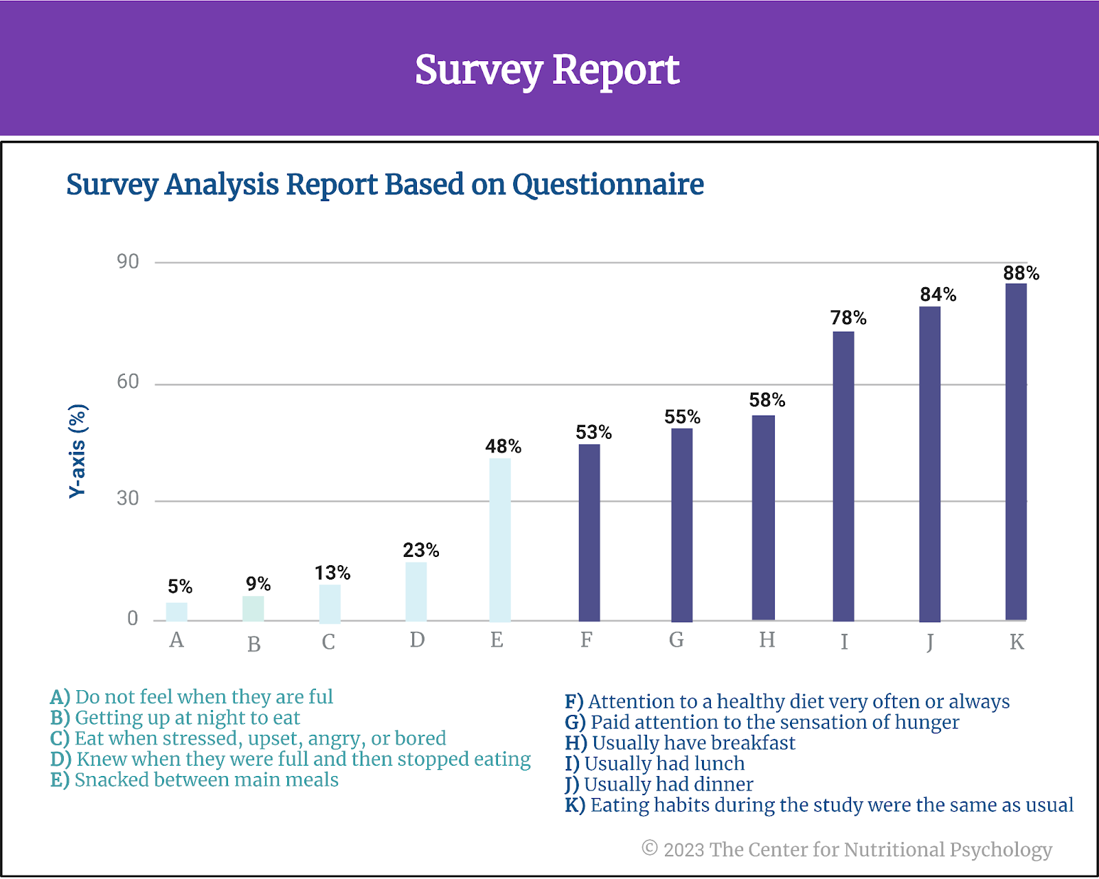
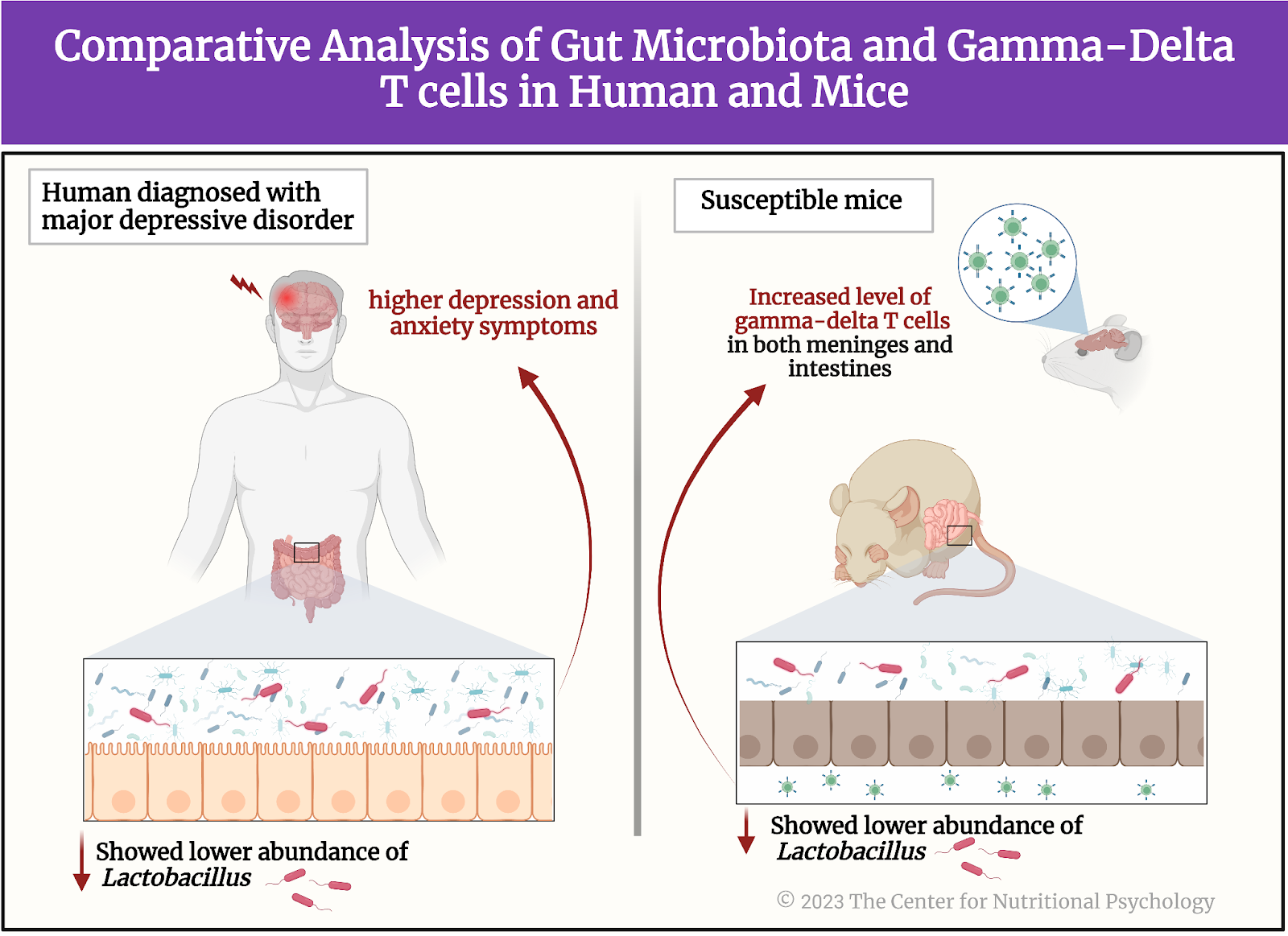
Figure 5. Humans + Mouse experiment showing a lower abundance of lactobacillus in gut bacteria = with higher depression and anxiety


A comparison of the gut microbiota of susceptible mice, resilient mice, and the control group showed that susceptible mice had less diverse microbial populations in the gut. The gut microbiota of susceptible mice differed from the gut microbiota of resilient mice and the control group on a number of bacterial species. As researchers expected, one of these species was Lactobacillus Johsonii. Their concentration was reduced in susceptible mice’s guts compared to resilient mice and the control group.

Susceptible mice had increased concentrations of gamma-delta T cells in both meninges and intestines

Given the previously described relationship between Lactobacillus bacteria and immune responses, researchers examined whether concentrations of gamma-delta T lymphocytes were increased in mice exposed to the chronic social defeat stress treatment. Results showed that susceptible mice had increased concentrations of gamma-delta T lymphocytes in their colons and meninges (membranes surrounding the brain) compared to resilient and healthy mice.


Susceptible mice had increased concentrations of gamma-delta T lymphocytes both in their colons and their meninges 


In susceptible mice, many of the gamma-delta T cells in brain membranes came from the gut!

Researchers then wanted to know whether the gamma-delta T cells found in the meninges of susceptible mice were cells that differentiated there or those cells that traveled from the gut. They identified differences between these two types of gamma-delta T cells and measured their concentrations. Results showed that, in resilient mice and the control group, gamma-delta T lymphocytes found in the meninges were indeed differentiated. However, in susceptible mice, there were fewer such cells, but many gamma-delta T lymphocytes came from the gut.


In susceptible mice, there were fewer such cells, but there were lots of gamma-delta T lymphocytes that came from the gut


Social avoidance after the chronic social defeat stress does not develop in mice without gamma-delta T lymphocytes.

Finally, researchers wanted to test whether the gamma-delta T lymphocytes were responsible for the social avoidance behavior after exposure to chronic social defeat. They repeated the procedure on a new group of special mice that did not have the gamma-delta T cells. As researchers expected, these mice did not develop social avoidance behavior after exposure to the chronic social defeat stress protocol.

Conclusion

The study showed that a certain type of white blood cells – gamma-delta T lymphocytes and their accumulation play a key role in changes to behavior induced by stress. In the context of MGBA-DMHR, it also demonstrates interactions between gut microbiota, the body’s immune responses, and the brain when an organism is stressed. Given that many of the physiological processes in mice are similar to those in humans, these findings contribute to the scientific understanding of physiological mechanisms of behavioral changes that chronic stress and related disorders in humans. These insights can help develop novel ways to treat and prevent major depressive disorder and other stress-related disorders. They can also open new approaches to diagnosing individual susceptibility to stress and increasing resilience.

The paper “Dectin-1 signaling on colonic gamma-delta T cells promotes psychosocial stress responses” was authored by Xiaolei Zhu, Shinji Sakamoto, Chiharu Ishii, Matthew D. Smith, Koki Ito, Mizuho Obayashi, Lisa Unger, Yuto Hasegawa, Shunya Kurokawa, Taishiro Kishimoto, Hui Li, Shinya Hatano, Tza-Huei Wang, Yasunobu Yoshikai, Shin-ichi Kano, Shinji Fukuda, Kenji Sanada, Peter A. Calabresi, and Atsushi Kamiya.

For more research in the Microbiota Gut-Brain Axis — Diet-Mental Health Relationship (MGBA-DMHR), visit CNP’s Nutritional Psychology Research Library (NPRL) Microbiota Gut-Brain Axis—Diet-Mental Health Relationship research category, or enroll in NP 120: Microbiota Gut-Brain Axis and the DMHR (available in May 2023).

References

Armon, G., Melamed, S., Toker, S., Berliner, S., & Shapira, I. (2014). Joint Effect of Chronic Medical Illness and Burnout on Depressive Symptoms Among Employed Adults. Health Psychology, 33(3), 264–272. https://doi.org/10.1037/a0033712

Golden, S. A., Covington, H. E., Berton, O., & Russo, S. J. (2011). A standardized protocol for repeated social defeat stress in mice. Nature Protocols, 6(8), 1183–1191. https://doi.org/10.1038/nprot.2011.361

Goodman, L., Saxe, L., & Harvey, M. (1991). Homelessness as psychological trauma. American Psychologist, 46(11), 1219.

Hodes, G. E., Pfau, M. L., Leboeuf, M., Golden, S. A., Christoffel, D. J., Bregman, D., Rebusi, N., Heshmati, M., Aleyasin, H., Warren, B. L., Lebonté, B., Horn, S., Lapidus, K. A., Stelzhammer, V., Wong, E. H. F., Bahn, S., Krishnan, V., Bolaños-Guzman, C. A., Murrough, J. W., … Russo, S. J. (2014). Individual differences in the peripheral immune system promote resilience versus susceptibility to social stress. Proceedings of the National Academy of Sciences of the United States of America, 111(45), 16136–16141. https://doi.org/10.1073/pnas.1415191111

Tsukerman, D., Leger, K. A., & Charles, S. T. (2020). Work-family spillover stress predicts health outcomes across two decades. Social Science & Medicine, 265, 113516. https://doi.org/10.1016/j.socscimed.2020.113516

Zhao, Y., Niu, C., & Cui, J. (2018). Gamma-delta (γδ) T Cells: Friend or Foe in Cancer Development. Journal of Translational Medicine, 16(1), 1–13. https://doi.org/10.1186/s12967-017-1378-2 

Zhu, X., Sakamoto, S., Ishii, C., Smith, M. D., Ito, K., Obayashi, M., Unger, L., Hasegawa, Y., Kurokawa, S., Kishimoto, T., Li, H., Hatano, S., Wang, T. H., Yoshikai, Y., Kano, S. ichi, Fukuda, S., Sanada, K., Calabresi, P. A., & Kamiya, A. (2023). Dectin-1 signaling on colonic γδ T cells promotes psychosocial stress responses. Nature Immunology. https://doi.org/10.1038/s41590-023-01447-8


The Diet-Mental Health Relationship in Astronaut Performance

The discipline of nutritional psychology is consolidating evidence worldwide on how dietary intake can impact various aspects of psychological, behavioral, cognitive, and functional performance in work environments (see the Diet and Human Performance research category within the NPRL). This includes how we think, behave, and perform in many workplace environments — from shift workers in hospitals (Leedo et al., 2017) and long-haul truck drivers (Ge et al., 2021) to students in the classroom (Barchitta, 2019), pilots in the cockpit (Lindseth, et al., 2011), and astronauts aboard space stations (Douglass, 2022).

NASA’s Human Exploration Research Analog (HERA) experiment compared the psychological and health effects of the standard diet currently served to astronauts on the International Space Station (ISS) with a new, enhanced diet formula. Results showed that study participants who ate a diet containing more fruits, vegetables, fish, and foods rich in flavonoids and omega-3 fatty acids had lower levels of cholesterol, reduced stress (as indicated by the hormone cortisol), better cognitive speed, accuracy, attention, and a more stable composition of gut microorganisms compared to participants who ate the standard International Space Station diet. The study was published in Scientific Reports.


Adding more fruit, vegetables, fish, and foods rich in flavonoids and omega-3 fatty acids to astronauts’ diets improves psychological, cognitive, and health-related outcomes.


What do astronauts eat?

Since the start of spaceflight over half a century ago, the food available to astronauts during space missions has improved dramatically. Early astronauts had very limited dietary choices, typically consisting of prepackaged foods served in either squeeze tubes or bite-sized cubes coated in gelatin to prevent the formation of crumbs. This was crucial in the confined environment of a space vessel, where loose food particles could pose a serious long-term health threat (Perchonok & Bourland, 2002).

Food options for astronauts have dramatically increased, as have the number of days astronauts routinely spend in orbit during a single mission. Longer duration missions, like those on the International Space Station (ISS), have increased the importance of spaceflight menus, allowing astronauts’ diets to be as Earth-like as possible. Today’s space foods include various food items packed in single-serving containers.


Longer space missions require more earth-like food items and menus to address astronauts’ psychosocial and psychological needs.


Modern spaceflight menus are typically organized into 6 or 7-day rotations, consisting of four meals per day – breakfast, lunch, dinner, and a snack. Food menus are planned based on the astronauts’ weight to provide the calories and nutrients required to maintain their health.

In the early days of spaceflight, experts believed they could calculate the ideal food composition for astronauts based solely on nutritional requirements for survival in space. However, the current perspective recognizes that “ideal food cannot ensure psychosocial comfort, while a grandma-style pie can” (Bychkov et al., 2021). Longer space missions have highlighted the importance of food beyond just nutritional survival needs, taking into account the diet-mental health relationship. This includes considering the psychological, cognitive, behavioral, and psychosocial value of food for the well-being of astronauts.

With this in mind, modern astronauts can select around 20% of their food items and beverages, while around 80% of their diet comes from a shared standard set of foods (Douglas et al., 2020).


Consistent with nutritional psychology-related factors, “Ideal food cannot ensure psychosocial comfort, while a grandma-style pie can” (Bychkov et al., 2021).


How is the food for astronauts prepared?

Food items are specially prepared on Earth before a flight to make them easy to use in a weightless environment and to minimize the risk of spillage. Spillage is dangerous in weightlessness as spilled material will not drop to the ground but disperse around the room or form free-floating bubbles. Crumbs or very small granules can also be dangerous and difficult to collect. For example, salt and pepper are available to astronauts in liquid form.

The food items need to be shelf-stable, either in their natural form or preserved by removing water and sterilized using heat or radiation. On the International Space Station, resupply vehicles arrive several times a year, bringing fresh fruits and vegetables and some semi-shelf-stable specialty items. Astronauts report that these deliveries provide profound psychological benefits (Douglas et al., 2020).


Astronauts report that these deliveries [fresh produce] provide profound psychological benefits (Douglas et al., 2020).


While NASA has successfully provided food for missions of up to 11 months in low Earth orbit (Douglas et al., 2020), plans for future deep-space manned exploration missions make space nutrition a topic of intense scientific research.

What is HERA?

The Human Exploration and Research Analog or HERA is a two-story building at the NASA Johnson Space Center (JSC) in Houston. Its purpose is to simulate the isolation, confinement, and remote conditions of space exploration scenarios. The building contains four habitats that simulate a space vessel with its simulated airlock and hygiene module.

HERA is used for studies that consist of 4 crew members living in the enclosed space of the simulated vessel for up to 45 days. One such stay is called a mission. During a mission, study participants stay in the habitat, spending their time in the way they would spend on a real space mission (with the difference that they are not weightless).

The simulation vessel crewmembers did not have access to the internet, social media, or communication outside mission control. They could communicate privately with their family, medical personnel, or a psychologist once a week. There was a limited selection of movies or music through a controlled account. HERA research studies last up to 68 study days (NASA ROI – Flight Analogs Human Research Program, 2019) and involve 16 days of preparation before entering the habitat and 7 days of follow-up after leaving it.


Plans for future deep-space manned exploration missions make space nutrition a topic of intense scientific research.


The study of health and psychological effects of an enhanced menu

A new study by Grace L. Douglas — an experienced researcher on the health effects of spaceflight working at the Human Health and Performance Directorate of the NASA Johnson Space Center in Houston — and her colleagues explored whether the diet of astronauts can be further improved (Douglas et al., 2022). They reasoned that studies investigating the effects of spaceflight nutrition conducted thus far have focused only on a limited number of nutrients and outcomes. They wanted to test how further increasing the variability and availability of healthy, shelf-stable, space flight-compatible foods that included fruits, vegetables, fish, and other foods rich in flavonoids and omega-3 fatty acids would affect the immune system, gut microbiome, nutritional status, and cognitive outcomes of future astronauts.

Study participants and procedure

The study participants were sixteen individuals with an average age of 40 and body mass indexes that placed them in the healthy weight range. Ten of the study participants were men, and 6 were women. These sixteen individuals participated in four 45-day HERA missions (four people per mission) conducted in 2017 and 2018. Each participant participated in a single mission. They were instructed to keep logs of the food and beverages they ate and drank 15 days before the mission.

After they entered the HERA habitat and started the mission, participants were not allowed to select any menu components. Trading food items was also not permitted. This is because the researchers wanted to make the study as consistent as possible with real-life situations in future space exploration missions. Food and beverages that the participants were expected to consume during the mission were stowed in the habitat at the beginning of the mission. All participants received the same base menu in each mission.

The standard International Space Station menu and the new enhanced menu

The menu was designed to provide the nutrients required to stay healthy with approximately 2300 calories daily. An additional 100 to 200 calories per person were provided “to ensure adequate provisioning with minimal choice.” Participants were instructed to consume the base menu before consuming additional calories.

Researchers randomly selected two missions to receive the new enhanced menu they designed, while the other two missions received the standard International Space Station menu. Study participants were not told which menu they were receiving during the mission they participated in.

Measures and assessments

After each meal, participants recorded actual food consumption using the International Space Station Food Intake Tracker iPad App. Researchers recorded the use of antibiotics and medications. Weight and height were measured before the mission and weight measurements were taken before breakfast each day during the mission.

Researchers collected blood, stool, urine, and saliva samples from each subject at 5-time points – twice before and 3 times during the mission. The collection of samples during the mission was done in a special HERA chamber and transferred out through a pass with minimal to no contact between the study participants (HERA “crew”) and the support team.

From these samples, researchers assessed levels of vitamins, flavonoids, calcium, and bone health, cardiovascular and fluid regulation, metabolic, oxidative stress, reproductive hormones, and iron and blood cell concentrations. They also conducted immunological analysis and assessed levels of the hormone cortisol and viruses from saliva. Researchers determined the composition of the gut microbiome from stool samples of participants and conducted a study of gene expressions of these microorganisms. The latter procedure is called metatranscriptomic analysis.

As part of a broader Cognition test battery (see Figure 1), participants completed vigilant attention assessments using the Psychomotor Vigilance Test (PVT) twice before the mission and three times per week during the mission. 

%learn about nutrition mental health %The Center for Nutritional Psychology

Figure 1. Crewmember measures and assessments protocol


Nutritional intake was improved for participants consuming the enhanced diet

Results showed that study participants in both groups did not fully consume their menu items. However, participants in the enhanced menu missions consumed more servings of fruits and vegetables per day, and ate more fish per week and more servings of tomato-based foods.

The participants receiving the enhanced diet took in more calcium, potassium, daily fiber, and omega-3 fatty acids than participants on the two standard menu missions (see Figure 2). The importance of omega-3 fatty acids comes from the fact that they are an integral part of cell membranes in the human body. They are also starting points for producing hormones that regulate blood clotting, contraction and relaxation of artery walls, and inflammation.  Intakes of protein, sodium, and iron were similar in the two groups.

Crewmembers on the enhanced diet took in more calcium, potassium, fiber per day, and omega-3 fatty acids than participants on the standard menu missions. 

%learn about nutrition mental health %The Center for Nutritional Psychology

 Figure 2. Enhanced menu missions consumption


The enhanced menu and health indicators

General health status was similar in participants consuming the two diets. However, cholesterol status was improved in participants consuming the enhanced diet. They also had higher physiological flavonoid concentrations in urine and more stable fatty acid concentrations in the blood (see Figure 3).

The concentration of the hormone cortisol in the blood was higher in participants on standard diets. Cortisol is a hormone produced by adrenal glands. It regulates the body’s response to stress. Higher concentrations are indicative of higher stress levels.

The diversity and richness of gut microbiota were reduced in participants consuming the standard diet. Researchers also detected changes in the abundance of several species of gut microorganisms that were associated with the differences in the diet. Additionally, gene expression profiles, i.e., metatranscriptomic profiles of the gut microbiome, were more stable for subjects consuming the enhanced diet.

Cognitive speed, accuracy, and attention were better for subjects consuming the enhanced diet

Psychological assessments showed that cognitive speed, accuracy, and attention were better for subjects consuming the enhanced diet. The reaction speeds in the tests of study participants consuming the enhanced diet were higher than the average reaction speeds of participants on the standard diet and improved compared to their pre-mission results.

%learn about nutrition mental health %The Center for Nutritional Psychology

 Figure 3. Enhanced vs Standard diet


Conclusion

The study showed that a specific diet rich in fruits, vegetables, and omega-3 fatty acids produces significant health and performance benefits in a simulated space exploration mission environment. Countering the physiological deterioration of astronauts’ health during long space missions is an ongoing goal for researchers and mission planners. These results can help achieve this goal by better planning food resources and astronauts’ menus and lend credence to the importance of the diet-mental health relationship on human performance and within work environments.

The paper “Impact of diet on human nutrition, immune response, gut microbiome, and cognition in an isolated and confined mission environment” was authored by Grace L. Douglas, Diane DeKerlegand, Holly Dlouhy, Nathan DumontLeblond, Eden Fields, Martina Heer, Stephanie Krieger, Satish Mehta, Bridgette V. Rooney, ManolitoG.Torralba, Sara E. Whiting, Brian Crucian, Hernan Lorenzi, Scott M. Smith, Millennia Young, and Sara R. Zwart.


References

Barchitta, M., Maugeri, A., Agrifoglio, O., Favara, G., La Mastra, C., La Rosa, M. C., Magnano San Lio, R., & Agodi, A. (2019). Dietary patterns and school performance: evidence from a sample of adolescents in Sicily, Italy. Annali di igiene : medicina preventiva e di comunita, 31(2 Supple 1), 72–80.

Bychkov, A., Reshetnikova, P., Bychkova, E., Podgorbunskikh, E., & Koptev, V. (2021). The current state and future trends of space nutrition from a perspective of astronauts’ physiology. International Journal of Gastronomy and Food Science, 24, 100324. https://doi.org/10.1016/J.IJGFS.2021.100324

Douglas, G. L., DeKerlegand, D., Dlouhy, H., Dumont-Leblond, N., Fields, E., Heer, M., Krieger, S., Mehta, S., Rooney, B. V., Torralba, M. G., Whiting, S. E., Crucian, B., Lorenzi, H., Smith, S. M., Young, M., & Zwart, S. R. (2022). Impact of diet on human nutrition, immune response, gut microbiome, and cognition in an isolated and confined mission environment. Scientific Reports, 12(1), 1–22. https://doi.org/10.1038/s41598-022-21927-5

Douglas, G. L., Zwart, S. R., & Smith, S. M. (2020). Space food for thought: Challenges and considerations for food and nutrition on exploration missions. Journal of Nutrition, 150(9), 2242–2244. https://doi.org/10.1093/JN/NXAA188

Ge, Y., He, S., Xu, Y., & Qu, W. (2021). Effects of dietary patterns on driving behaviours among professional truck drivers: the mediating effect of fatigue. Occupational and environmental medicine, 78(9), 669–675. https://doi.org/10.1136/oemed-2020-107206

Leedo, E., Beck, A. M., Astrup, A., & Lassen, A. D. (2017). The effectiveness of healthy meals at work on reaction time, mood and dietary intake: a randomised cross-over study in daytime and shift workers at a university hospital. The British journal of nutrition, 118(2), 121–129. https://doi.org/10.1017/S000711451700191X

Lindseth, G. N., Lindseth, P. D., Jensen, W. C., Petros, T. V., Helland, B. D., & Fossum, D. L. (2011). Dietary Effects on Cognition and Pilots’ Flight Performance. The International journal of aviation psychology, 21(3), 269–282. https://doi.org/10.1080/10508414.2011.582454

NASA ROI – Flight Analogs Human Research Program. (2019). Human Research Program Human Exploration Research Analog (HERA) Facility and Capabilities Information (Issue July). https://www.nasa.gov/sites/default/files/atoms/files/2019_hera_facility_capabilities_information.pdf

Perchonok, M., & Bourland, C. (2002). NASA Food Systems: Past, Present, and Future. Nutrition, 18(10), 913–920. https://doi.org/https://doi.org/10.1016/S0899-9007(02)00910-3

Binge Drinking in Adolescents Linked to Changed Composition of Bacteria in the Gut

If we drink multiple alcoholic beverages at once, this will lead to intoxication. But when we sober up, are the alcoholic effects gone or are there changes to our body that linger? A new study by Carbia and colleagues done at the University College Cork in Ireland aimed to identify changes in our body that can be used to detect binge drinking, even before this drinking habit leads to alcohol use disorder. These researchers studied potential links between binge drinking, changes to the gut microbiome, social cognition, and impulsivity. The second goal of the study was to identify the changes in our body that happen when we experience alcohol cravings by examining the associations of the microbiome composition and craving for alcohol.

This study was published in eBioMedicine and explores whether changes in the gut microbiome are associated with cravings for alcohol, and whether alterations in the composition of the gut microbiome due to binge drinking have detrimental effects on cognitive functioning, particularly in relation to social and emotional processing. 

Let’s begin by learning more about binge drinking.

What is binge drinking?

Binge drinking is defined as the consumption of excessive amounts of alcoholic beverages in a short period of time. It is the most frequent type of alcohol misuse during adolescence in Western countries (Carbia et al., 2023), and is most frequent in late adolescence, up to around 24 years of age (Sawyer et al., 2018). Binge drinking is associated with an increased risk of developing alcohol use disorder, but also other types of psychiatric disorders later in life.

The World Health Organization (WHO) defines a binge drinking episode as consuming at least 60 grams of pure alcohol on a single occasion, which leads to a blood alcohol concentration of 0.8 grams/liter or higher (Rolland et al., 2017). A standard shot of whiskey is 1.5 ounces, equaling 42 grams. Given 40% alcohol content, a standard shot of whiskey contains .6 oz or a bit less than 17 grams of alcohol. A binge drinking episode would thus be any single occasion on which a person consumes 4 or more standard shots of whiskey (or of a similar alcoholic beverage) (see Figure 1).


%learn about nutrition mental health %The Center for Nutritional Psychology

Figure 1. Binge drinking episode per WHO 


The microbiome-gut-brain axis 

The human gut microbiome consists of a plethora of microorganisms (predominantly bacteria, but also archaea, viruses, and fungi) that inhabit the human gastrointestinal tract, primarily the colon or large intestine. These microorganisms have co-evolved with the human host over millions of years, forming a complex ecosystem that plays a critical role in human health and disease (Cryan et al., 2019).

The MGBA is a bi-directional communication system existing between our gut, its microbiome, and our brain. This communication system is facilitated by various signaling pathways and molecules which together perform a wide range of functions essential for human health — including digesting complex carbohydrates, producing essential vitamins and nutrients, regulating the immune system, and maintaining the integrity of the intestinal barrier (which serves as a physical and biochemical barrier between the gut and the rest of the body’s organs).

The microbiome gut-brain axis regulates processes beyond the gut, including metabolism, body weight, and brain functions involving mood, emotion, craving, behavior, and cognition. And because this communication system is bidirectional, just as the gut microbiome can influence mood and stress states, mood and stress states can, in turn, influence the gut microbiomeleading to gut dysbiosis, intestinal permeability, and other gut-related health problems (see Figure 2). 


%learn about nutrition mental health %The Center for Nutritional Psychology

Figure 2. The Microbiome gut-brain axis and stress 


The gut microbiome in individuals suffering from binge drinking and alcohol use disorder

Several studies have explored the link between altered gut microbiome in the development of alcohol use disorder and binge drinking in young people (Rodríguez-González et al., 2021; Gorky & Schwaber, 2016). 

In the scope of alcohol use disorder treatment, for example, studies demonstrate that patients who abruptly cease drinking alcohol show increased gut permeability and changes in the composition of their gut microbiome. They also show higher levels of cytokines and cortisol (Adams et al., 2020). The increased levels of cytokines and cortisol in patients’ circulation are associated with the severity of their alcohol use disorder, and the intensity of their craving for alcohol.

Since binge drinking can negatively impact the microbiome-gut-brain axis and contribute to the development of alcohol-related health problems, targeting the gut microbiome holds promise as a potential method for reducing alcohol-related harm in this population.

The gut microbiome composition might be associated with social behavior

Numerous studies demonstrate that gut microbiota composition appears to be connected to social behavior (Johnson, et. al, 2022; Sylvia, et al., 2018). An experiment where the gut microbiome from patients suffering from alcohol use disorder was transplanted into the guts of mice, showed that this action impaired the sociability of the mice and also lead to certain disturbances in their brain function (Leclercq et al., 2020) such as neuroinflammation (see Figure 3). 


%learn about nutrition mental health %The Center for Nutritional Psychology

Figure 3. Gut microbiome composition association with social behavior


This and other findings have led researchers to postulate that the gut microbiome might play a role in the development of alcohol use disorder, and also in other psychiatric disorders characterized by problems in social cognition and functioning.


The gut microbiome might play a role in the development of alcohol use disorder. 


The study of young Irish binge drinkers

A 2023 study by Carbia and colleagues aimed to identify early biomarkers of alcohol misuse by exploring the gut microbiome and neurocognitive correlates in young binge drinkers who do not have an alcohol use disorder (Carbia et al., 2023). 

They reasoned that because of the intense development occurring during adolescence, people have increased vulnerability to developing alcohol use disorder (and other psychiatric disorders) during this stage of life. They purport that it is possible that factors creating this vulnerability go beyond the developing brain of a young person and extend to specificities of the gut microorganism composition.

As previously mentioned, these authors wanted to study potential links between binge drinking, changes to the gut microbiome, social cognition, and impulsivity. Social cognition and impulsivity are two cognitive domains most often linked to increase in alcohol drinking. The second goal of the study was to identify the biomarkers of alcohol craving by examining the associations between changes to the microbiome composition and craving for alcohol.


Because of the intense development during adolescence, people have increased vulnerability to developing alcohol use disorder (and other psychiatric disorders) during this stage of life. 


Study participants and procedure

Study participants were 71 healthy persons, between 18 and 25 years of age, living in Cork, Ireland. Those excluded from the study were prospective participants who never drank alcohol, whose scores on an alcohol use assessment test indicated alcohol use disorder and those who had a family history of alcoholism. Participants were also excluded if they had any other mental disorder, or were using drugs or any medications.

After recruitment, participants underwent a clinical interview in which researchers collected their demographic information, made clinical assessments, and collected a range of other data, such as information about their diets.  During their second visit, participants underwent neuropsychological evaluation and researchers collected their biological samples (saliva, hair, blood, stool). Three months later, participants reported their alcohol use and craving. Participants were paid up to 50 EUR for their participation in the study.


It is possible that factors creating vulnerability in young people for alcohol disorders go beyond the developing brain and extend to specificities of the gut microorganism composition.


Measures and assessments

In the scope of the study, participants completed assessments of alcohol consumption patterns (the AUDIT questionnaire and the Alcohol Timeline Follow-Back, TFLB), alcohol craving (the Alcohol Craving Questionnaire – Short Form, ACQ), emotional processing (the Emotion Recognition Task, ERT and the Affective Go/No-Go task from the Cambridge Neuropsychological Test Automated Battery, CANTAB), and impulsivity (The Barratt Impulsiveness Scale, BIS-11). 

Researchers also made assessments of participants’ inflammatory markers from blood samples of kynurenine and tryptophan (metabolites suspected to be a part of the microbiome-gut-brain axis communication pathway) and cortisol. Genomic sequencing of the stool samples was conducted for the purposes of identifying composition of the gut microbiome and short-chain fatty acid contents — another suspected component of the microbiome-gut-brain communication axis (see Figure 4).


%learn about nutrition mental health %The Center for Nutritional Psychology

Figure 4. Research assessment protocol


Binge drinkers have higher alcohol cravings and are worse at recognizing emotions.


Results showed that participants who had more binge drinking episodes had higher cravings for alcohol. Participants who had a higher number of drinks within a drinking episode were also less able to recognize sadness and disgust. People who drank more were slower to respond when they gave correct answers to these tests. They also scored higher on impulsivity.

Binge drinking and the gut microbiome

Recent binge drinking episodes were associated with higher microbiome alterations, including changes to how widespread multiple species of bacteria were (referred to as microbial diversity). Higher numbers of drinks per binge drinking session were associated with lower levels of isovalerate, one type of branched-chain saturated fatty acid, thought to be involved in how the gut microbiome interacts with body processes outside the gut. Greater craving for alcohol was found to be associated with reductions in numbers of the gut bacteria Ruthenibacterium lactiformans. It was also associated with changes in levels of a number of compounds thought to be involved in the microbiome-gut-brain axis as well as those involved in the gut-metabolic interactions. Notably, reduced butyrate and inositol synthesis and increased acetate, glutamate, and tryptophan synthesis were associated with higher cravings. 

Let’s see this visually in Figure 5. 


%learn about nutrition mental health %The Center for Nutritional Psychology

Figure 5.  Link of binge drinking with gut microbiota alteration, craving, and poor emotional processing


Conclusion

This study showed that binge drinking, the most common pattern of alcohol misuse in adolescence, is linked with changes to the gut microbiome and specific cognitive impairments. The study also revealed that changes in the gut microbiome are linked with increased cravings for alcoholic beverages. This was shown to happen in participants without alcohol addiction (i.e., before the alcohol use disorder has developed). These findings open the possibility to develop methods for predicting alcohol drinking habits and possibly alcohol-related social functioning impairments by analyzing gut microbiota composition from stool samples. They could also lead to novel dietary or pre/probiotic interventions directed at improving early alcohol-related alterations in the gut microbiota of young drinkers.

More about the gut microbiome and its connection to psychological, cognitive, behavioral and psychosocial functioning and mental health can be found in NP 120 (scheduled to be published in May 2023). NP 120 is part of the Introductory Certificate in Nutritional Psychology through the Center for Nutritional Psychology.  

The paper “The Microbiome-Gut-Brain axis regulates social cognition & craving in young binge drinkers” was authored by Carina Carbia, Thomaz F. S. Bastiaanssen, Luigi Francesco Iannone, Rubén García-Cabrerizo, Serena Boscaini, Kirsten Berding, Conall R. Strain, Gerard Clarke, Catherine Stanton, Timothy G. Dinan, and John F. Cryan.

References

Adams, C., Conigrave, J. H., Lewohl, J., Haber, P., & Morley, K. C. (2020). Alcohol use disorder and circulating cytokines: A systematic review and meta-analysis. Brain, Behavior, and Immunity, 89, 501-512. https://doi.org/10.1016/j.bbi.2020.08.002

Carbia, C., Bastiaanssen, T. F. S., Iannone, F., García-cabrerizo, R., Boscaini, S., Berding, K., Strain, C. R., Clarke, G., Stanton, C., Dinan, T. G., & Cryan, J. F. (2023). The Microbiome-Gut-Brain axis regulates social cognition & craving in young binge drinkers. EBioMedicine, In press), 104442. https://doi.org/10.1016/j.ebiom.2023.104442

Gorky, J., & Schwaber, J. (2016). The role of the gut–brain axis in alcohol use disorders. Progress in Neuro-Psychopharmacology and Biological Psychiatry, 65, 234-241. https://doi.org/10.1016/j.pnpbp.2015.06.013

Johnson, K. V., Watson, K. K., Dunbar, R. I. M., & Burnet, P. W. J. (2022). Sociability in a non-captive macaque population is associated with beneficial gut bacteria. Frontiers in microbiology, 13, 1032495. https://doi.org/10.3389/fmicb.2022.1032495

Leclercq, S., Le Roy, T., Furgiuele, S., Coste, V., Bindels, L. B., Leyrolle, Q., Neyrinck, A. M., Quoilin, C., Amadieu, C., Petit, G., Dricot, L., Tagliatti, V., Cani, P. D., Verbeke, K., Colet, J. M., Stärkel, P., de Timary, P., & Delzenne, N. M. (2020). Gut Microbiota-Induced Changes in β-Hydroxybutyrate Metabolism Are Linked to Altered Sociability and Depression in Alcohol Use Disorder. Cell Reports, 33(2). https://doi.org/10.1016/J.CELREP.2020.108238

Rodríguez-González, A., Vitali, F., Moya, M., De Filippo, C., Passani, M. B., & Orio, L. (2021). Effects of Alcohol Binge Drinking and Oleoylethanolamide Pretreatment in the Gut Microbiota. Frontiers in Cellular and Infection Microbiology, 1134. https://doi.org/10.3389/fcimb.2021.731910

Rolland, B., Chazeron, I. de, Carpentier, F., Moustafa, F., Viallon, A., Jacob, X., Lesage, P., Ragonnet, D., Genty, A., Geneste, J., Poulet, E., Dematteis, M., Llorca, P. M., Naassila, M., & Brousse, G. (2017). Comparison between the WHO and NIAAA criteria for binge drinking on drinking features and alcohol-related aftermaths: Results from a cross-sectional study among eight emergency wards in France. Drug and Alcohol Dependence, 175, 92–98. https://doi.org/10.1016/J.DRUGALCDEP.2017.01.034

Sawyer, S. M., Azzopardi, P. S., Wickremarathne, D., & Patton, G. C. (2018). The age of adolescence. The Lancet Child & Adolescent Health, 2(3), 223–228. https://doi.org/10.1016/S2352-4642(18)30022-1

Sylvia, K. E., & Demas, G. E. (2018). A gut feeling: Microbiome-brain-immune interactions modulate social and affective behaviors. Hormones and behavior, 99, 41–49. https://doi.org/10.1016/j.yhbeh.2018.02.001


Why are Hyper-Palatable Foods so Alluring? New Type of Brain Cell Lends Insight

Our food preferences have changed significantly over the last decade, with processed, hyperpalatable foods being increasingly available and consumed. A substantial percentage of foods in the US food system (62%) is considered to be hyperpalatable, including foods not previously conceptualized as hyperpalatable, such as foods labeled “reduced” or “low fat” and sauces, trail mixes, vegetables cooked in creams, etc. (Fazzino et al., 2019).


Sixty-two percent of the foods in the US food system may be hyperpalatable, including foods not previously conceptualized as hyperpalatable.


We know that the highly palatable sensations and interoceptive experiences associated with these foods can activate the brain’s reward system, providing us with an experience of pleasure and reward. Activation of the reward system occurs in other activities such as achieving a goal or engaging in addictive behaviors like substance abuse. In truth, there are many factors involved in our choosing of highly palatable foods, including the availability of these foods, and the social environment in which we live.

Have you ever wondered why hyperpalatable foods are so desirable, and why they have been increasingly making it into our shopping carts despite the knowledge that they lack nutrients? Certainly, it is because hyperpalatable foods are cheap, convenient, and taste good, not to mention they give you a rewarding and memorable experience. However, what you might not know is that there’s a type of neuron in our brain that influences our dietary intake patterns.

In truth, many factors influence our dietary intake behavior and food cravings (learn more in NP 110) – both of which are important topics in the study of the diet-mental health relationship (DMHR) and within nutritional psychology. In this article, we focus on the new discovery of a type of brain cell (neuron) which may play a role in our desire to consume these highly processed foods.


Neurotensin neurons are a new structure that might impact our eating behavior.


Alessandro Furlan and his research team explored a new structure within the brain that can modulate dietary intake behavior and increase our preference for fatty foods. This structure is a group of neurons called neurotensin neurons, located in a part of the amygdala known as the interstitial nucleus of the posterior limb of the anterior commissure (IPAC) (Furlan et al., 2022). Let’s uncover the crucial role of these neurotensin neurons within the IPAC and see what Furlan and his team discovered about their role in orchestrating our dietary intake behaviors and metabolic health.


Activation of neurotensin neurons can promote hedonic feeding and weight gain.


This experiment consisted of four steps. First, they examined how activation of the neurotensin neurons can change the food behavior of the animals (i.e., whether they will be more interested in palatable food). They then examined how different foods can activate neurotensin neurons (i.e., fat vs. water). They also tested whether these cells play a role in food-seeking behavior by testing whether they are activated by merely food stimuli. Finally, they examined how inhibiting these cells can alter the food behavior of animals. 

Furlan found many things in their study, including:

  • Activation of neurotensin neurons can promote hedonic feeding. Hedonic feeding and hunger involve the consumption of food uniquely because of its rewarding properties (pleasure) rather than eating for homeostatic energy balance (Monteleone, 2012). 
  • When rats had their neurotensin neurons activated, they exhibited increased dietary intake. The effect was more pronounced for more palatable high-fat diets than for chow.
  • When mice were fed with either fat or water, they found that immediately following ingestion, neurotensin neurons were more strongly activated by fat than by water in a concentration-dependent manner. 
  • They also tried to see the effect when they compared two palatable foods (with one of them being more palatable). They found that these cells were activated by sucrose more than by sucralose (a palatable, sweet food but non-caloric sugar analog).
  • Neurotensin neurons can be activated by sensory cues that anticipate meals (i.e., odors). This means that they also play a role in food-seeking behaviors.

As shown in Figure 1, these findings show that neurotensin neurons are not only influenced by the type of food consumed (i.e., highly palatable), but that they also activate our response to food stimuli, and their activity is scaled by the palatability of food. They affect our hedonic feeding behavior and increase our intake of highly palatable foods.

%learn about nutrition mental health %The Center for Nutritional Psychology Figure 1. Summary of how neurotensin neurons can affect our food behavior


When neurotensin neurons are inhibited, the preference for highly palatable food decreases.


Conversely, acute inhibition of neurotensin neurons reduces feeding on a high-fat diet in sated mice and reduces feeding in hungry mice. In addition, prolonged inactivation of neurotensin neurons leads to a dramatic increase in aerobic locomotion and long-term weight loss.

The inactivation of these cells protected mice from obesity and the long-term negative consequences of chronic feeding. Mice who had their neurotensin neurons inhibited exhibited higher energy expenditure, higher lipid oxidation rate, low blood sugar levels, and engaged in more activity. A summary of the research findings by Furlan and his team is shown in Figure 2.

%learn about nutrition mental health %The Center for Nutritional Psychology

Figure 2. Summary of findings by Furlan and his team


What are the implications of this in everyday life?

The desire to consume highly palatable foods more frequently than healthier, nutrient-dense foods is a major cause of the current obesity pandemic. Exercise programs are known to be the first-line intervention when treating obesity, however, body weight is often restored through metabolic processes (Petridou et al., 2019).

Because these neurons seem to play a crucial role in promoting metabolic responses via behavior alteration, this knowledge may play a critical therapeutic role in future obesity interventions. Inhibiting these neurons can change our food choices, promote long-term weight loss, and protect us from obesity. Neurotensin neurons can be a piece of the puzzle in unraveling the complex mechanisms underlying our feeding behavior and metabolic health.

Of course, this research is not free of limitations. The main limitation is that this study was a mouse study, and it takes time to do translational research on human subjects. Furthermore, because this was found in animals, this does not necessarily mean that studies on human subjects would come up with the same findings.

Conclusion

The amygdala is a complex neurological structure that helps us to regulate our emotions. It is not surprising that this structure may play a role in dietary intake behavior. The discovery of neurotensin neurons in this area of the brain can contribute to our understanding the behavioral aspects of obesity and support the development of novel interventions for treating obesity. While more research is needed, this new insight makes critical strides in our understanding of the alure of hyperpalatable foods.

More about the science of the Diet-Behavior relationship can be found in NP 110: Introduction to Nutritional Psychology Methods.


References

Fazzino, T. L., Rohde, K., & Sullivan, D. K. (2019). Hyper-palatable foods: Development of a quantitative definition and application to the US Food System Database. Obesity, 27(11), 1761–1768. https://doi.org/10.1002/oby.22639

Furlan, A., Corona, A., Boyle, S., Sharma, R., Rubino, R., Habel, J., Gablenz, E. C., Giovanniello, J., Beyaz, S., Janowitz, T., Shea, S. D., & Li, B. (2022). Neurotensin neurons in the extended amygdala control dietary choice and energy homeostasis. Nature Neuroscience, 25(11), 1470–1480. https://doi.org/10.1038/s41593-022-01178-3

Monteleone, P., Piscitelli, F., Scognamiglio, P., Monteleone, A. M., Canestrelli, B., Di Marzo, V., & Maj, M. (2012). Hedonic eating is associated with increased peripheral levels of ghrelin and the endocannabinoid 2-arachidonoyl-glycerol in healthy humans: a pilot study. The Journal of Clinical Endocrinology and Metabolism, 97(6), E917–E924. https://doi.org/10.1210/jc.2011-3018

Petridou, A., Siopi, A., & Mougios, V. (2019). Exercise in the management of obesity. Metabolism: Clinical and Experimental, 92, 163–169. https://doi.org/10.1016/J.METABOL.2018.10.009


Blue Zones and Their Role in the Diet-Mental Health Relationship (DMHR): A Three Part Series Exploring the Interplay of Diet, Longevity, and Mental Health.

Editor’s Note: This article, the second in this three-part series, dives deeper into the dietary habits of Blue Zone regions and highlights the physiological impact of nutrition on aging bodies. The next and final article will explore the interplay of diet and mental health in the context of abnormally aging populations. 

As introduced in What is a Blue Zone, Blue Zones are geographic territories that produce abnormal numbers of centenarians: their residents are ten times more likely to reach age 100 than the average US citizen (Buettner & Skemp, 2016). These five regions Ikaria, Greece; Okinawa, Japan; Sardinia, Italy; Loma Linda, California; and Nicoya, Costa Rica encompass nine shared lifestyle characteristics that help explain exceptional longevity. These are called the Power 9 (Buettner & Skemp, 2016), and were reviewed in the previous article. 


Blue Zone residents are ten times more likely to reach age 100 than the average US citizen.


Of these nine characteristics, food is involved in three the 80% rule, plant slant, and wine at 5 two of which speak to the actual dietary composition of Blue Zone meals. Evidence supports a correlation between healthy dietary patterns and a decreased risk of early death, begging the further inspection of diet as a factor in the atypical longevity of Blue Zone residents (Harmon et al., 2015). 

When evaluating diet and its impact on longevity, nutritional epidemiologic studies have begun to assess populations through overall dietary patterns instead of tracking a single nutrient or food (Cespedes & Hu, 2015). Human beings do not habitually consume nutrients or food in isolation, and tracking dietary patterns produces a more realistic idea of how diet influences lifespan and health outcomes (Cespedes & Hu, 2015). Therefore, examining  Blue Zones, we explore whole dietary patterns and their commonalities. 


%learn about nutrition mental health %The Center for Nutritional Psychology

The traditional village of Evdilos, in Ikaria island, Greece. Courtesy of Shutterstock Images.


Ikarian cuisine largely revolves around Mediterranean diet principles (Buettner & Skemp, 2016). The Mediterranean diet is characterized by a high intake of fruits, vegetables, whole grains, legumes, herbs, and olive oil, a moderate intake of fish, poultry, dairy products, and red wine, and a low intake of sweets and red meat (Davis et al., 2015). Green vegetables and wild plants cooked in olive oil act as main meals rather than side dishes, complimenting the finding that Ikarian individuals’ fruit and vegetable consumption often exceeds dietary recommendations (Panagiotakos et al., 2011) 


Green vegetables and wild plants cooked in olive oil act as main meals rather than side dishes.


Given their Mediterranean-inspired dietary habits, the high life expectancy of Ikarian individuals is not shocking. Adherence to a Mediterranean diet has been significantly associated with a reduced risk of mortality: 9% from cardiovascular disease, 6% from cancer, and 13% from Parkinson’s disease and Alzheimer’s disease (Sofi et al., 2008). Additionally, in an Ikarian-focused study, long-term fish consumption was independently associated with the improved kidney function of elders (Chrysohoou et al., 2013). 

By growing much of their own diet, Ikarian individuals have been able to keep up with traditional Mediterranean eating habits into the 21st century, which may play a central role in their longevity (Legrand et al., 2021).


%learn about nutrition mental health %The Center for Nutritional Psychology

Okinawa, Japan. Courtesy of Shutterstock Images.


The traditional Okinawan dietary pattern is anchored majorly by root vegetables (specifically sweet potatoes), yellow and green leafy vegetables, legumes, and soy-based foods (Willcox et al., 2014). It emphasizes the intake of low-GI grains and high fiber-rich foods while stressing minimal consumption of meat and dairy products (Willcox et al., 2014). A typical meal includes miso-soup topped with tofu, fish, pork, or vegetables paired with fresh jasmine tea (medicinal plants hold significant importance to their diet) (Willcox et al., 2014).  

Okinawan cuisine is largely plant-based, as staple foods there have always been locally grown due to financial strains (Willcox et al., 2014). Sweet potatoes in particular became a main source of caloric consumption due to their ability to survive severe climates (Willcox et al., 2014).

The high phytonutrient nature of the Okinawan diet may play a protective role in longevity, as antioxidants have the ability to buffer against oxidative stress: a set of chain reactions that impact the development of diseases such as arthritis, diabetes, dementia, and cancer (Tan et al., 2018; Willcox et al., 2013). The diet’s low glycemic load and low saturated fat content further reduce the known effects of oxidative stress (i.e., cardiovascular disease and other chronic illnesses), potentially aiding in the extension of Okinawan lifespans (Willcox et al., 2013). 


%learn about nutrition mental health %The Center for Nutritional Psychology

Alley of Castelsardo old city in Sardinia, Italy. Courtesy of Shutterstock Images.


The classic Sardinian diet is considered a variant of the Mediterranean diet, as it stresses the consumption of carbohydrate foods and olive oil. However, it differs in its emphasis on meat (Pes et al., 2022). Sardinian dietary patterns, influenced by the city’s history of animal husbandry, involve the high consumption of animal products such as cheese, pork fat, and poultry meat (Pes et al., 2022). As sheep and goats contributed to Sardinia’s economic stability, dairy products such as soft sour cheeses are also prevalent (Pes et al., 2021). 


Sardinian dietary patterns involve the high consumption of animal products such as cheese, pork fat, and poultry meat. 


Positive associations between daily functioning and eating sheep, goat, and poultry meat have been found. The consumption of poultry alongside vegetables, for instance, is associated with a risk reduction of cardiovascular diseases, obesity, and type 2 diabetes mellitus (Marangoni et al., 2015; Pes et al., 2021). Additionally, the diet’s high antioxidant content, stemming largely from red wine, may impact lower cardiovascular mortality through compounds in the wine  — resveratrol, and proanthocyanidins  — that counteract age-related inflammation (Corder et al., 2006). 


%learn about nutrition mental health %The Center for Nutritional Psychology

Costa Rica scenery, looking towards the Nicoya Peninsula from Monteverde. Courtesy of Shutterstock Images.


The general dietary pattern in Nicoya revolves around four foods eaten with high frequency: Gallo Pinto, tortillas de maiz, Cuajada, and Galla pinto con huevos (Nieddu et al., 2020). Popular dishes incorporate legumes, rice, potato, and cassava (a nutty-flavored, starchy root vegetable) (Link & Elliott, 2021; Pies et al., 2022). These carbohydrate-based foods have a low glycemic index, possibly promoting a delayed onset of various metabolic diseases (Rosero-Bixby et al., 2014). The frequent consumption of traditional foods such as rice and beans showcases an adherence to whole foods (Rosero-Bixby et al., 2013). While overall the residents of Nicoya eat a plant-based diet, the consumption of animal proteins is still relatively high. Nieddu et al. (2020) found that at least half of the oldest Nicoyans ate three to five servings of meat per week, and about 25% consumed meat daily. 


While overall the residents of Nicoya eat a plant-based diet, the consumption of animal proteins is still relatively high.


Something unique to the Nicoya diet is the high calcium and magnesium content of their drinking water (Buettner & Skemp, 2016). This not only helps protect against heart disease but also promotes strong bones, a crucial component of healthy aging (Buettner & Skemp, 2016). Also notable is their high fruit consumption — mango and papaya specifically are known to promote high levels of antioxidants (Pes et al., 2022).


%learn about nutrition mental health %The Center for Nutritional Psychology

Loma Linda, California on a map. Courtesy of Shutterstock Images.


Loma Linda’s large Adventist community takes their diet directly from the Bible: they consume a largely vegetarian diet filled with leafy greens, nuts, and legumes (Buettner & Skemp, 2016). Fraser (1999) found that Adventist vegetarians eat an average of 3.5 servings of meat substitutes per week, often derived from soy, as well as copious amounts of fruit, which increases antioxidant and dietary fiber intake (Fraser, 1999).    

This surge in dietary fiber through the consumption of vegetables, fruits, grains, and nuts is associated with lowered blood cholesterol and may offer protection against colon and rectal cancer, potentially explaining Loma Linda Adventists’ longevity (Howe et al., 1992; Soliman, 2019). The consumption of these foods combined with reduced meat intake may also decrease the risk of diabetes mellitus, hypertension, and arthritis (Fraser, 1999), facilitating healthier aging.  

It is important to note that abstinence from smoking and generally limited alcohol intake might serve as confounding variables in exploring the longevity of Adventists (Montgomery et al., 2007). In other words, diet alone may not explain the above-average longevity of Adventists, as various lifestyle factors also impact healthful aging. Nonetheless, the lifespan for California Adventists is 7.3 years longer for men and 4.4 years longer for women than the general population in California, calling for the continued exploration of how diet influences abnormal aging (Fraser & Shavlik, 2001). 


Overall Themes 

What commonalities can we extract from the five independent Blue Zones? Each diet is influenced by unique religious, historical, cultural, and environmental factors, making it hard to combine them all under a single pattern (Pes et al., 2022). Nonetheless, there are a few takeaways we can make. 


Each diet is influenced by unique religious, historical, cultural, and environmental factors, making it hard to combine them all under a single pattern. 


First: the self-produced nature of the food consumed. Whether it be the fishermen of Ikaria catching the evening’s dinner or Okinawan elders growing yellow and green vegetables, much of the food prepared comes from local sources. This may be due to most of the Blue Zones’ more geographically isolated nature as well as financial constraints.  

Second: the antioxidant-rich tendencies of the diets. Though from different sources (i.e., fruit in Nicoya, vegetation in Loma Linda, or wine in Sardinia), all the diets offer high amounts of antioxidants. 

Third: the consumption of potatoes and carbohydrates with a low glycaemic index. If not low in the glycaemic index to begin with, the carbohydrates consumed are prepared in specific ways to lower the overall index (i.e., potatoes are boiled in Sardinia as opposed to fried). 

Fourth: the inclusion of legumes and nuts. Though not all vegetarian, there are many plant-based themes among the five Blue Zone diets, including their use of legumes and nuts as protein sources.


What now? 

It is clear that diet impacts longevity, as foods hold the power to either protect against or increase the risk of morbidity as well as health issues such as cardiovascular diseases, cancers, and arthritis. Knowing that nutrition impacts longevity from a physiological standpoint begs the question of how it indirectly influences longevity through its connection with mental health. The diet-mental health relationship within Blue Zone territories will be explored in this series’s third and final article.  


References
Buettner, D., Skemp, S. (2016). Blue zones: lessons from the world’s longest lived. American Journal of Lifestyle Medicine, 10(5), 318-321. https://doi.org/10.1177/1559827616637066

Cespedes, E.M., Hu, F.B. (2015). Dietary patterns: from nutritional epidemiologic analysis to national guidelines. The American Journal of Clinical Nutrition, 101(5), 899-900.
https://doi.org/10.3945/ajcn.115.110213

Chrysohoou, C., Pitsavos, C., Panagiotakos, D., Skoumas, J., Lazaros, G., Oikonomou, E., Galiatsatos, N., Striggou, M., Xynogala, M., Stefanadis, C. (2013). Long-term fish intake preserves kidney function in elderly individuals: the Ikaria study. Journal of Renal Nutrition, 23(4), e75-e82. https://doi.org/10.1053/j.jrn.2012.09.002

Corder, R., Mullen, W., Khan, N.Q., Marks, S.C., Wood, E.G., Carrier, M.J., Crozier, A. (2006). Red wine procyanidins and vascular health. Nature, 556. https://doi.org/10.1038/444566a

Davis, C., Bryan, J., Hodgson, J., Murphy, K. (2015). Definition of the Mediterranean diet; a literature review. Nutrients, 7(11), 9139-9153. https://doi.org/10.3390/nu7115459

Fraser, G.E. (1999). Associations between diet and cancer, ischemic heart disease, and all-cause mortality in non-Hispanic white California seventh-day Adventists. The American Journal of Clinical Nutrition, 70(3), 532s-538s. https://doi.org/10.1093/ajcn/70.3.532s

Fraser, G.E., Shavlik, D.J.

(2001). Ten years of life: is it a matter of choice? Arch Intern Med, 161(13), 1645-1652. doi: 10.1001/archinte.161.13.1645

Harmon, B.E., Boushey, C.J., Shvetsov, Y.B., Ettienne, R., Reed, J., Wilkens, L.R., Marchand, L.L., Henderson, B.E., Kolonel, L.N. (2015). Associations of key diet-quality indexes with mortality in the multiethnic cohort: the dietary patterns methods project. The American Journal of Clinical Nutrition, 101(3), 587-597. https://doi.org/10.3945/ajcn.114.090688

Howe, G.R., Benito, E., Castelleto, R., Cornee, J., Esteve, J., Gallagher, R.P., Iscovich, J.M., Deng-ao, J., Kaaks, R., Kune, G.A., Kune, S., L’Abbe, K.A., Lee, H.P., Lee, M., Miller, A.B., Peters, R.K., Potter, J.D., Riboli, E., Slattery, M.L., Trichopoulos, D., Tuyns, A., Tzonou, A., Whittemore, A.S., Wu-Williams, A.H., Shu, Z. (1992). Dietary intake of fiber and decreased risk of cancers of the colon and rectum: evidence from the combined analysis of 13 case-control studies. Journal of the National Cancer Institute, 84(24), 1887-1896. https://doi.org/10.1093/jnci/84.24.1887

Larrson, S.C., Orsini, N. (2013). Red meat and processed meat consumption and all-cause mortality: a meta-analysis. American Journal of Epidemiology, 179(3), 282-289. https://doi.org/10.1093/aje/kwt261

Legrand, P., Nuemi, G., Poulain, M., Manckoundia, P. (2021). Description of lifestyle, including social life, diet and physical activity, of people ≥ 90 years living in Ikaria, a longevity blue zone. International Journal of Environmental Research and Public Health, 18(12), 6602. https://doi.org/10.3390/ijerph18126602

Link, R., Elliott, B. (2021). What is cassava? Health benefits and how to prepare it. Healthline. https://www.healthline.com/nutrition/tapioca#TOC_TITLE_HDR_5

Nieddu, A., Vindas, L., Errigo, A., Vindas, J., Pes, G.M., Dore, M.P. (2020). Dietary habits, anthropometric features and daily performance in two independent long-lived populations from Nicoya Peninsula (Costa Rica) and Ogliastra (Sardinia). Nutrients, 12(6), 1621. https://doi.org/10.3390/nu12061621

Marangoni, F., Corsello, G., Cricelli, C., Ferrara, N., Ghiselli, A., Lucchin, L., Poli, A. (2015). Role of poultry meat in a balanced diet aimed at maintaining health and wellbeing: an Italian consensus document. Food and Nutrition Research, 59(1). https://doi.org/10.3402/fnr.v59.27606

Montgomery, S., Herring, P., Beeson, L., Butler, T., Knutsen, S., Sabate, J., Chan, J., Fraser, G., Yancey, A., Preston-Martin, S. (2007). Comparing self-reported disease outcomes, diet, and lifestyles in a national cohort of Black and White seventh-day Adventists. Preventing Chronic Disease, 4(3), A62. PMCID: PMC1955428

Panagiotakos, D.B., Chrysohoou, C., Siasos, G., Zisimos, K., Skoumas, J., Pitsavos, C., Steganadis, C. (2011). Sociodemographic and lifestyle statistics of oldest old people (>80 years) living in Ikaria island: the Ikaria study. Cardiology Research and Practice, 2011: 679187. doi:10.4061/2011/679187

Passeri, G., Pini, G., Troiano, L., Vescovini, R., Sansoni, P., Passeri, M., Gueresi, P., Delsignore, R., Pedrazzoni, M., Franceschi. (2003). Low vitamin D status, high bone turnover, and bone fractures in centenarians. The Journal of Clinical Endocrinology & Metabolism, 88(11), 5109-5155. https://doi.org/10.1210/jc.2003-030515

Pes, G.M., Dore, M.P., Tsofliou, F., Poulain, M. (2022). Diet and longevity in the blue zones: a set-and-forget issue? Maturitas, 164, 31-37. https://doi.org/10.1016/j.maturitas.2022.06.004

Pes, G.M., Poulain, M., Errigo, A., Dore, M.P. (2021). Evolution of the dietary patterns across nutrition transition in the Sardinian longevity blue zone and association with health indicators in the oldest old. MDPI, 13(5), 1495. https://doi.org/10.3390/nu13051495

Rosero-Bixby, L., Dow, W.H., Rehkoph, D.H. (2013). The Nicoyan region of Costa Rica: a high longevity island for elderly males. Vienna Yearb Population Research, 11, 109-136. doi:10.1553/populationyearbook2013s109

Sofi, F., Cesari, F., Abbate, R., Gensini, G.F., Casini, A. (2008). Adherence to Mediterranean diet and health status: meta-analysis. British Medical Journal, 337, a1344. doi:10.1136/bmj.a1344

Soliman, G.A. (2019). Dietary fiber, atherosclerosis, and cardiovascular disease. Nutrients, 11(5), 1155. doi: 10.3390/nu11051155

Sotos-Prieto, M., Bhupathiraju, S.N., Mattei, J., Fung, T.T., Li Y., Pan, A., Willett, W.C., Rimm, E.B., Hu, F.B. (2017). Association of changes in diet quality with total and cause-specific mortality. The New England Journal of Medicine, 337, 143-153. DOI: 10.1056/NEJMoa1613502

Tan, B.L., Norhaizan, M.E., Liew, W.P.P., Rahman, H.S. (2018). Antioxidant and oxidative stress: a mutual interplay in age-related diseases. Frontiers in Pharmacology, 9, 1162. https://doi.org/10.3389/fphar.2018.01162

Willcox, D.C., Scapagnini, G., Willcox, B.J. (2014). Healthy aging diets other than the Mediterranean: a focus on the Okinawan diet. Mechanisms of Ageing and Development, 136-137, 148-162. https://doi.org/10.1016/j.mad.2014.01.002

Willcox, D.C., Willcox, B.J., Todoriki, H., Suzuki, M. (2013). The Okinawan diet: health implications of a low-calorie, nutrient-dense, antioxidant-rich dietary pattern low in glycemic load. Journal of the American College of Nutrition, 28(sup4), 500S-516S. https://doi.org/10.1080/07315724.2009.10718117


Recent Articles

SUPPORT THE FIELD

CNP is a non-profit that relies on our small team of staff and our many dedicated volunteers.

If you find nutritional psychology meaningful, please consider supporting our mission in one of the following ways:

We would also love to connect with you on social media!

CONTINUING EDUCATION