Neuroimagine Study Identifies Patterns Of Neural Activity In Obese Persons
A neuroimaging study identified neural activity patterns specific to obese persons. These patterns allowed researchers to differentiate between obese and non-obese individuals based on the characteristics of their brain activity and psychological test scores with 77% accuracy. It was also possible to differentiate between male and female obese individuals (75% accuracy), indicating that the cortical mechanisms underlying obesity are unequal (Bhatt et al., 2023). The study was published in Brain Communications.
What is obesity?
Obesity is a medical condition characterized by excess body fat. It is usually defined through a person’s body mass index. Body mass index is a person’s weight in kilograms (or pounds) divided by the square of the person’s height in meters (or feet). Persons whose body mass index (BMI) is between 25 and 29.9 are considered overweight. A body mass index of 30 or over usually indicates obesity.
Body mass index is a person’s weight in pounds divided by the square of the person’s height in feet
Obesity is a major risk factor for developing type 2 diabetes and several other diseases, including heart disease, high blood pressure, and certain types of cancer. Over 42% of adults in the United States are obese. The percentage of obese individuals has increased dramatically in recent decades worldwide (Bhatt et al., 2023; Wong et al., 2022). Existing obesity treatments mostly do not result in lasting weight loss because most patients regain their lost weight within five years (Gearhardt et al., 2011).
Over 42% of adults in the United States are considered obese
Causes and consequences of obesity
Obesity develops when the energy intake of a person’s body exceeds energy expenditure over a prolonged period, leading to body fat accumulation. This can happen due to various causes, including genetics, environmental factors, lifestyle choices, diet, and medical conditions. For example, researchers have identified a rare defect of a single gene that causes severe obesity that develops early in life. This happens due to the deficiency of the hormone leptin (Wilding, 2001). Leptin regulates the balance between food intake and energy expenditure.
Obesity can be caused by altered functioning of certain regions of the brain
Other studies have indicated that obesity can be caused by altered functioning of certain brain regions. The central nervous system regulates energy intake and expenditure, and the brain’s hypothalamus region plays a vital role in this process. Damage to the hypothalamus caused by tumors or by surgery often results in increased appetite and weight gain (Wilding, 2001).
Food addiction as a cause of obesity
Studies of brain activity patterns in response to food have found these patterns to be similar to those found in drug users reacting to drugs. This made scientists propose food addiction as one of the causes of obesity (Gearhardt et al., 2011).
Food addiction is defined as a constant obsession with what and when to eat and how to obtain more food. This is typically paired with overeating behaviors, hiding, or hoarding food. A food-addicted person feels great pleasure from eating and cannot stop overeating.
A food-addicted person feels great pleasure from eating and is unable to stop overeating
Studies have linked food addiction with altered activation patterns in certain brain areas when food is anticipated. These areas include the anterior cingulate cortex, medial orbitofrontal cortex, and amygdala regions of the brain (Gearhardt et al., 2011) (see Figure 1).
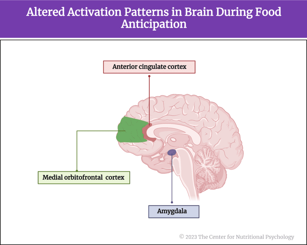
Figure 1. Areas in the brain linked with addiction
Due to this, the study of brain activity and structure may be key to better understanding obesity. One way to approach this is through multimodal magnetic resonance imaging.
Studies have linked food addiction with altered activation patterns in certain brain areas when food is anticipated
What is multimodal magnetic resonance imaging?
Multimodal magnetic resonance imaging (MRI) involves using multiple imaging techniques to create a more comprehensive picture of a certain part of the body, the brain. In multimodal brain MRI, different MRI scans are combined to provide information about the structure, function, and chemistry of examined brain regions.
The current study
The author of this study Ravi R. Bhatt and his colleagues wanted to understand the differences in brain structure and function between men and women who are obese and non-obese. Previous studies have shown that obese individuals show heightened activation in brain reward and salience networks when shown images associated with food, i.e., visual food cues (Rothemund et al., 2007). However, although there are many studies about differences in neural functioning between males and females, few studies have explored the differences in brain functioning between obese men and women.
What is the reward network of the brain?
The brain’s reward network is a group of interconnected brain regions active when we are involved in pleasurable activities. Under normal circumstances, this reward network controls an individual’s responses to natural rewards, such as food, sex, and social interactions. Activation of this network releases dopamine, a neurotransmitter that is associated with feelings of pleasure and motivation. This reinforces the behavior that led to the network’s activation and encourages the individual to repeat it in the future. Subjectively, it creates a rewarding experience, telling the individual to memorize what he/she just did to repeat it again in the future so that the rewarding experience would be repeated as well.
The brain’s reward network is a group of interconnected brain regions that are active when we are involved in pleasurable activities
The reward network includes the ventral tegmental area, the nucleus accumbens, and the prefrontal cortex regions of the brain. It is also called the mesolimbic dopamine system. Evolutionary, is a very old system found in many different organisms, including very simple ones such as worms and flies (see Figure 2).
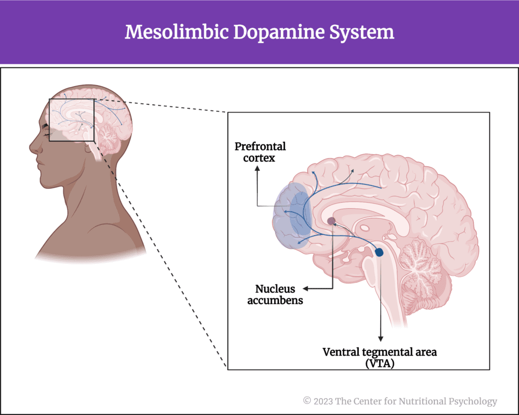
Figure 2. Reward network of the brain (aka mesolimbic dopamine system)
The salience network regulates our attention and responses to stimuli
The brain’s salience network is a group of connected brain regions that select which stimuli deserve our attention. It plays a critical role in detecting and responding to important sensory stimuli and regulates our attention and behavior in response to these stimuli.
The salience network of the brain is a group of connected brain regions that select which stimuli are deserving of our attention
Its key nodes are the anterior cingulate cortex and ventral anterior insular cortex regions of the brain, but it also has nodes in the amygdala, hypothalamus, ventral striatum, and thalamus regions of the brain, as well as in certain parts of the brainstem (see Figure 3).

Figure 3. Salience network of the brain
The study participants
The study participants were 183 persons between 18 and 55 years of age. They were recruited through advertisements between 2015 and 2021. Participants were divided into two groups. Those with body mass indexes above 25 represented the overweight/obese group. Those with body mass indexes of 19-20 were considered the non-obese groups. Participants were excluded if they had any major medical condition or current or past psychiatric illness.
Brain imaging
All participants completed multimodal magnetic resonance imaging. These consisted of a structural scan to create a detailed image of the brain’s internal structure, a resting-state blood-oxygen-level-dependent (or BOLD) scan, and a diffusion-weighted scan (Bhatt et al., 2023).
A blood-oxygen-level-dependent scan is a functional magnetic resonance imaging scan that measures changes in blood oxygenation in the brain. When a brain region is active, it uses oxygen, creating an increased demand for oxygenated blood that this type of scan detects. In this way, activity levels in different brain regions are measured.
A diffusion-weighted scan visualizes the movement of water molecules in brain tissues. It is particularly sensitive to the movements of these molecules along axons of nerve fibers. This is used to provide information about the microstructure of white matter in specific brain areas. Images are created indicating areas of high and low water molecule movement. Regions, where water molecule movement is high are regions with high white brain matter density, and regions where water molecule movement is low are regions of low white matter density.
Psychological assessments
Participants also completed a battery of self-report questionnaires. These included assessments of food addiction (the Yale Food Addictions Scale), childhood trauma (the Early Trauma Inventory and Childhood Traumatic Events Scale), anxiety and depression (Hospital Anxiety/Depression Scale and the State-Trait Anxiety Inventory, STAI), visceral sensitivity (the Visceral Sensitivity Index), stress and resilience to stress (the Perceived Stress, Scale, Brief Resilience Scale, Connor-Davidson Resilience Scale), personality traits (an international personality item pool derived questionnaire), perceptions of various aspects of one’s own health (Short-Form Health Survey) and others.
Obese individuals had worse scores across several psychological assessments
Results showed that participants from the obese group, on average, had worse scores on several psychological assessments compared to the non-obese group. They had higher scores on anxiety and depression assessments and higher average food addiction scores. They reported more childhood traumatic experiences, gastrointestinal problems, and higher sensitivity to them (see Figure 4).
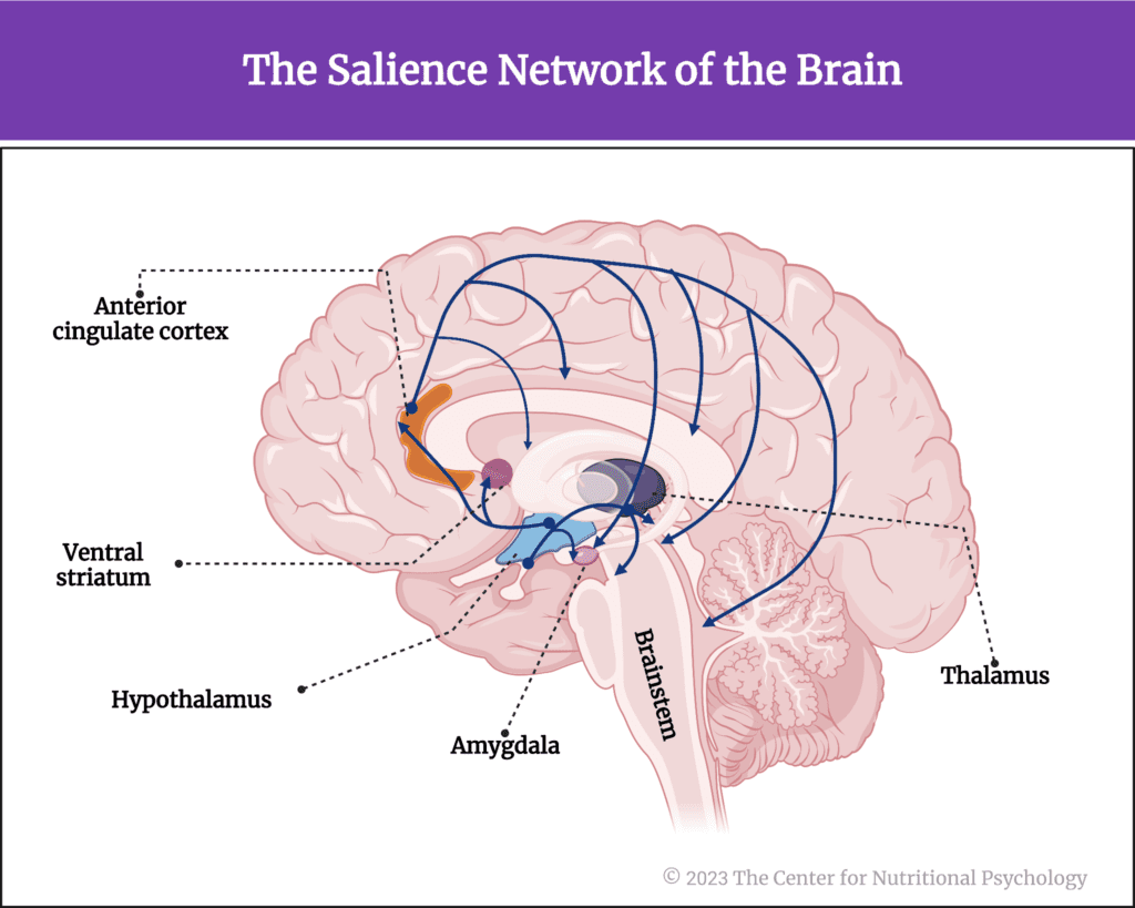
Figure 4. Results from the obese group
On average, they rated their physical and mental health worse than the participants in the non-obese group, i.e., they had lower scores on physical and mental health components of the Short-Form Health Survey.
The obese group, on average, had worse scores on several psychological assessments than the non-obese group
Obese individuals had lower connectivity in key parts of the reward network
Researchers created a statistical model to differentiate between obese and non-obese participants based on the results of brain scans and psychological assessments. This model was 77% accurate in classifying study participants into obese and non-obese groups.
Researchers found that participants from the obese group had lower connectivity in the cortico-basal-ganglia-thalamo-cortico loop. This loop is a key part of the reward network of the brain. In obese persons, it is known to receive inputs from the ventral tegmental area and substantia nigra regions of the brain to regulate the motivational and incentive properties of food. Lower connectivity indicates a lower count of white matter tracts within that part of the network. This, in turn, means that the network is less efficient in what it does, i.e., less able to regulate food intake beyond what the body needs.
Researchers found that participants in the obese group had lower connectivity in their cortico-basal-ganglia-thalamo-cortico loop, a key part of the brain’s reward network
Neuroimaging scans also showed that the brain’s choroid plexus and ventricle regions have greater volume in obese individuals. Changes in many other regions of the brain of obese individuals were also detected. Higher levels of these changes were found in participants who reported greater childhood trauma experiences.
Higher levels of these changes were found in participants who reported greater childhood trauma
Obese females had lower connectivity between the amygdala and the sensorimotor network of the brain
Obese females reported lower mental health compared to obese males. They also had lower connectivity between the amygdala and various regions of the sensorimotor network of the brain while the person was resting. The sensorimotor network is the neural pathway involved in processing sensory data and controlling motor reactions of the body. This lower connectivity was strongly associated with lower mental health. It has also been associated with increased motivational rewards placed on food-related stimuli.
Obese females reported lower mental health compared to obese males
Additionally, obese females had a lower surface area of the anterior cingulate cortex, which is a key hub of the brain’s salience network, compared to obese men. This difference was stronger in obese females who reported more physical symptoms and were generally more focused on their bodies.
The statistical model researchers developed was able to differentiate between obese males and females with 75% accuracy (Bhatt et al., 2023).
Conclusion
The study showed that there are differences in both brain structure and functioning between obese and non-obese individuals. It also reported differences in brain structures and functioning between obese males and females. This means that neural mechanisms causing and maintaining obesity might differ in males and females. Given this, treatments for obesity that consider these differences might be more effective than the existing ones. These findings contribute to the scientific understanding of the neural correlates and mechanisms of obesity. They may also contribute to developing novel approaches to treating this condition.
The paper “Integrated multimodal brain signatures predict sex-specific obesity status” was authored by Ravi R. Bhatt, Svetoslav Todorov, Riya Sood, Soumya Ravichandran, Lisa A Kilpatrick, Newton Peng, Cathy Liu, Priten P. Vora, Neda Jahanshad, and Arpana Gupta.
References
Bhatt, R. R., Todorov, S., Sood, R., Ravichandran, S., Kilpatrick, L. A., Peng, N., Liu, C., Vora, P. P., Jahanshad, N., Gupta, A., & Bhatt, R. R. (2023). Integrated multi-modal brain signatures predict sex-specific obesity status. Brain Communications, 5(2), 1–14. https://doi.org/10.1093/BRAINCOMMS/FCAD098
Gearhardt, A. N., Yokum, S., Orr, P. T., Stice, E., Corbin, W. R., & Brownell, K. D. (2011). Neural Correlates of Food Addiction. Archives of General Psychiatry, 68(8), 808–816. https://doi.org/10.1001/ARCHGENPSYCHIATRY.2011.32
Rothemund, Y., Preuschhof, C., Bohner, G., Bauknecht, H. C., Klingebiel, R., Flor, H., & Klapp, B. F. (2007). Differential activation of the dorsal striatum by high-calorie visual food stimuli in obese individuals. NeuroImage, 37(2), 410–421. https://doi.org/10.1016/J.NEUROIMAGE.2007.05.008
Wilding, J. P. H. (2001). Causes of obesity. Practical Diabetes International, 18(8), 288–292. https://doi.org/10.1002/PDI.277
Wong, M. C., Mccarthy, C., Fearnbach, N., Yang, S., Shepherd, J., & Heymsfield, S. B. (2022). Emergence of the obesity epidemic: 6-decade visualization with humanoid avatars. The American Journal of Clinical Nutrition, 115(4), 1189–1193. https://doi.org/10.1093/AJCN/NQAC005