Gut Microbiota Play Crucial Role in Mediating Effects of Western Diet

Introduction

The past several decades have seen the rise of an obesity pandemic that is ongoing worldwide. While obese individuals were quite rare just a century ago, 2015-2018 estimates for the U.S. state that more than two-thirds of the adult population is overweight or obese (Wong et al., 2022). Determining the causes of this increase in obesity rates has attracted much research attention. Studies have revealed a complex interplay between diet components, environmental factors, and previously unknown psychological and physiological mechanisms resulting in overeating and obesity in the long term. These novel studies on the intersection of nutrition and psychology are part of a developing field of science called nutritional psychology (The Center for Nutritional Psychology, 2023) (see Figure 1).


%learn about nutrition mental health %The Center for Nutritional Psychology

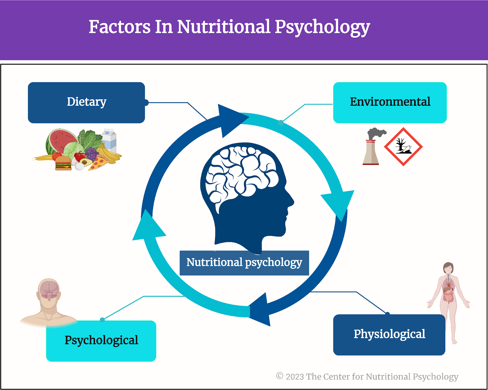
Figure 1. Diet, environment, psychological, and physiological factors in nutritional psychology 


There is a complex interplay between diet, environmental factors, and psychological and physiological mechanisms resulting in overeating and obesity


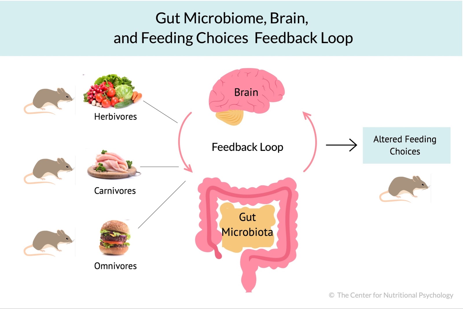
Gut microbiota and the microbiota-gut-brain axis

The human gut microbiome consists of trillions of microorganisms that live in the human intestinal tract. These microorganisms play a key role in digesting the food we eat. However, their influence extends beyond the gut, encompassing crucial roles in metabolic regulation, body weight maintenance, and immune system modulation. 

This growing body of evidence suggests that these gut microorganisms also profoundly impact brain functions, mood, cognition, and emotional well-being  (Zhu et al., 2023). This topic is explored in continuing education curricula within nutritional psychology — particularly how the gut microbiota and the gut-brain axis interconnect with the diet-mental health relationship to influence psychological functioning and experience, shedding light on its potential therapeutic implications for mental health outcomes.


This growing body of evidence suggests that gut microorganisms profoundly impact brain functions, mood, cognition, and emotional well-being


Scientists have recently discovered a communication pathway connecting the gut microbiome and the brain. This pathway is called the microbiota-gut-brain axis. It is based on small proteins called cytokines and a number of other biomolecules, including the hormone cortisol, short-chain fatty acids (SCFAs), tryptophan, and others.

The Western diet

The Western diet is a modern dietary pattern prevalent in Western societies, characterized by a high intake of processed and hyperpalatable foods with increased contents of fat, sugary snacks, and refined grains. It typically includes low consumption of fruits, vegetables, unprocessed-high-quality proteins, nuts, and seeds. This diet’s excessive reliance on added sugars and unhealthy fats has been linked to an increased risk of obesity, metabolic syndrome, and various chronic diseases.

Studies have indicated that feeding mice a Western diet causes inflammation in the region of the brain called the hypothalamus (Heiss et al., 2021; Thaler et al., 2013). Inflammation of the hypothalamus damages the neurons and leads to the formation of scars made of glial cells. This is called gliosis. Inflammation of the hypothalamus often happens before a mouse starts gaining weight. Due to this, scientists believe it might cause weight gain by causing leptin resistance.


Studies have indicated that feeding mice a Western diet causes inflammation of the region of the brain called the hypothalamus


Leptin and leptin resistance

Leptin is a hormone produced by fat cells during eating. It regulates appetite and body weight and is produced in proportion to the amount of fat in the body. Leptin concentrations inform the brain of how much fat is stored. Increased leptin concentrations (normally caused by an abundance of body fat) “tell” the brain to decrease food intake and increase energy expenditure.


Leptin is a hormone produced by fat cells that regulate appetite and body weight


Factors such as chronic inflammation or eating high-fat diets (HFDs) may cause the body to be less receptive to leptin. This is called leptin resistance. Leptin resistance results in disrupted appetite and energy regulation, i.e., the brain does not reduce food intake in spite of the abundance of body fat. This can contribute to obesity and cause difficulty controlling body weight (Thaler et al., 2013) (see Figure 2).


%learn about nutrition mental health %The Center for Nutritional Psychology

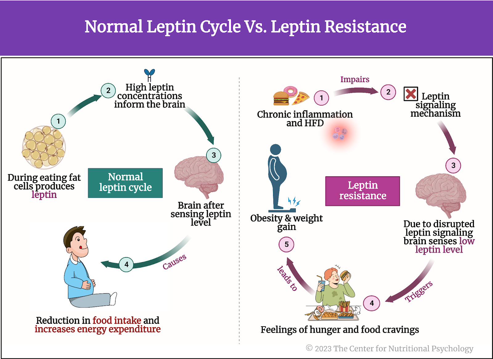
Figure 2. Normal leptin cycle versus leptin resistance.


Gliosis, leptin resistance, and gut microbiota

Microglia are a type of immune cell in the central nervous system that helps protect and maintain the brain and spinal cord by detecting and responding to potential threats or damage. Studies have shown that activation of microglia cells that happens during inflammation of the hypothalamus might be causing leptin resistance. Removing these microglia cells from the hypothalamus has improved sensitivity to leptin. Improved sensitivity to leptin allows the brain to recognize when enough fat is stored in the body and reduce food intake. 


Studies have shown that the activation of microglia cells that happens during inflammation of the hypothalamus might be causing leptin resistance


Intriguingly, according to the scientific evidence presented in our recent NP 120 course, it has been discovered that the gut microbiota plays a significant role in regulating the development and maturation of microglia cells and influencing their function. Although the mechanism of this action remains unknown, it has led scientists to believe there might be a link between hypothalamus inflammation and gut microbiota (Heiss et al., 2021) (see Figure 3).


%learn about nutrition mental health %The Center for Nutritional Psychology

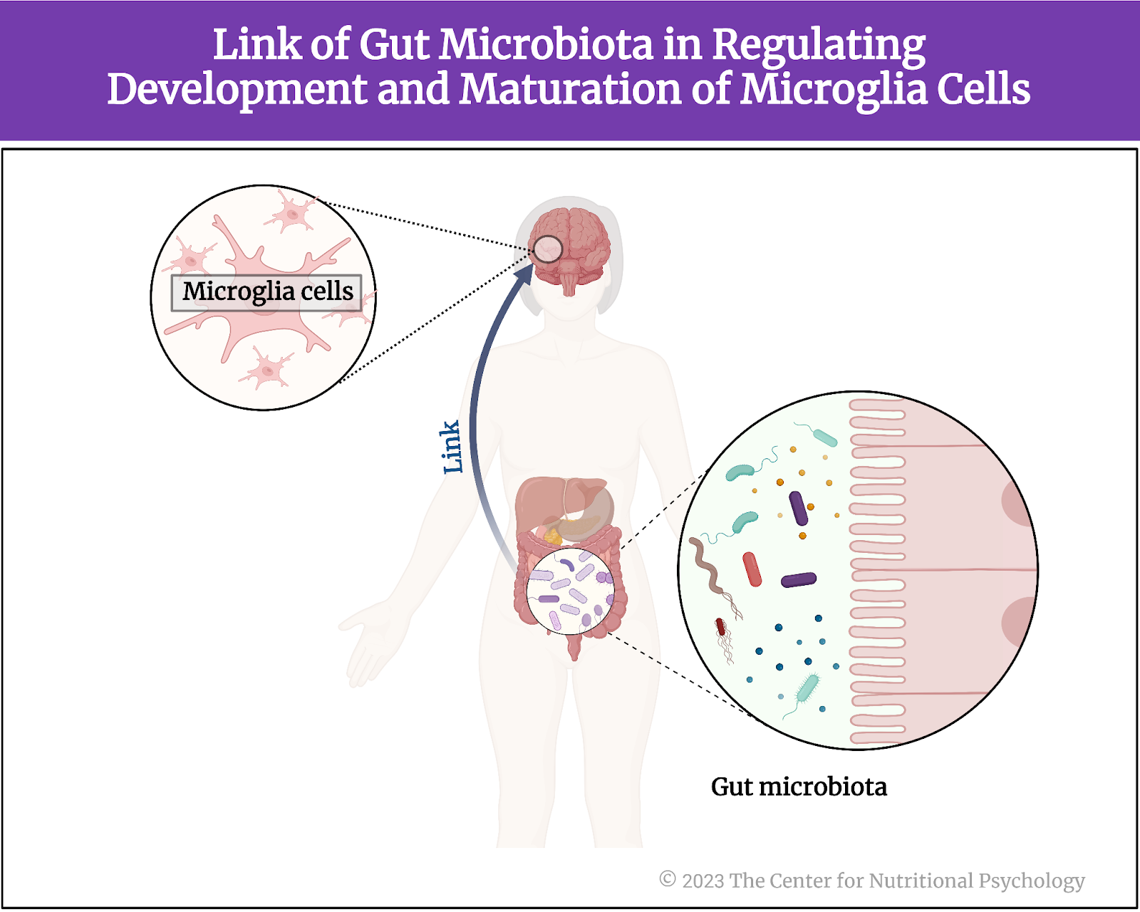
Figure 3. Link of Gut Microbiota in regulating development and maturation of microglia cells


There might be a link between hypothalamus inflammation and gut microbiota (Heiss et al., 2021)


The current study

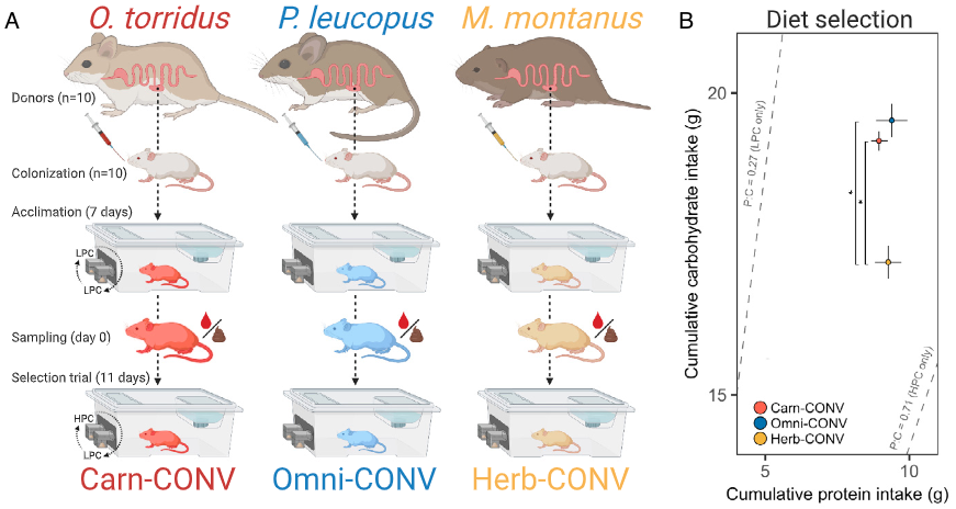
Study author Christina N. Heiss and her colleagues wanted to know whether mice that lack gut microbiota are protected against diet-induced inflammation of the hypothalamus. They noted previous studies’ results showing mice with depleted gut microbiota and those treated with antibiotics to be protected from diet-induced obesity.

In other words, they noted that mice that consume a Western diet, a diet that normally leads to obesity in mice, do not become obese if microbiota are not present in their guts. This might be because the absence of microbiota prevents inflammation of the hypothalamus, which would, in turn, prevent leptin resistance from developing. If this is the case, the mechanism for preventing overeating based on leptin would remain intact, preventing mice from becoming obese. Alternatively, it could be that, without microbiota, the guts of mice could not digest complex nutrients from the food they eat, thus substantially reducing the amount of nutrition they can derive from food. In this case, obesity would be avoided because their bodies cannot use their food. But which of these is the case?

The procedure

The study authors used three groups of male mice, 10-13 weeks old – conventionally raised mice, germ-free mice, and antibiotic-fed mice. They used several genetic groups of mice, including a strain of genetically engineered mice that allow for controlled and regulated manipulation of specific genes in specific tissues (Tamoxifen-inducible Cre mice).

In the scope of the experiments, researchers fed mice either a chow diet or a Western diet. Western diet was given for either 2 days, 1 week, or 4 weeks, depending on the experiment conducted in the scope of the study.

The chow diet for mice is a nutritionally balanced and standardized diet formulated to provide essential nutrients required for the health and growth of laboratory mice. It typically consists of a combination of proteins, carbohydrates, fats, vitamins, and minerals in pellet or block form. The Western diet used in this study was high in fat and sucrose, with 40% of calories coming from fat.

All food for mice was sterilized, i.e., underwent procedures that killed all microorganisms in the food before mice ate it. The chow diet was sterilized in an autoclave, which uses high pressure and steam to kill microorganisms. The Western diet food was irradiated, i.e., radiation was used to kill microorganisms.

The mice

Conventionally raised mice were laboratory mice kept in regular conditions and fed a normal diet for laboratory mice. They have normal gut microbiota.

Germ-free mice are created through techniques that ensure they do not acquire gut microbiota from birth through their entire lifetimes. They are typically born using cesarean section deliveries of pregnant mice in a sterile environment. This is done to prevent the transfer of microbes during birth. After that, they are kept in specialized sterile isolation spaces called isolators or bubbles that maintain a controlled germ-free environment.

These isolators provide filtered air, sterile food, and autoclaved water to prevent microbial contamination. Researchers raising these mice take special care to maintain strict barrier measures, including specialized clothing and equipment, to prevent the unintentional introduction of microorganisms. They regularly monitor these mice’s bodily fluids and tissues through special techniques to ensure the absence of any detectable microorganisms. Germ-free mice are typically leaner than conventionally raised mice and, consequently, have lower leptin levels in circulation.

Antibiotic-fed mice in this study had 1g of ampicillin and 0.5g of neomycin added per liter of their drinking water. Ampicillin and neomycin are antibiotics. Researchers kept the drinking water with antibiotics added in bottles protected from light. They prepared a new solution every second day. Researchers started giving this water with antibiotics to mice three days before changing their regular diet to a Western diet.


Mice without gut microbiota are protected from diet-induced inflammation of the hypothalamus


Results showed that conventionally raised mice fed a Western diet for 1 week developed gliosis in the hypothalamus. Inflammation indicators were increased in these mice in the part of the hypothalamus called the arcuate nucleus compared to conventionally raised mice fed regular mice food (chow) (see Figure 4).


%learn about nutrition mental health %The Center for Nutritional Psychology

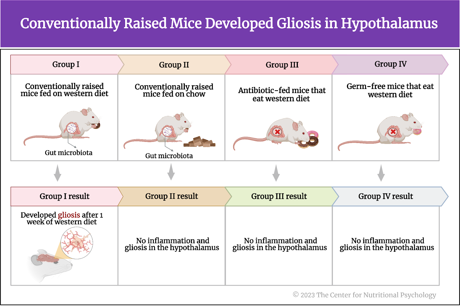
Figure 4. Conventionally raised mice fed a Western diet developed gliosis in the hypothalamus


When researchers examined germ-free mice and mice whose gut microbiota were depleted using antibiotic treatment (antibiotic-fed mice), results showed that these mice also showed no increase in inflammation indicators or the proliferation of microglia cells after eating a Western diet for a week.


Gliosis in the hypothalamus leads to greater gain in body weight and fat mass


The study authors also wanted to know whether gliosis in the hypothalamus caused by a Western diet leads, in turn, to increased body weight and fat mass accumulation in mice. To test that, they fed conventionally raised mice and antibiotic-fed mice a Western diet for 4 weeks.

Results showed that conventionally raised mice fed a Western diet gained more body weight and fat mass than antibiotic-fed mice. Compared to antibiotic-fed mice, they also had increased hypothalamus inflammation indicators and increased numbers of a specific type of microglia cells (iba1-positive microglia).

There was no association between the number of microglia in the arcuate nucleus region of the hypothalamus and changes in body weight or fat mass at the end of the 4 weeks. However, fat mass and the relative increase in fat mass during the study were associated with the number of a specific type of glial cells called astrocytes.

Further analysis showed that germ-free and antibiotic-fed mice are more sensitive to leptin than conventionally raised mice. When researchers gave them leptin injections, the first two types of mice reduced their food intake more than conventionally raised mice did.


Glucagon-like-peptide 1 (GLP-1) seems to be crucial for protection against diet-induced inflammation of the hypothalamus


Germ-free and antibiotic-fed mice had higher levels of the hormone called glucagon-like peptide 1 (GLP-1) when they were fed a regular diet. This hormone secreted in the small intestine’s intestinal lining cells (L cells) is important in regulating blood sugar levels. It also helps reduce inflammation and protect neurons.

Study authors believed it might also be crucial for the protection from inflammation of the hypothalamus induced by the Western diet. After mice were fed a Western diet for a week, antibiotic-treated and germ-free mice had higher levels of GLP-1 than conventionally raised mice. These mice did not gain weight or develop hypothalamic inflammation after this diet. However, when researchers measured these same things in antibiotic-fed and germ-free mice whose GLP-1 signaling pathway was disabled, they also gained weight and developed inflammation, similar to conventionally raised mice. This indicated that the functional signaling path of GLP-1 is crucial for countering the inflammation of the hypothalamus induced by a Western diet.


Just a week on a Western diet led to inflammation of the hypothalamus that, in turn, disrupted the body’s mechanism for regulating food intake


Conclusion

These findings in mice show that gut microbiota changes how the organism, of mice in this case, reacts to a Western diet. When gut microbiota was intact, just a week on a Western diet led to inflammation of the hypothalamus that, in turn, disrupted the body’s mechanism for regulating food intake. However, when gut microbiota was depleted or absent, this inflammation did not happen, provided that the signaling pathway of one specific hormone (GLP-1) was intact.

While the study was done on mice, similar physiological mechanisms exist in humans. Due to this, these findings on mice help scientists better understand how and through which physiological mechanisms changes in the human diet that occurred in the last century disrupted food intake regulation in the human body leading to the current obesity pandemic.

The paper “The gut microbiota regulates hypothalamic inflammation and leptin sensitivity in Western diet-fed mice via a GLP-1R-dependent mechanism” was authored by Christina N. Heiss, Louise Manneras-Holm, Ying Shiuan Lee, Julia Serrano-Lobo, Anna Hakansson Gladh, Randy J. Seeley, Daniel J. Drucker, Fredrik Backhed, and Louise E. Olofsson.

References

Heiss, C. N., Mannerås-Holm, L., Lee, Y. S., Serrano-Lobo, J., Håkansson Gladh, A., Seeley, R. J., Drucker, D. J., Bäckhed, F., & Olofsson, L. E. (2021). The gut microbiota regulates hypothalamic inflammation and leptin sensitivity in Western diet-fed mice via a GLP-1R-dependent mechanism. Cell Reports, 35(8). https://doi.org/10.1016/j.celrep.2021.109163

Thaler, J. P., Guyenet, S. J., Dorfman, M. D., Wisse, B. E., & Schwartz, M. W. (2013). Hypothalamic inflammation: Marker or mechanism of obesity pathogenesis? Diabetes, 62(8), 2629–2634. https://doi.org/10.2337/DB12-1605

The Center for Nutritional Psychology. (2023). What is Nutritional Psychology? https://www.nutritional-psychology.org/what-is-nutritional-psychology/

Wong, M. C., Mccarthy, C., Fearnbach, N., Yang, S., Shepherd, J., & Heymsfield, S. B. (2022). Emergence of the obesity epidemic: 6-decade visualization with humanoid avatars. The American Journal of Clinical Nutrition, 115(4), 1189–1193. https://doi.org/10.1093/AJCN/NQAC005

Zhu, X., Sakamoto, S., Ishii, C., Smith, M. D., Ito, K., Obayashi, M., Unger, L., Hasegawa, Y., Kurokawa, S., Kishimoto, T., Li, H., Hatano, S., Wang, T. H., Yoshikai, Y., Kano, S. ichi, Fukuda, S., Sanada, K., Calabresi, P. A., & Kamiya, A. (2023). Dectin-1 signaling on colonic γδ T cells promotes psychosocial stress responses. Nature Immunology. https://doi.org/10.1038/s41590-023-01447-8


Researchers Discover Immune Mechanism Linking Changes in Gut Microorganisms and Behavior after Chronic Stress

Stress is inevitable, but it can disturb our body’s physiological signaling mechanisms when it becomes chronic. These mechanisms are interlinked with the Microbiota Gut-brain axis (MGBA) and influence the diet-mental health relationship (DMHR) in many ways. Being a major immune organ and highly colonized with the microbiome, our gut experiences certain immune inflammatory responses due to stress, which affect the gut microbiome composition and contribute to the onset of depression and anxiety. 

A new study led by researchers at Johns Hopkins has strengthened our understanding of the role of specific gut immune cells in microbiota composition and influencing the brain’s responsiveness to stress. This early-stage experiment on mice found that a specific type of white blood cells, gamma-delta T lymphocytes, play a key role in the cellular mechanism leading to adverse behavioral changes under chronic stress. After chronic stress, some mice in the experiment developed social avoidance behavior, i.e., they started avoiding contact with other mice. These mice had reduced diversity of microorganisms in their guts and increased concentrations of gamma-delta T lymphocytes in their intestines and in the membranes surrounding their brains. Under equal chronic stress conditions, mice without gamma-delta T lymphocytes did not develop social avoidance behavior (Zhu et al., 2023). The study was published in Nature Immunology.


After chronic stress, some mice in the experiment started avoiding contact with other mice. They had reduced microbial diversity and increased concentrations of gamma-delta T lymphocytes in their gut and surrounding their brain


Chronic stress

Chronic stress is a consistent sense of feeling pressured and overwhelmed over a long period of time. There are many possible sources of chronic stress in modern society. These include bad living conditions and homelessness, bad family and social relations, negative interactions between work and family, adverse work conditions, illness, and many others (Armon et al., 2014; Goodman et al., 1991; Tsukerman et al., 2020). Chronic stress slowly drains a person’s psychological energy and has damaging effects on both health and well-being.

Chronic stress and the gut microbiota

Physiologically, chronic stress induces immune changes and inflammation, leading to psychiatric disorders such as depression and anxiety (Hodes et al., 2014). These immune changes include changes to the gut microbiota – the trillions of microorganisms that live in the human intestinal tract (Figure 1). 


%learn about nutrition mental health %The Center for Nutritional Psychology

Figure 1. Chronic stress leads to immune changes, inflammation, depression, and anxiety


Gut microorganisms play a key role in digesting food, but they are also incredibly important for various other processes, such as the differentiation of certain immune cells (Zhu et al., 2023). Differentiation is when immature and unspecialized cells transform into specialized, mature cells that perform specific bodily functions. It is one of the critical processes of life. When a person is under stress, the body reacts with inflammation, that in turn affects the composition of microorganisms in the gut but also creates physiological changes that reach the brain and affect cognition and behavior.

Due to this, studying physiological changes associated with chronic stress is very important for understanding the development of the most common psychiatric disorders and finding effective ways to treat them. However, research ethics and practical considerations impose very strict limits on what types of studies can be conducted on humans. That is why studies of the physiology and biochemistry of chronic stress are often done on animals, particularly mice, using specific research protocols. One research protocol used to induce chronic stress in mice for research purposes is the chronic social defeat stress protocol.

What is chronic social defeat stress?

Chronic social defeat stress is a protocol (procedure) in which a mouse is exposed to a larger aggressive mouse in an enclosed space. This is followed by a confrontation between the two mice in which the mouse undergoing this treatment is defeated and forced into a subordinate position (social defeat). Typically, this procedure is repeated daily over ten days (Figure 2). 


%learn about nutrition mental health %The Center for Nutritional Psychology

Figure 2. Chronic Social Defeat Stress Protocol


The chronic social defeat stress protocol produces effects similar to depression in exposed mice. It also produces a number of other easily detectable effects such as increased weight of spleen of these mice, lower preference for sucrose, and others. That is why it is extensively used in research on mice (Golden et al., 2011).


The chronic social defeat stress protocol produces effects similar to depression in exposed mice. 


The current study

Study author Xiaolei Zhu and his colleagues wanted to explore the cellular mechanisms behind social avoidance behaviors caused by chronic stress. They were particularly interested in the role a specific type of white blood cell called the gamma-delta T lymphocyte has in these changes and in the changes in the composition of gut microorganisms caused by stress.

What are gamma-delta T lymphocytes?

Gamma-delta T lymphocytes (γδ T-cells) are a specific type of white blood cells in the body. Still, they are found in high concentrations in various mucosal tissues called meninges, including intestines and membranes surrounding the brain. Lymphocytes are involved in the body’s immune responses. Studies have shown that gamma-delta T lymphocytes located in the meninges regulate anxiety-like behaviors and memory. Furthermore, research indicated that gamma-delta T lymphocytes in the intestines could travel to the meninges under certain conditions. This has led scientists to assume that these gamma-delta T lymphocytes from the gut may be involved in brain function changes when inflammation occurs (Zhao et al., 2018; Zhu et al., 2023) (Figure 3).


%learn about nutrition mental health %The Center for Nutritional Psychology

Figure 3. Gamma-delta T lymphocytes are in intestinal mucosal tissue and membranes around the meninges. 


The experiment

The study authors applied the chronic defeat stress protocol on a group of mice. Afterward, researchers examined the social behavior of these mice (towards other mice, using a social interaction test). They noticed that some of these mice started avoiding contact with other mice in a test situation, i.e., manifested social avoidance behavior, while others did not. They named the group of mice that showed social avoidance behavior the susceptible group. In contrast, the group of mice that did not show social avoidance behavior was named the resilient group. Researchers kept a third group of the same genetic strain of mice as controls and did not expose them to the chronic social defeat protocol (see Figure 4).


%learn about nutrition mental health %The Center for Nutritional Psychology

Figure 4. Susceptible vs. resilient vs. no exposure


After the procedure, researchers collected stool samples from the mice and conducted their genetic analysis in order to identify the compositions of microorganisms present in the guts of these mice. This procedure is called the metagenomic sequencing of the gut microbiota. They also used a procedure called flow cytometry to determine the number and characteristics of gamma-delta T lymphocytes in the gut and in the meninges of these mice.

Comparison with humans

Parallel with this experiment, researchers investigated the differences in the composition of gut microbiota in humans diagnosed with major depressive disorder and healthy individuals by analyzing their stool samples. They found that a lower abundance of the Lactobacillus group of bacteria in the gut was associated with higher depression and anxiety symptoms. They confirmed this using three different assessments of depression and anxiety symptoms (the Montgomery-Asberg depression scale, the Hamilton Depression scale, and the Hamilton Anxiety Scale)(Zhu et al., 2023). Based on this, study authors assumed that concentrations of these bacteria in the gut might play a role in the vulnerability to stress in mice and humans. They decided to look for differences in the abundance of Lactobacillus bacteria in different groups of mice in their experiment.

Susceptible mice had a reduced abundance of Lactobacillus johnsonii bacteria in the gut! (See Figure 5).


%learn about nutrition mental health %The Center for Nutritional Psychology 
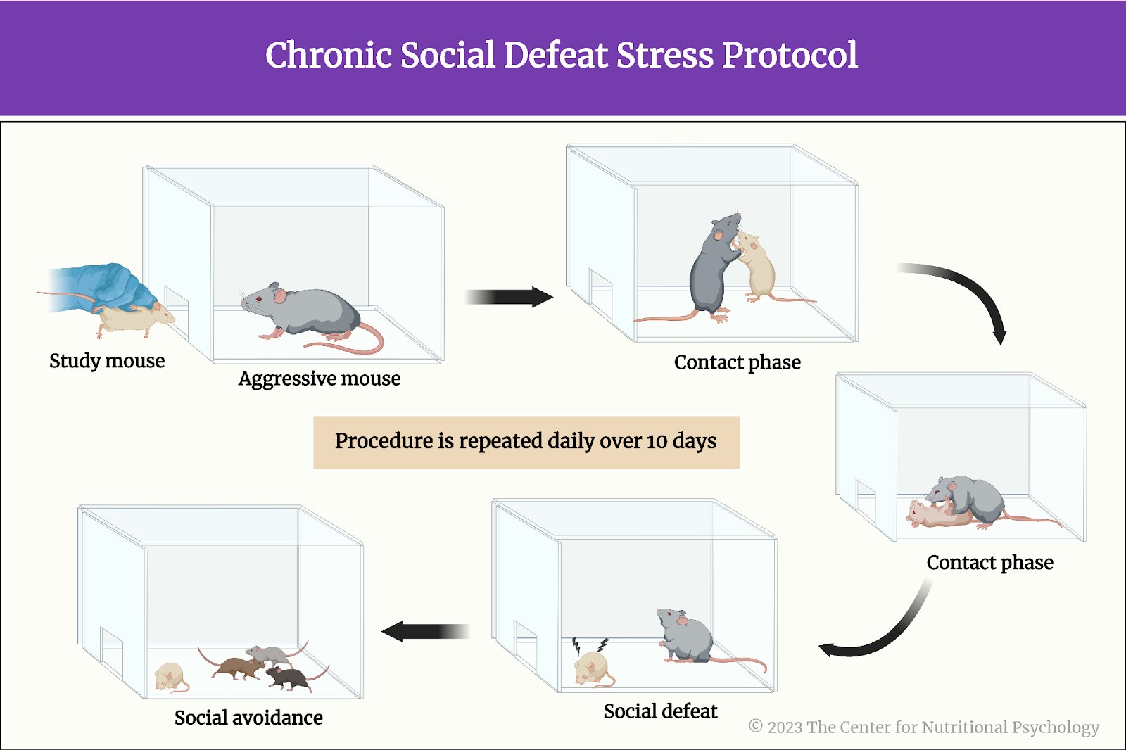
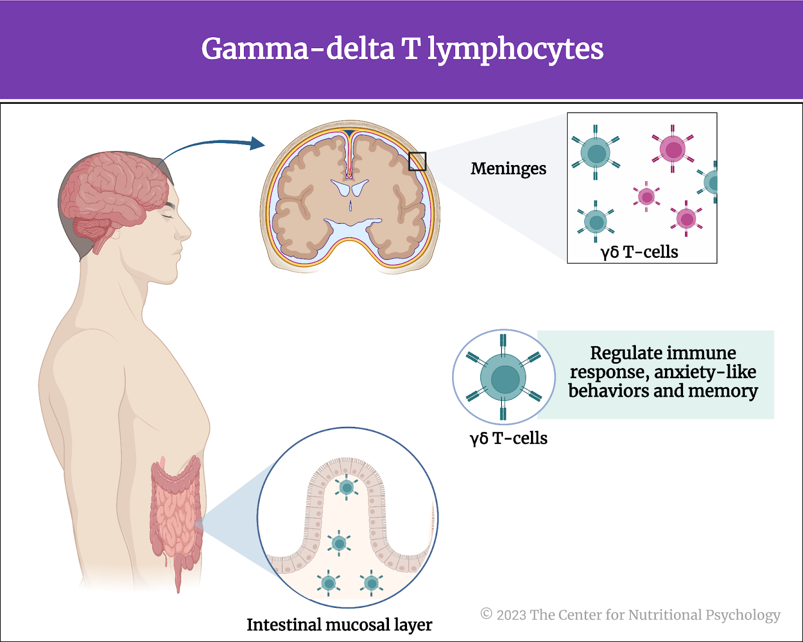
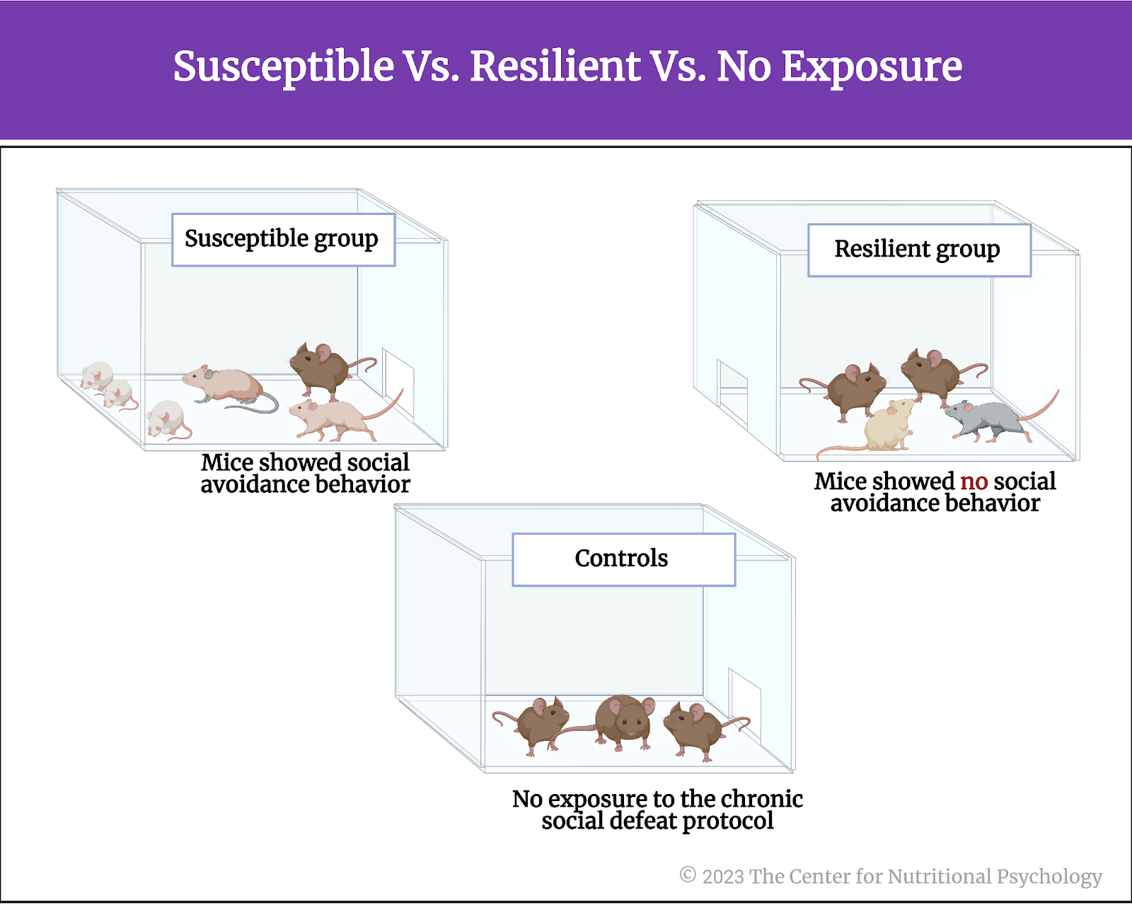
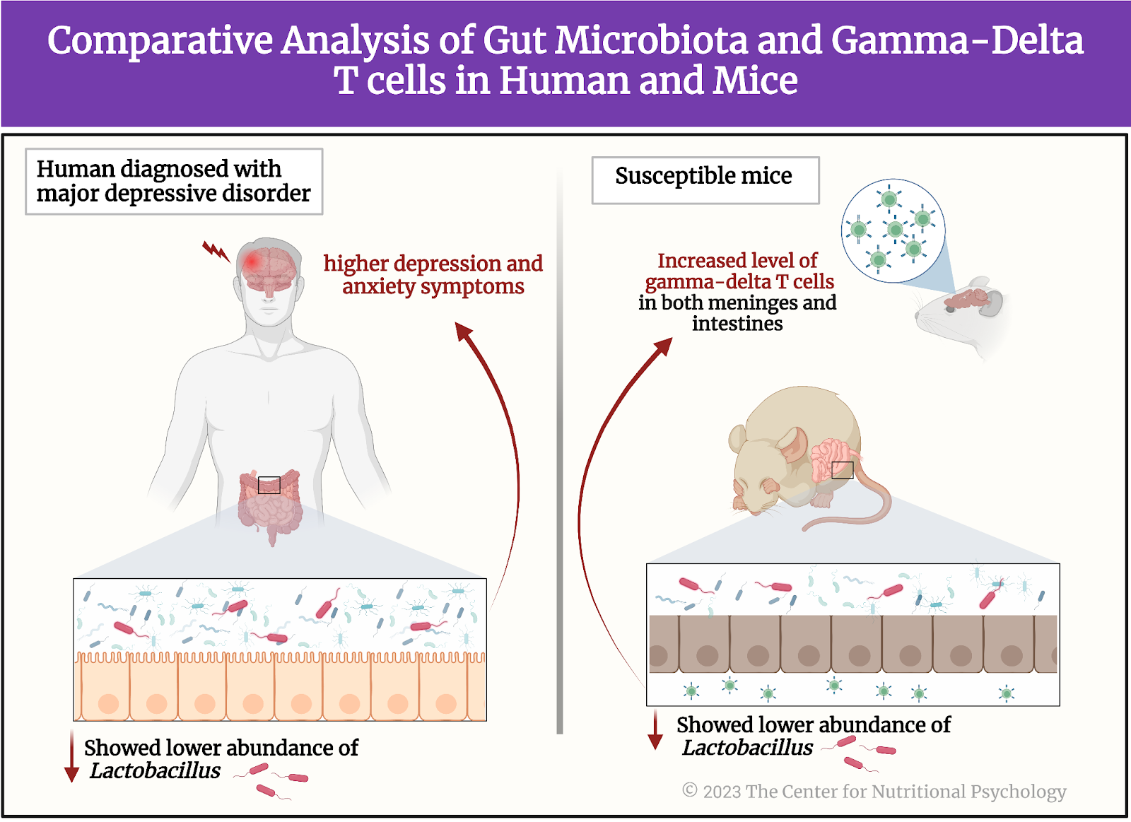
Figure 5. Humans + Mouse experiment showing a lower abundance of lactobacillus in gut bacteria = with higher depression and anxiety


A comparison of the gut microbiota of susceptible mice, resilient mice, and the control group showed that susceptible mice had less diverse microbial populations in the gut. The gut microbiota of susceptible mice differed from the gut microbiota of resilient mice and the control group on a number of bacterial species. As researchers expected, one of these species was Lactobacillus Johsonii. Their concentration was reduced in susceptible mice’s guts compared to resilient mice and the control group.

Susceptible mice had increased concentrations of gamma-delta T cells in both meninges and intestines

Given the previously described relationship between Lactobacillus bacteria and immune responses, researchers examined whether concentrations of gamma-delta T lymphocytes were increased in mice exposed to the chronic social defeat stress treatment. Results showed that susceptible mice had increased concentrations of gamma-delta T lymphocytes in their colons and meninges (membranes surrounding the brain) compared to resilient and healthy mice.


Susceptible mice had increased concentrations of gamma-delta T lymphocytes both in their colons and their meninges 


In susceptible mice, many of the gamma-delta T cells in brain membranes came from the gut!

Researchers then wanted to know whether the gamma-delta T cells found in the meninges of susceptible mice were cells that differentiated there or those cells that traveled from the gut. They identified differences between these two types of gamma-delta T cells and measured their concentrations. Results showed that, in resilient mice and the control group, gamma-delta T lymphocytes found in the meninges were indeed differentiated. However, in susceptible mice, there were fewer such cells, but many gamma-delta T lymphocytes came from the gut.


In susceptible mice, there were fewer such cells, but there were lots of gamma-delta T lymphocytes that came from the gut


Social avoidance after the chronic social defeat stress does not develop in mice without gamma-delta T lymphocytes.

Finally, researchers wanted to test whether the gamma-delta T lymphocytes were responsible for the social avoidance behavior after exposure to chronic social defeat. They repeated the procedure on a new group of special mice that did not have the gamma-delta T cells. As researchers expected, these mice did not develop social avoidance behavior after exposure to the chronic social defeat stress protocol.

Conclusion

The study showed that a certain type of white blood cells – gamma-delta T lymphocytes and their accumulation play a key role in changes to behavior induced by stress. In the context of MGBA-DMHR, it also demonstrates interactions between gut microbiota, the body’s immune responses, and the brain when an organism is stressed. Given that many of the physiological processes in mice are similar to those in humans, these findings contribute to the scientific understanding of physiological mechanisms of behavioral changes that chronic stress and related disorders in humans. These insights can help develop novel ways to treat and prevent major depressive disorder and other stress-related disorders. They can also open new approaches to diagnosing individual susceptibility to stress and increasing resilience.

The paper “Dectin-1 signaling on colonic gamma-delta T cells promotes psychosocial stress responses” was authored by Xiaolei Zhu, Shinji Sakamoto, Chiharu Ishii, Matthew D. Smith, Koki Ito, Mizuho Obayashi, Lisa Unger, Yuto Hasegawa, Shunya Kurokawa, Taishiro Kishimoto, Hui Li, Shinya Hatano, Tza-Huei Wang, Yasunobu Yoshikai, Shin-ichi Kano, Shinji Fukuda, Kenji Sanada, Peter A. Calabresi, and Atsushi Kamiya.

For more research in the Microbiota Gut-Brain Axis — Diet-Mental Health Relationship (MGBA-DMHR), visit CNP’s Nutritional Psychology Research Library (NPRL) Microbiota Gut-Brain Axis—Diet-Mental Health Relationship research category, or enroll in NP 120: Microbiota Gut-Brain Axis and the DMHR (available in May 2023).

References

Armon, G., Melamed, S., Toker, S., Berliner, S., & Shapira, I. (2014). Joint Effect of Chronic Medical Illness and Burnout on Depressive Symptoms Among Employed Adults. Health Psychology, 33(3), 264–272. https://doi.org/10.1037/a0033712

Golden, S. A., Covington, H. E., Berton, O., & Russo, S. J. (2011). A standardized protocol for repeated social defeat stress in mice. Nature Protocols, 6(8), 1183–1191. https://doi.org/10.1038/nprot.2011.361

Goodman, L., Saxe, L., & Harvey, M. (1991). Homelessness as psychological trauma. American Psychologist, 46(11), 1219.

Hodes, G. E., Pfau, M. L., Leboeuf, M., Golden, S. A., Christoffel, D. J., Bregman, D., Rebusi, N., Heshmati, M., Aleyasin, H., Warren, B. L., Lebonté, B., Horn, S., Lapidus, K. A., Stelzhammer, V., Wong, E. H. F., Bahn, S., Krishnan, V., Bolaños-Guzman, C. A., Murrough, J. W., … Russo, S. J. (2014). Individual differences in the peripheral immune system promote resilience versus susceptibility to social stress. Proceedings of the National Academy of Sciences of the United States of America, 111(45), 16136–16141. https://doi.org/10.1073/pnas.1415191111

Tsukerman, D., Leger, K. A., & Charles, S. T. (2020). Work-family spillover stress predicts health outcomes across two decades. Social Science & Medicine, 265, 113516. https://doi.org/10.1016/j.socscimed.2020.113516

Zhao, Y., Niu, C., & Cui, J. (2018). Gamma-delta (γδ) T Cells: Friend or Foe in Cancer Development. Journal of Translational Medicine, 16(1), 1–13. https://doi.org/10.1186/s12967-017-1378-2 

Zhu, X., Sakamoto, S., Ishii, C., Smith, M. D., Ito, K., Obayashi, M., Unger, L., Hasegawa, Y., Kurokawa, S., Kishimoto, T., Li, H., Hatano, S., Wang, T. H., Yoshikai, Y., Kano, S. ichi, Fukuda, S., Sanada, K., Calabresi, P. A., & Kamiya, A. (2023). Dectin-1 signaling on colonic γδ T cells promotes psychosocial stress responses. Nature Immunology. https://doi.org/10.1038/s41590-023-01447-8


Why are Hyper-Palatable Foods so Alluring? New Type of Brain Cell Lends Insight

Our food preferences have changed significantly over the last decade, with processed, hyperpalatable foods being increasingly available and consumed. A substantial percentage of foods in the US food system (62%) is considered to be hyperpalatable, including foods not previously conceptualized as hyperpalatable, such as foods labeled “reduced” or “low fat” and sauces, trail mixes, vegetables cooked in creams, etc. (Fazzino et al., 2019).


Sixty-two percent of the foods in the US food system may be hyperpalatable, including foods not previously conceptualized as hyperpalatable.


We know that the highly palatable sensations and interoceptive experiences associated with these foods can activate the brain’s reward system, providing us with an experience of pleasure and reward. Activation of the reward system occurs in other activities such as achieving a goal or engaging in addictive behaviors like substance abuse. In truth, there are many factors involved in our choosing of highly palatable foods, including the availability of these foods, and the social environment in which we live.

Have you ever wondered why hyperpalatable foods are so desirable, and why they have been increasingly making it into our shopping carts despite the knowledge that they lack nutrients? Certainly, it is because hyperpalatable foods are cheap, convenient, and taste good, not to mention they give you a rewarding and memorable experience. However, what you might not know is that there’s a type of neuron in our brain that influences our dietary intake patterns.

In truth, many factors influence our dietary intake behavior and food cravings (learn more in NP 110) – both of which are important topics in the study of the diet-mental health relationship (DMHR) and within nutritional psychology. In this article, we focus on the new discovery of a type of brain cell (neuron) which may play a role in our desire to consume these highly processed foods.


Neurotensin neurons are a new structure that might impact our eating behavior.


Alessandro Furlan and his research team explored a new structure within the brain that can modulate dietary intake behavior and increase our preference for fatty foods. This structure is a group of neurons called neurotensin neurons, located in a part of the amygdala known as the interstitial nucleus of the posterior limb of the anterior commissure (IPAC) (Furlan et al., 2022). Let’s uncover the crucial role of these neurotensin neurons within the IPAC and see what Furlan and his team discovered about their role in orchestrating our dietary intake behaviors and metabolic health.


Activation of neurotensin neurons can promote hedonic feeding and weight gain.


This experiment consisted of four steps. First, they examined how activation of the neurotensin neurons can change the food behavior of the animals (i.e., whether they will be more interested in palatable food). They then examined how different foods can activate neurotensin neurons (i.e., fat vs. water). They also tested whether these cells play a role in food-seeking behavior by testing whether they are activated by merely food stimuli. Finally, they examined how inhibiting these cells can alter the food behavior of animals. 

Furlan found many things in their study, including:

  • Activation of neurotensin neurons can promote hedonic feeding. Hedonic feeding and hunger involve the consumption of food uniquely because of its rewarding properties (pleasure) rather than eating for homeostatic energy balance (Monteleone, 2012). 
  • When rats had their neurotensin neurons activated, they exhibited increased dietary intake. The effect was more pronounced for more palatable high-fat diets than for chow.
  • When mice were fed with either fat or water, they found that immediately following ingestion, neurotensin neurons were more strongly activated by fat than by water in a concentration-dependent manner. 
  • They also tried to see the effect when they compared two palatable foods (with one of them being more palatable). They found that these cells were activated by sucrose more than by sucralose (a palatable, sweet food but non-caloric sugar analog).
  • Neurotensin neurons can be activated by sensory cues that anticipate meals (i.e., odors). This means that they also play a role in food-seeking behaviors.

As shown in Figure 1, these findings show that neurotensin neurons are not only influenced by the type of food consumed (i.e., highly palatable), but that they also activate our response to food stimuli, and their activity is scaled by the palatability of food. They affect our hedonic feeding behavior and increase our intake of highly palatable foods.

%learn about nutrition mental health %The Center for Nutritional Psychology Figure 1. Summary of how neurotensin neurons can affect our food behavior


When neurotensin neurons are inhibited, the preference for highly palatable food decreases.


Conversely, acute inhibition of neurotensin neurons reduces feeding on a high-fat diet in sated mice and reduces feeding in hungry mice. In addition, prolonged inactivation of neurotensin neurons leads to a dramatic increase in aerobic locomotion and long-term weight loss.

The inactivation of these cells protected mice from obesity and the long-term negative consequences of chronic feeding. Mice who had their neurotensin neurons inhibited exhibited higher energy expenditure, higher lipid oxidation rate, low blood sugar levels, and engaged in more activity. A summary of the research findings by Furlan and his team is shown in Figure 2.

%learn about nutrition mental health %The Center for Nutritional Psychology

Figure 2. Summary of findings by Furlan and his team


What are the implications of this in everyday life?

The desire to consume highly palatable foods more frequently than healthier, nutrient-dense foods is a major cause of the current obesity pandemic. Exercise programs are known to be the first-line intervention when treating obesity, however, body weight is often restored through metabolic processes (Petridou et al., 2019).

Because these neurons seem to play a crucial role in promoting metabolic responses via behavior alteration, this knowledge may play a critical therapeutic role in future obesity interventions. Inhibiting these neurons can change our food choices, promote long-term weight loss, and protect us from obesity. Neurotensin neurons can be a piece of the puzzle in unraveling the complex mechanisms underlying our feeding behavior and metabolic health.

Of course, this research is not free of limitations. The main limitation is that this study was a mouse study, and it takes time to do translational research on human subjects. Furthermore, because this was found in animals, this does not necessarily mean that studies on human subjects would come up with the same findings.

Conclusion

The amygdala is a complex neurological structure that helps us to regulate our emotions. It is not surprising that this structure may play a role in dietary intake behavior. The discovery of neurotensin neurons in this area of the brain can contribute to our understanding the behavioral aspects of obesity and support the development of novel interventions for treating obesity. While more research is needed, this new insight makes critical strides in our understanding of the alure of hyperpalatable foods.

More about the science of the Diet-Behavior relationship can be found in NP 110: Introduction to Nutritional Psychology Methods.


References

Fazzino, T. L., Rohde, K., & Sullivan, D. K. (2019). Hyper-palatable foods: Development of a quantitative definition and application to the US Food System Database. Obesity, 27(11), 1761–1768. https://doi.org/10.1002/oby.22639

Furlan, A., Corona, A., Boyle, S., Sharma, R., Rubino, R., Habel, J., Gablenz, E. C., Giovanniello, J., Beyaz, S., Janowitz, T., Shea, S. D., & Li, B. (2022). Neurotensin neurons in the extended amygdala control dietary choice and energy homeostasis. Nature Neuroscience, 25(11), 1470–1480. https://doi.org/10.1038/s41593-022-01178-3

Monteleone, P., Piscitelli, F., Scognamiglio, P., Monteleone, A. M., Canestrelli, B., Di Marzo, V., & Maj, M. (2012). Hedonic eating is associated with increased peripheral levels of ghrelin and the endocannabinoid 2-arachidonoyl-glycerol in healthy humans: a pilot study. The Journal of Clinical Endocrinology and Metabolism, 97(6), E917–E924. https://doi.org/10.1210/jc.2011-3018

Petridou, A., Siopi, A., & Mougios, V. (2019). Exercise in the management of obesity. Metabolism: Clinical and Experimental, 92, 163–169. https://doi.org/10.1016/J.METABOL.2018.10.009


Are You What Your Gut-Microbiome Wants You To Eat?

We’ve all heard the saying “you are what you eat,” but new microbiome research is shedding light on this old adage, with a more modern-day update being “you are what your gut-microbiome wants you to eat.” Let’s look at why this is the case. 

First, we know that our food choices significantly impact our physical and mental health. As far back as the 1800s and 1900s, scientists hypothesized an apparent correlation between our food intake and the subsequent effects on appetite, body image, and brain function (Tzameli, 2013). Though biomedical research has already established the endocrine responses that regulate hunger and satiety in the gut-brain axis signaling, little attention has been paid to the mechanisms that influence an individual’s choice of food and nutrition.


Microorganisms that live in our gut may influence what we eat!


A growing body of evidence indicates that our gut microbiome may be one of the factors influencing our food choices. From Nutritional Psychology conceptualization, we are beginning to understand that eating behavior and food preferences are dependent on many aspects of the diet-mental health relationship (DMHR), such as our psychosocial environment, interoceptive experiences, sensory perception, cognitive processes, and psychological state. However, emerging research in the Microbiota-Gut Brain Axis (MGBA) suggests that the microorganisms residing within our gut may also influence what we eat. Therefore, the classic expression, “you are what you eat,” may soon be reframed as “you are [also] what your microbiome wants you to eat.”  


A feedback loop between our gut microbiome, brain, and food choices.


To explore the influence of the gut microbiome on diet selection behavior, Trevelline and Kohl conducted an experiment in 2022 to study the influence of gut microbes on the diet selection behaviors in mice. 


The classic expression, “you are what you eat,” may soon be reframed as “you are [also] what your microbiome wants you to eat.”  


To achieve this, intestinal microbiota from three “donor” mouse species, each with distinct foraging behavior, were transplanted into germ-free “host” mice to colonize their intestinal tracts.  

Following that, the donor germ-free mice were randomly divided into three treatment groups, each based on the donor species:

  • Carnivore (i.e., predatory-based)
  • Herbivore (i.e., plant-based)
  • Omnivore (i.e., inclusive-based)

The mice were then given a choice between a low protein-carbohydrate (LPC) diet and a high protein-carbohydrate (HPC) diet, and their diet preferences were tracked for 11 days. To assess the impact of the donor microbiome on host diet selection behavior, the researchers compared the microbiomes of mice in three treatment groups: predatory (carnivores), inclusive (omnivores), and plant-based (herbivores) (Fig 1A).

%learn about nutrition mental health %The Center for Nutritional Psychology

Figure 1A. Experimental design to assess host diet selection behaviors across different microbiomes. From Trevelline and Kohl, Proceedings of the National Academy of Sciences, 2022.


Strikingly, the authors discovered that when mice have given a choice of selected diets varying in macronutrient composition, each microbiome had a distinct effect on food choice behavior (Fig 1B). For example, host mice that received microbiota from herbivorous donors voluntarily ate fewer carbohydrates, evidenced by a higher protein:carbohydrate (P:C) ratio diet intake. On the other hand, omnivore and carnivore treatment groups chose a lower P:C ratio diet intake.

Given that these host mice had no microbiome prior to transplantation, the change in diet selection behavior is evidence of the microbiome influencing food choice (Alcock, 2014). Moreover, through an in-depth analysis of blood and fecal samples, the authors discovered the microbial release of essential amino acids (EAAs) from the gut microbiome of host mice, including tryptophan. Tryptophan is an important food choice driver because it is a precursor to serotonin, the happiness hormone that has been shown to regulate feeding behavior, metabolism, and diet selection (Harrold, 2012; Cryan, 2019; Kaur & Bose, 2019; Yabut, 2019; Gao, 2020; Trevelline & Kohl, 2022). Together, these findings show that the gut microbiome can influence host diet selection behavior by mediating the availability of essential amino acids (EAAs).


The gut microbiome can influence host diet selection behavior by mediating the availability of Essential Amino Acids (EAAs).


Finally, the findings discussed here are of great interest to Nutritional Psychology. Together with other studies, they show us that what we eat can be influenced by our microbiota’s ‘bottom up’ connection. And in turn, this connection affects our food choices and dietary intake, which cycles back to influence our microbiota.

%learn about nutrition mental health %The Center for Nutritional Psychology

Figure 1B. Gut microbiome of donor mice altering feeding choices in host mice.


This reciprocal feedback loop is partly caused by the gut microbiome’s ability to synthesize essential amino acids (EAAs), which interact with the gut-brain axis and, in turn, influence dietary habits. Depending on the food choices made, the body’s response to those choices can be beneficial or detrimental. Therefore, increasing awareness of the factors influencing dietary intake may help us to impact both our physical and mental health positively.


References 

Alcock, J., Maley, C. C., & Aktipis, C. A. (2014). Is eating behavior manipulated by the gastrointestinal microbiota? Evolutionary pressures and potential mechanisms. BioEssays : news and reviews in molecular, cellular and developmental biology, 36(10), 940–949. https://doi.org/10.1002/bies.201400071 

Cryan, J. F., O’Riordan, K. J., Cowan, C., Sandhu, K. V., Bastiaanssen, T., Boehme, M., Codagnone, M. G., Cussotto, S., Fulling, C., Golubeva, A. V., Guzzetta, K. E., Jaggar, M., Long-Smith, C. M., Lyte, J. M., Martin, J. A., Molinero-Perez, A., Moloney, G., Morelli, E., Morillas, E., O’Connor, R., … Dinan, T. G. (2019). The microbiota-gut-brain axis. Physiological reviews, 99(4), 1877–2013. https://doi.org/10.1152/physrev.00018.2018 

Gao, K., Mu, C. L., Farzi, A., & Zhu, W. Y. (2020). Tryptophan metabolism: A link between the gut microbiota and brain. Advances in nutrition (Bethesda, Md.), 11(3), 709–723. https://doi.org/10.1093/advances/nmz127 

Harrold, J. A., Dovey, T. M., Blundell, J. E., & Halford, J. C. (2012). CNS regulation of appetite. Neuropharmacology, 63(1), 3–17. https://doi.org/10.1016/j.neuropharm.2012.01.007 

Kaur, H., Bose, C., & Mande, S. S. (2019). Tryptophan metabolism by gut microbiome and gut-brain-axis: An in silico analysis. Frontiers in Neuroscience, 13, 1365. https://doi.org/10.3389/fnins.2019.01365 

Trevelline, B. K., & Kohl, K. D. (2022). The gut microbiome influences host diet selection behavior. Proceedings of the National Academy of Sciences of the United States of America, 119(17), e2117537119. https://doi.org/10.1073/pnas.2117537119 

Tzameli I. (2013). Appetite and the brain: You are what you eat. Trends in Endocrinology and Metabolism: TEM, 24(2), 59–60. https://doi.org/10.1016/j.tem.2012.12.001 

Yabut, J. M., Crane, J. D., Green, A. E., Keating, D. J., Khan, W. I., & Steinberg, G. R. (2019). Emerging roles for serotonin in regulating metabolism: New implications for an ancient molecule. Endocrine reviews, 40(4), 1092–1107. https://doi.org/10.1210/er.2018-00283 


Can Food Cues and Stress Influence What We Eat?

Today’s food landscape is full of sensory-perceptual cues that can drive us to consume high-calorie, energy-dense foods (Ravussin & Ryan, 2018). The abundance of these food cues is believed to be one of the main drivers of food overconsumption (Charbonnier, 2018), as is the availability of high-calorie, energy-dense foods. Exposure and availability are two concepts within nutritional psychology shown to influence our dietary intake behaviors and patterns.

Exposure to high-calorie, energy-dense food cues invoke a host of psychological, cognitive, behavioral, sensory-perceptual, and interoceptive processes that affect our response to these foods. This can be particularly true for those with obesity (Chao et al., 2020). The cognitive process associated with learning and memory involve the hippocampus — which is one of the structures in our brain found to be particularly vulnerable to the influence of high-calorie, energy-dense foods.

What does memory have to do with our response to food cues in our environment? Studies show that memories can influence our future food seeking for highly palatable and remembered food experiences. Let’s explore how these memories influence our response to food cues in our environment, particularly when related to memories of events we find stressful, or foods we remember as preferred.


Many situations that cause people to recall memories from their past are related to food.


Humans become conditioned to respond to food-related cues that are informed by past memory associations with previous food experiences. For example, an individual may start to associate stressful events with the feeling of reward experienced after eating high-calorie foods. Once this association is learned, encountering that same food stimulus can induce the same physiological and behavioral responses as previously experienced, including salivation, hunger, and ultimately repeated food intake (Chao et al. 2020).

A study by Chao et al. in 2020 examined whether briefly exposing individuals to their personal favorite foods or to an event they personally find stressful, would impact their hunger, anxiety, and food intake, compared with exposing them to cues that are considered ‘neutral’ to them.

Since it was hypothesized that the obese participants would have greater responses to cues than ‘normal weight’ participants, the researchers also investigated whether cue responses of hunger and food intake differed by weight status. Participants recruited were 18 to 45 years old and scored less than 40 on the BMI scale (30 and above is classified as obese).


It was hypothesized that the obese participants would have greater responses to cues than ‘normal weight’ participants.


‘Scene imagination’ questionnaires were used to find out more about the participants’ recent life events, helping to create personalized imagery scripts indicating participant’s personal stressors, favorite foods, and the cues they deemed neutral. Audiotapes were recorded using these structured and personalized scripts, and were played during the 3-day laboratory experiments to reproduce the same stressful, food, or neutral situation. In these sessions, participants were given headphones to listen to different audio recordings of these cues each day.

After each scene imagination session, each person was given free access to a buffet consisting of high-calorie snack foods such as chips, cookies, and brownies, as well as low-calorie snack alternatives including carrots and grapes. After an hour, the snack tray was carried away and examined to measure how much the person had eaten.

The results showed that food cues induced hunger to a significantly greater extent than the neutral and stress stimuli. But the weight class of the individual did not have an impact on the level of hunger evoked by food cues. A similar number of calories were consumed across the three stimuli. However, a difference was observed in the type of snacks mostly eaten by certain individuals after listening to the food and stress cue audiotapes.

In response to the food cue, those with obesity sought 81% of their calories from high-calorie snacks, which was significantly higher than ‘normal weight’ participants (63.5%). The obese subjects also recorded a significantly higher percentage intake of calories from calorie-rich snacks than their ‘normal weight’ counterparts following exposure to stress cues. Weight status, however, did not predict how much calorie-rich food a person ate following the neutral cue condition.


Interventions that decrease cue reactivity to food and stress may help obese individuals to cut down on calorie-rich foods.


This study found that obese adults obtained a greater proportion of their calories from high-calorie foods relative to ‘normal weight’ adults in response to food cues and stress, which is in accordance with previously conducted research. While these findings represent the efforts in this study only, they support the notion that people with obesity can be more vulnerable to food cues and stress, leading them to seek out more high-calorie and energy-dense foods. The study authors note that interventions that decrease cue reactivity to food and stress may help obese individuals to cut down on their intake of calorie-rich foods, and in turn, improve their diet-mental health relationship.


References

Chao, A. M., Fogelman, N., Hart, R., Grilo, C. M., & Sinha, R. (2020). A laboratory-based study of the priming effects of food cues and stress on hunger and food intake in individuals with obesity. Obesity (Silver Spring, Md.)28(11), 2090–2097. https://doi.org/10.1002/oby.22952

Charbonnier, L., van Meer, F., Johnstone, A. M., Crabtree, D., Buosi, W., Manios, Y., Androutsos, O., Giannopoulou, A., Viergever, M. A., Smeets, P., & Full4Health consortium (2018). Effects of hunger state on the brain responses to food cues across the life span. NeuroImage171, 246–255. https://doi.org/10.1016/j.neuroimage.2018.01.012

Ravussin, E., & Ryan, D. H. (2018). Three New Perspectives on the Perfect Storm: What’s behind the obesity epidemic?. Obesity (Silver Spring, Md.)26(1), 9–10. https://doi.org/10.1002/oby.22085

Stevenson, R., Francis, H., Attuquayefio, T., Gupta, D., Yeomans, M., Oaten, M., & Davidson, T. (2020) Hippocampal-dependent appetitive control is impaired by experimental exposure to a Western-style diet. Royal Society Open Science, 7(2).


Diet and Sport Psychology in Nutritional Psychology

Gatorade, Muscle Milk, Protein Powders, and Carb-Loading — all things elite athletes know well. Nutrition is not a new topic as it relates to sports performance. It’s no secret to athletes, coaches, and trainers that diet impacts an athlete’s physical health and their ability to physically train, perform, and recover. But what is a newer, more novel concept is how diet can impact an athlete’s mental health and their ability to perform. 

The field of Sport Psychology has been helping athletes to develop psychological skills that allow them to unlock their potential for years. Sport Psychology can be defined as “the scientific study of the psychological factors that are associated with participation and performance in sport, exercise, and other types of physical activity.” (APA, 2021). Professionals in this field are trained in techniques such as mindfulness, Cognitive Behavioral Therapy (CBT), and counseling to empower athletes to develop the focus, confidence, and motivation they need to perform optimally in their sport. Only now are we beginning to establish the evidence base showing how diet can influence the mental performance of athletes.


Improving an athlete’s mindset through mental training can help improve their athletic performance.


It has well been established that improving an athlete’s mindset through mental training can help improve their athletic performance. Research is now showing that a person’s diet plays a strong role in the cognitive processes that are important to peak performance, including maintaining focus (Baker et al., 2014), learning and remembering (Hepsomali et al., 2021), controlling emotions (Dorthy, 2019), and even handling pressure in high-stress situations. In fact, one study found that adding probiotics in the form of yogurt to an elite diver’s diet actually decreased the risk of “choking” under the pressure of competition (Dong et al., 2020). Choking is a phenomenon that occurs often in sports, one that Mental Performance Coaches and Sport Psychologists work with athletes to regulate, and a circumstance that we now know can be improved through dietary changes. 


In sport, athletes face intense physical and cognitive demands.


In sport, athletes face intense physical and cognitive demands. These demands require unique nutritional needs to support optimal athletic performance, as well as physical and mental health. Current dietary guidelines for athletes take their physical performance into consideration but fail to account for how dietary habits may impact one’s overall physical and mental well-being. For example, it has been shown that endurance athletes may be at higher risk for intestinal permeability (Mach & Fuster-Botella, 2017). Intestinal lining permeability has recently been implicated in several mental disorders and cognitive processes (Mohajeri et al., 2018).


The Diet and Sport Psychology research category has been created in CNP’s Nutritional Psychology Research Library (NPRL).


The Diet and Sport Psychology research category has been created in CNP’s Nutritional Psychology Research Library (NPRL) to bring awareness of current research to coaches, trainers, athletes, and sport psychologists regarding the connection between athletic performance and nutrition. This research category is contributing to the field of Sport Psychology by making the connection between an athlete’s diet and their ability to perform psychologically, cognitively, and behaviorally. 


References 

Baker, L. B., Nuccio, R. P., & Jeukendrup, A. E. (2014). Acute effects of dietary constituents on motor skill and cognitive performance in athletes. Nutrition Reviews, 72(12), 790–802. https://doi.org/10.1111/nure.12157

Clark, A., Mach, N. Exercise-induced stress behavior, gut-microbiota-brain axis and diet: A systematic review for athletes. J Int Soc Sports Nutr, 13, 43 (2016). https://doi.org/10.1186/s12970-016-0155-6

Defining the practice of Sport and … – APA divisions. (n.d.). Retrieved October 15, 2021, from https://www.apadivisions.org/division-47/about/resources/defining.pdf. 

Dong, W., Wang, Y., Liao, S., Lai, M., Peng, L., & Song, G. (2020). Reduction in the Choking Phenomenon in Elite Diving Athletes Through Changes in Gut Microbiota Induced by Yogurt Containing Bifidobacterium animalis subsp. lactis BB-12: A Quasi-Experimental Study. Microorganisms, 8(4), 597. https://doi.org/10.3390/microorganisms8040597

Du, Dorothy. (2019). You may be what you eat, can you be violent due to your food?. European Journal of Biomedical and Phramaceutical Sciences, 6(7), 20-28. 

Hepsomali P, Greyling A, Scholey A and Vauzour D (2021) Acute Effects of Polyphenols on Human Attentional Processes: A Systematic Review and Meta-Analysis. Frontiers in Neuroscience, 15, 678769. DOI: 10.3389/fnins.2021.678769

Mach, N., Fuster-Botella, D. (2017). Endurance exercise and gut microbiota: A review. Journal of Sport and Health Science, 6 (2), 179-197. https://doi.org/10.1016/j.jshs.2016.05.001

Mohajeri, M. H., La Fata, G., Steinert, R. E., & Weber, P. (2018). Relationship between the gut microbiome and brain function. Nutrition Reviews, 76(7), 481–496. https://doi.org/10.1093/nutrit/nuy009


A New Path Forward: Introduction to Nutritional Psychology Methods (NP 110)

Thirty years ago, there existed no university-level education in how diet and nutrients relate to human behavior and psychological functioning. Over time, an increasing number of students, professionals, practitioners, and researchers realized that there is indeed a connection between what we eat and how we feel. They sought to educate and train themselves in the science of this connection, wondering why their respective fields hadn’t formally acknowledged it, nor provided an educational curriculum on this subject.

Today, an abundance of research supports and strengthens our understanding of this “food-mood” link, but it is spread across many countries, disciplines, and scientific journals. nutritional psychology (NP) is the field of study that unites these findings and recognizes the elements needed to define the relationship between nutrition and psychological functioning. 

Here at The Center for Nutritional Psychology, we are consolidating the wealth of NP-related research and information. We are using it to support the development of methods, education, and training in NP. We are committed to paving the path for professionals, educators, parents, researchers, and everyone else wishing to learn to use NP to improve psychological functioning.

Launching March 2021, NP 110: Introduction to Nutritional Psychology Methods will be the first in a series of educational courses based on the latest scientific findings relating to NP. This course will…

  • Introduce conceptualization, methods, and research structures informing the field
  • Introduce the six elements of the field of NP: the psychological, behavioral, cognitive, perceptual, interoceptive, and psychosocial
  • Provide administrative information helpful in taking this course 
  • Introduce preliminary Scope of Practice Guidelines for implementing this knowledge in a professional setting

While designed primarily for students and mental health, nutrition, and allied health professionals, this course is open to anyone wishing to learn the science of how dietary intake affects mood, behavior, and psychological functioning. 

NP 110 Introduction to Nutritional Psychology Methods will be the first of many courses examining the elements that inform our relationship with food and its nutrients. 

Welcome to the beginning of your formal educational journey in nutritional psychology.

Recent Articles

SUPPORT THE FIELD

CNP is a non-profit that relies on our small team of staff and our many dedicated volunteers.

If you find nutritional psychology meaningful, please consider supporting our mission in one of the following ways:

We would also love to connect with you on social media!

CONTINUING EDUCATION