Chronic Stress Alters Gut Microbiome Composition by Reducing the Production of a Protein Called Mucin 13

  • A series of experiments published in Brain Behavior and Immunity revealed that exposing mice to chronic stress reduced the levels of a protein called mucin 13.
  • Deleting the gene that encodes mucin 13 was sufficient to create the changes in the gut microbiome seen after exposure to chronic stress and to produce and produce depression-like symptoms.
  • Mice without the gene that encodes mucin 13 were more susceptible to stress.

Depression and anxiety affect millions of people worldwide. Despite this, treatments for these disorders are often not very effective. Estimates state that at least 30% of individuals suffering from depression do not experience symptom withdrawal even after multiple treatment procedures (McIntyre et al., 2023). The situation is similar to anxiety – only about 60% of patients respond to treatments to any significant degree (Bystritsky, 2006).

One of the reasons for the low effectiveness of treatments for these widespread disorders probably lies in the fact that their causes are not fully understood by researchers. However, the recent discovery of the microbiota-gut-brain axis (MGBA) and studies of the biochemical pathways involved in depression and anxiety show promise to change this situation (Bonaz et al., 2018; Hedrih, 2023).

The microbiota-gut-brain axis and mental health


The gut microbiota is a community of trillions of microorganisms living in our gut. These organisms help us digest the food we eat, allowing us to extract nutrients from foods that we would not be able to use without their help. However, their role in our organisms far exceeds these digestive processes.

Researchers discovered a bidirectional communication pathway that allows the gut microbiota to influence processes in the brain and vice versa (Valles-Colomer et al., 2019). This pathway was named the microbiota-gut-brain axis. Studies show that individuals with specific mental health issues have altered gut microbiota composition.


Studies show that individuals with specific mental health issues have altered gut microbiota composition


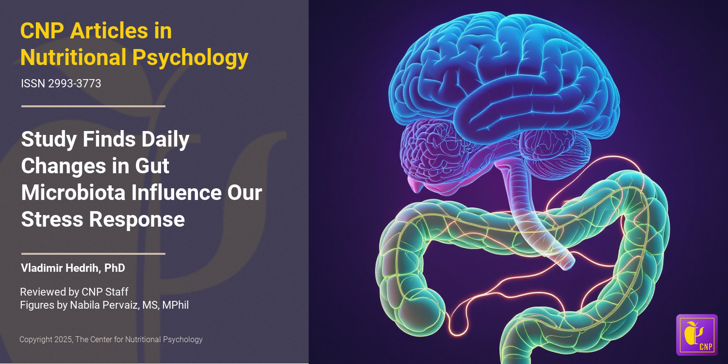
Additionally, studies on rodents revealed that it is possible to transfer serious mental health symptoms, such as those of social anxiety or even cognitive deficits associated with Alzheimer’s disease, by simply transplanting gut microbiota from humans suffering from these disorders into rodents (Hedrih, 2024; Kim et al., 2021; Ritz et al., 2024). Recent studies also identified specific biochemicals regulated by gut microbiota that affect processes such as inflammation in the brain or that induce changes in the brain, resulting in behavioral alterations (Heiss et al., 2021; Ritz et al., 2024). An important finding was that exposure to chronic stress alters gut microbiota composition (Ritz et al., 2024; Rivet-Noor et al., 2024).


It is possible to transfer serious mental health symptoms by simply transplanting gut microbiota from humans suffering from these disorders into rodents


These results created hope that novel ways to treat anxiety and depression, as well as other mental health issues, might be developed after we better understand the interplay between gut microbiota and processes in the brain.

The current study


Study author Courtney R. Rivet-Noor and her colleagues noted that the mucus layer in the gut is crucial for regulating microbiome composition. This mucus layer is a protective barrier that covers the gut lining, preventing damage from digestive enzymes, pathogens, and mechanical stress. The main component of this layer is a class of proteins called mucins.

The authors of this study hypothesized that exposure to chronic stress alters this mucus layer, thus initiating microbiome changes (see Figure 1). To test this, they conducted a study on mice.


%learn about nutrition mental health %The Center for Nutritional Psychology

Figure 1. The mucus layer of the gut


The study procedure


The study was conducted on two widely used strains of inbred laboratory mice (C57BL/6J and BALB/cJ). Mice were divided into two groups – one was exposed to chronic mild restraint stress by keeping them restrained in conical tubes for 2 hours and exposing them to one overnight stressor per day. The overnight stressors were 45-degree cage tilt, wet bedding, or 2x cage change. After a certain period, this treatment produces symptoms akin to depression and anxiety in humans. Mice not exposed to stress were used as controls.

The authors conducted a series of behavioral tests to test whether the stress treatment produced the expected effects (the forced swim, tail suspension, sucrose preference, open field, elevated plus maze, and nestlet shred tests). They also analyzed the tissues of these mice and conducted microbiota transfer experiments (see Figure 2).


%learn about nutrition mental health %The Center for Nutritional Psychology

Figure 2. Study Procedure (Rivet-Noor et al., 2024)


Chronic stress modified microbiome composition and reduced mucin 13 levels


As expected, exposing mice to chronic stress increased depression and anxiety-like behaviors in exposed mice. It also altered their gut microbiota composition. These mice had reduced expression of the Muc13 gene, resulting in lower levels of one of the mucin proteins – mucin 13. Other mucin proteins were unaffected.

Transferring gut microbiota from stressed mice to mice not exposed to chronic stress did not affect mucin 13 levels in the recipient mice. However, transplanting gut microbiota from stressed mice into mice without gut microbiota made the latter group of mice develop depression and anxiety-like symptoms, although they were not exposed to stress. This meant that mucin 13 reductions were not driven by changes in gut microbiota, but gut microbiota changes did lead to depression- and anxiety-like symptoms (see Figure 3).


%learn about nutrition mental health %The Center for Nutritional Psychology

Figure 3. Chronic stress modified microbiome composition and reduced mucin 13 levels


Deleting the Muc13 gene leads to microbiome changes similar to those resulting from chronic stress


The reduction in the expression of the Muc13 gene was caused by a protein called hepatocyte nuclear factor 4 or HNF4. This protein regulates the expression of specific genes by binding to particular DNA sequences. Analysis showed that chronic stress reduced the production of HNF4, leading to lower expression of the Muc13 gene. This was independent of changes to the microbiome.

Finally, the study authors created a line of mice without the Muc13 gene. Without being exposed to chronic stress, these mice had microbiota composition resembling regular mice exposed to chronic stress. This suggested that reductions in mucin 13 protein drive the changes in microbiota after experiencing chronic stress.

Behavioral tests showed that mice without the Muc13 gene displayed depression-like but not anxiety-like behaviors without being exposed to stress. However, after being exposed to stress, mice without the Muc13 gene developed anxiety-like symptoms much faster (after only one week) than regular mice. This indicated that the lack of this gene and the consequent lack of mucin 13 made them more susceptible to the effects of stress (see Figure 4).


%learn about nutrition mental health %The Center for Nutritional Psychology

Figure 4. Effects of absence of Muc13 gene in Mice


Conclusion


The study showed that chronic stress’s effects on behavior are mediated by a protein in the mucus layer of the gut called mucin 13. Although the study was done on mice, mucin 13 also exists in the human gut. This discovery could potentially open new ways to treat the consequences of chronic stress by targeting the production or level of mucin 13 in the gut.

The paper “Stress-induced mucin 13 reductions drive intestinal microbiome shifts and despair behaviors” was authored by Courtney R. Rivet-Noor, Andrea R. Merchak, Caroline Render, Naudia M. Gay, Rebecca M. Beiter, Ryan M. Brown, Austin Keeler, G. Brett Moreau, Sihan Li, Deniz G. Olgun, Alexandra D. Steigmeyer, Rachel Ofer, Tobey Phan, Kiranmayi Vemuri, Lei Chen, Keira E. Mahoney, Jung-Bum Shin, Stacy A. Malaker, Chris Deppmann, Michael P. Verzi, and Alban Gaultier.


References

Bonaz, B., Bazin, T., & Pellissier, S. (2018). The vagus nerve at the interface of the microbiota-gut-brain axis. Frontiers in Neuroscience, 12(FEB). https://doi.org/10.3389/fnins.2018.00049

Bystritsky, A. (2006). Treatment-resistant anxiety disorders. Molecular Psychiatry, 11(9), 805–814. https://doi.org/10.1038/sj.mp.4001852

Hedrih, V. (2023, September 2). Gut Microbiota’s Role in Mental Health: The Positives and Negatives. CNP Articles in Nutritional Psychology. https://www.nutritional-psychology.org/how-your-gut-microbiota-is-linked-to-both-positive-and-negative-aspects-of-mental-health/

Hedrih, V. (2024, March 12). Can Social Anxiety From Humans be Transmitted to Mice? CNP Articles in Nutritional Psychology. https://www.nutritional-psychology.org/can-social-anxiety-from-humans-be-transmitted-to-mice/

Heiss, C. N., Mannerås-Holm, L., Lee, Y. S., Serrano-Lobo, J., Håkansson Gladh, A., Seeley, R. J., Drucker, D. J., Bäckhed, F., & Olofsson, L. E. (2021). The gut microbiota regulates hypothalamic inflammation and leptin sensitivity in Western diet-fed mice via a GLP-1R-dependent mechanism. Cell Reports, 35(8). https://doi.org/10.1016/j.celrep.2021.109163

Kim, N., Jeon, S. H., Ju, I. G., Gee, M. S., Do, J., Oh, M. S., & Lee, J. K. (2021). Transplantation of gut microbiota derived from Alzheimer’s disease mouse model impairs memory function and neurogenesis in C57BL/6 mice. Brain, Behavior, and Immunity, 98, 357–365. https://doi.org/10.1016/J.BBI.2021.09.002

McIntyre, R. S., Alsuwaidan, M., Baune, B. T., Berk, M., Demyttenaere, K., Goldberg, J. F., Gorwood, P., Ho, R., Kasper, S., Kennedy, S. H., Ly-Uson, J., Mansur, R. B., McAllister-Williams, R. H., Murrough, J. W., Nemeroff, C. B., Nierenberg, A. A., Rosenblat, J. D., Sanacora, G., Schatzberg, A. F., … Maj, M. (2023). Treatment-resistant depression: Definition, prevalence, detection, management, and investigational interventions. World Psychiatry, 22(3), 394–412. https://doi.org/10.1002/wps.21120

Ritz, N. L., Brocka, M., Butler, M. I., Cowan, C. S. M., Barrera-Bugueño, C., Turkington, C. J. R., Draper, L. A., Bastiaanssen, T. F. S., Turpin, V., Morales, L., Campos, D., Gheorghe, C. E., Ratsika, A., Sharma, V., Golubeva, A. V., Aburto, M. R., Shkoporov, A. N., Moloney, G. M., Hill, C., … Cryan, J. F. (2024). Social anxiety disorder-associated gut microbiota increases social fear. Proceedings of the National Academy of Sciences, 121(1), e2308706120. https://doi.org/10.1073/pnas.2308706120

Rivet-Noor, C. R., Merchak, A. R., Render, C., Gay, N. M., Beiter, R. M., Brown, R. M., Keeler, A., Moreau, G. B., Li, S., Olgun, D. G., Steigmeyer, A. D., Ofer, R., Phan, T., Vemuri, K., Chen, L., Mahoney, K. E., Shin, J.-B., Malaker, S. A., Deppmann, C., … Gaultier, A. (2024). Stress-induced mucin 13 reductions drive intestinal microbiome shifts and despair behaviors. Brain, Behavior, and Immunity, 119, 665–680. https://doi.org/10.1016/j.bbi.2024.03.028

Valles-Colomer, M., Falony, G., Darzi, Y., Tigchelaar, E. F., Wang, J., Tito, R. Y., Schiweck, C., Kurilshikov, A., Joossens, M., Wijmenga, C., Claes, S., Van Oudenhove, L., Zhernakova, A., Vieira-Silva, S., & Raes, J. (2019). The neuroactive potential of the human gut microbiota in quality of life and depression. Nature Microbiology, 4(4), 623–632. https://doi.org/10.1038/s41564-018-0337-x


Do Gut Microbiota Play an Important Role in Regulating Food Intake and Satiety?

  • A review published in the Journal of Physiological Sciences discussed how the human body regulates satiety and food intake.
  • GLP-1 encourages the development of various advantageous bacteria in the gut, making it easier to produce satiety-related microbial products.
  • Gut microorganisms that produce short-chain fatty acids stimulate cells in the colon lining to produce GLP-1 and other hormones.
  • Other types of gut microorganisms produce substances that can affect inflammatory processes in the hypothalamus, disrupting or restoring the functionality of the body’s food intake regulation mechanism.

Living beings need to eat to stay alive. Multiple times daily, processes in our body tell our brain that we need to eat. We feel hunger, prompting us to look for food and eat it. After we have eaten, we feel satiated. This cycle continues as long as we live. But how does this function on the neural and biochemical level?

Hunger and satiety


A complex interaction between the digestive system, the brain, and various hormones regulates hunger and satiety. Satiety is the feeling of being full or satisfied after eating. It’s a physiological state where the body senses that it has consumed enough food. This allows it to regulate how much and how often a person eats. Satiety is influenced by various factors, including the type and volume of food consumed, its nutrient content, and hormonal responses during and after a meal. 

When the stomach is empty, the hormone ghrelin is released, which signals the brain to trigger feelings of hunger. After eating, the stomach and intestines produce hormones like glucagon-like peptide-1 (or GLP-1), peptide YY (PYY), and others, signaling the brain to produce feelings of satiety. Leptin, a hormone primarily produced by fat tissue, acts in a similar fashion, signaling the brain to suppress appetite. The more fat tissue there is, the higher the production of leptin (Hedrih, 2023; Stevenson et al., 2023; Swami et al., 2022) (see Figure 1).


%learn about nutrition mental health %The Center for Nutritional Psychology

Figure 1. Complex interaction between the digestive system, brain, and hormones to regulate hunger and satiety


These hormones act on the hypothalamus, a region of the brain that plays a key role in regulating hunger and satiety. This structure of the brain contains groups of neurons that increase the feeling of satiety, such as the pro-opiomelanocortin (POMC) and cocaine–amphetamine-regulated transcript-containing (CART) neurons, but also those that trigger appetite and eating behaviors—neuropeptide Y (NPY) and agouti-related peptide (AgRP) (Barakat et al., 2024).

AgRP neurons are also known as “hunger neurons” because studies demonstrate that artificially triggering them (in rodents) initiates feeding behavior (Chen et al., 2016) (see Figure 2).


%learn about nutrition mental health %The Center for Nutritional Psychology

Figure 2. “Hunger and satiety neurons” in the hypothalamus


The sensation of hunger


Hunger is not only a physiological process but also a subjective sensation. This sensation makes us more sensitive to stimuli related to food, pay more attention to food items and to things we learned to associate with food (e.g., restaurant or food brand logos), and be willing to eat (Hedrih, 2023; Lazarus et al., 1953; McKiernan et al., 2008).


Hunger is not only a physiological process but also a subjective sensation


Scientists initially thought that hunger results from decreased levels of nutrients in the body (e.g., lower blood sugar levels, decreased fat contents, empty stomach), but more novel studies indicated that many other conditions can trigger this sensation. These include (but are not limited to) boredom, desire for sensory stimulation, lack of sleep, and chronic stress (Brondel et al., 2010; Hedrih, 2023; Levine & Morley, 1981; McKiernan et al., 2008; Swami et al., 2022). Also, humans and animals can develop eating habits at specific times. These habits make them feel hungry when what they perceive as mealtime arrives (Isherwood et al., 2023) (Nutritional Psychology Research Library, 2024) (see Figure 3).


%learn about nutrition mental health %The Center for Nutritional Psychology

Figure 3. Factors influencing our sense of hunger


The role of GLP-1 and gut microbiota in satiety


In their review, Ghinwa M. Barakat and her colleagues note that recent discoveries indicate that gut microbiota, the community of microorganisms living in our gut, play an important role in human metabolism. This became particularly evident with the discovery of the microbiota-gut-brain axis, a bidirectional communication pathway that allows gut microbiota to influence processes in the brain and vice versa (Barakat et al., 2024; Bonaz et al., 2018; Heiss et al., 2021).

Recent findings indicate that GLP-1 hormone might be particularly important for gut microbiota’s role in the feeling of satiety. GLP-1 is produced by specialized endocrine cells located in the lining of the small intestine and the colon called L cells. Receptors for this hormone, i.e., proteins on the surface of cells that react with it, are abundant in the hypothalamus, particularly in the arcuate nucleus region. Studies have demonstrated that injections of GLP-1 and medicines that behave like GLP-1 in the body reduce food intake (Barakat et al., 2024; Heiss et al., 2021) (see Figure 4).


%learn about nutrition mental health %The Center for Nutritional Psychology

Figure 4. GLP-1 hormone stimulates the feelings of satiety


Recent studies on mice indicated that Liraglutide, a substance commonly used to treat diabetes and obesity, mimics the effects of GLP-1 and can also affect the gut microbiota composition. High levels of GLP-1 or substances that act like it seems to help enrich microbiota strains more abundant in lean individuals and reduce the abundance of strains of microorganisms more common in obese individuals. For example, these injections increased the amounts of Akkermansia muciniphila, a species known to be more present in the gut when an individual loses weight and in lean individuals. On the other hand, it reduces the amount of Proteobacteria, which is more abundant in obese individuals.

Gut microbiota participate in satiety hormone regulation


Studies also indicate that gut microbiota might participate in regulating the release of satiety hormones. The authors of this review point to several studies indicating that some short-chain fatty acids (SFCAs) produced by gut bacteria increase the production and release of GLP-1 into the bloodstream. If more gut bacteria produce these substances, the digestive system lining will produce more GLP-1, sending a stronger satiety signal to the brain.

This effect is achieved through specific receptors on the intestine’s GLP-1-producing cells, which react to short-chain fatty acids, such as acetate, propionate, and butyrate, produced by gut bacteria (see Figure 5).


%learn about nutrition mental health %The Center for Nutritional Psychology

Figure 5. Role of gut microbiota in regulating satiety hormone secretions


Gut microbiota affect inflammatory processes


Another way in which gut microbiota can affect satiety is by affecting the inflammatory processes in the hypothalamus. Specific species of gut microorganisms can produce substances that increase inflammatory processes in the hypothalamus. This reduces its ability to regulate appetite (e.g., through reducing sensitivity to leptin). These types of bacteria tend to be more abundant in the guts of obese individuals. Studies on mice indicated that this could be countered by introducing bacteria into the gut that have the opposite effect, such as Lactobacillus rhamnosus, Lactobacillus acidophilus, and Bifidobacterium bifidum. Research results indicate that these bacteria restore sensitivity to leptin, thus helping reduce excessive weight (see Figure 6).


%learn about nutrition mental health %The Center for Nutritional Psychology

Figure 6. Role of gut microbiota in leptin sensitivity


Conclusion


The review discusses the various mechanisms through which the body regulates food intake and satiety. It points to the important role gut microorganisms play in this system and how it can be affected by gut microbiota.

The presented findings potentially open a new avenue of research to look for methods to prevent and treat obesity by influencing the gut microbiome. Future discoveries may lead to a new group of obesity treatments based on probiotics, devise ways to detect developing obesity, and allow effective prevention.

The paper “Satiety: a gut–brain–relationship” was authored by Ghinwa M. Barakat, Wiam Ramadan, Ghaith Assi, and Noura B. El Khoury.

For more information on mechanisms influencing dietary intake and food satiety, enroll in NP 150: Mechanisms in the Diet-Mental Health Relationship (DMHR). Find NP 150 and other courses in the DMHR through The world’s leader in nutritional psychology education here.  


References

Barakat, G. M., Ramadan, W., Assi, G., & Khoury, N. B. E. (2024). Satiety: A gut–brain–relationship. The Journal of Physiological Sciences, 74(1), 11. https://doi.org/10.1186/s12576-024-00904-9

Bonaz, B., Bazin, T., & Pellissier, S. (2018). The vagus nerve at the interface of the microbiota-gut-brain axis. Frontiers in Neuroscience, 12(FEB). https://doi.org/10.3389/fnins.2018.00049

Brondel, L., Romer, M. A., Nougues, P. M., Touyarou, P., & Davenne, D. (2010). Acute partial sleep deprivation increases food intake in healthy men. The American Journal of Clinical Nutrition, 91(6), 1550–1559. https://doi.org/10.3945/ajcn.2009.28523

Chen, Y., Lin, Y.-C., Zimmerman, C. A., Essner, R. A., & Knight, Z. A. (2016). Hunger neurons drive feeding through a sustained, positive reinforcement signal. eLife, 5, e18640. https://doi.org/10.7554/eLife.18640.001

Hedrih, V. (2023). Food and Mood: Is the Concept of ‘Hangry’ Real? CNP Articles in Nutritional Psychology. https://www.nutritional-psychology.org/food-and-mood-is-the-concept-of-hangry-real/

Heiss, C. N., Mannerås-Holm, L., Lee, Y. S., Serrano-Lobo, J., Håkansson Gladh, A., Seeley, R. J., Drucker, D. J., Bäckhed, F., & Olofsson, L. E. (2021). The gut microbiota regulates hypothalamic inflammation and leptin sensitivity in Western diet-fed mice via a GLP-1R-dependent mechanism. Cell Reports, 35(8). https://doi.org/10.1016/j.celrep.2021.109163

Isherwood, C. M., van der Veen, D. R., Hassanin, H., Skene, D. J., & Johnston, J. D. (2023). Human glucose rhythms and subjective hunger anticipate meal timing. Current Biology, 33(7), 1321-1326.e3. https://doi.org/10.1016/j.cub.2023.02.005

Lazarus, R. S., Yousem, H., & Arenberg, D. (1953). Hunger and Perception. Journal of Personality, 21(3), 312–328. https://doi.org/10.1111/J.1467-6494.1953.TB01774.X

Levine, A. S., & Morley, J. E. (1981). Stress-induced eating in rats. American Journal of Physiology – Regulatory, Integrative and Comparative Physiology, 241(1), R72–R76.

McKiernan, F., Houchins, J. A., & Mattes, R. D. (2008). Relationships between human thirst, hunger, drinking, and feeding. Physiology & Behavior, 94(5), 700. https://doi.org/10.1016/J.PHYSBEH.2008.04.007

Nutritional Psychology Research Library (NPRL). (n.d.). The Center for Nutritional Psychology. Retrieved May 1, 2024, from https://www.nutritional-psychology.org/np-research-library/

Stevenson, R. J., Bartlett, J., Wright, M., Hughes, A., Hill, B. J., Saluja, S., & Francis, H. M. (2023). The development of interoceptive hunger signals. Developmental Psychobiology, 65(2), 1–11. https://doi.org/10.1002/dev.22374

Swami, V., Hochstöger, S., Kargl, E., & Stieger, S. (2022). Hangry in the field: An experience sampling study on the impact of hunger on anger, irritability, and affect. PLOS ONE, 17(7), e0269629. https://doi.org/10.1371/JOURNAL.PONE.0269629


Recent Articles

SUPPORT THE FIELD

CNP is a non-profit that relies on our small team of staff and our many dedicated volunteers.

If you find nutritional psychology meaningful, please consider supporting our mission in one of the following ways:

We would also love to connect with you on social media!

CONTINUING EDUCATION