Chronic Stress Alters Gut Microbiome Composition by Reducing the Production of a Protein Called Mucin 13

  • A series of experiments published in Brain Behavior and Immunity revealed that exposing mice to chronic stress reduced the levels of a protein called mucin 13.
  • Deleting the gene that encodes mucin 13 was sufficient to create the changes in the gut microbiome seen after exposure to chronic stress and to produce and produce depression-like symptoms.
  • Mice without the gene that encodes mucin 13 were more susceptible to stress.

Depression and anxiety affect millions of people worldwide. Despite this, treatments for these disorders are often not very effective. Estimates state that at least 30% of individuals suffering from depression do not experience symptom withdrawal even after multiple treatment procedures (McIntyre et al., 2023). The situation is similar to anxiety – only about 60% of patients respond to treatments to any significant degree (Bystritsky, 2006).

One of the reasons for the low effectiveness of treatments for these widespread disorders probably lies in the fact that their causes are not fully understood by researchers. However, the recent discovery of the microbiota-gut-brain axis (MGBA) and studies of the biochemical pathways involved in depression and anxiety show promise to change this situation (Bonaz et al., 2018; Hedrih, 2023).

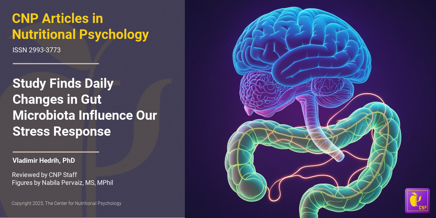
The microbiota-gut-brain axis and mental health


The gut microbiota is a community of trillions of microorganisms living in our gut. These organisms help us digest the food we eat, allowing us to extract nutrients from foods that we would not be able to use without their help. However, their role in our organisms far exceeds these digestive processes.

Researchers discovered a bidirectional communication pathway that allows the gut microbiota to influence processes in the brain and vice versa (Valles-Colomer et al., 2019). This pathway was named the microbiota-gut-brain axis. Studies show that individuals with specific mental health issues have altered gut microbiota composition.


Studies show that individuals with specific mental health issues have altered gut microbiota composition


Additionally, studies on rodents revealed that it is possible to transfer serious mental health symptoms, such as those of social anxiety or even cognitive deficits associated with Alzheimer’s disease, by simply transplanting gut microbiota from humans suffering from these disorders into rodents (Hedrih, 2024; Kim et al., 2021; Ritz et al., 2024). Recent studies also identified specific biochemicals regulated by gut microbiota that affect processes such as inflammation in the brain or that induce changes in the brain, resulting in behavioral alterations (Heiss et al., 2021; Ritz et al., 2024). An important finding was that exposure to chronic stress alters gut microbiota composition (Ritz et al., 2024; Rivet-Noor et al., 2024).


It is possible to transfer serious mental health symptoms by simply transplanting gut microbiota from humans suffering from these disorders into rodents


These results created hope that novel ways to treat anxiety and depression, as well as other mental health issues, might be developed after we better understand the interplay between gut microbiota and processes in the brain.

The current study


Study author Courtney R. Rivet-Noor and her colleagues noted that the mucus layer in the gut is crucial for regulating microbiome composition. This mucus layer is a protective barrier that covers the gut lining, preventing damage from digestive enzymes, pathogens, and mechanical stress. The main component of this layer is a class of proteins called mucins.

The authors of this study hypothesized that exposure to chronic stress alters this mucus layer, thus initiating microbiome changes (see Figure 1). To test this, they conducted a study on mice.


%learn about nutrition mental health %The Center for Nutritional Psychology

Figure 1. The mucus layer of the gut


The study procedure


The study was conducted on two widely used strains of inbred laboratory mice (C57BL/6J and BALB/cJ). Mice were divided into two groups – one was exposed to chronic mild restraint stress by keeping them restrained in conical tubes for 2 hours and exposing them to one overnight stressor per day. The overnight stressors were 45-degree cage tilt, wet bedding, or 2x cage change. After a certain period, this treatment produces symptoms akin to depression and anxiety in humans. Mice not exposed to stress were used as controls.

The authors conducted a series of behavioral tests to test whether the stress treatment produced the expected effects (the forced swim, tail suspension, sucrose preference, open field, elevated plus maze, and nestlet shred tests). They also analyzed the tissues of these mice and conducted microbiota transfer experiments (see Figure 2).


%learn about nutrition mental health %The Center for Nutritional Psychology

Figure 2. Study Procedure (Rivet-Noor et al., 2024)


Chronic stress modified microbiome composition and reduced mucin 13 levels


As expected, exposing mice to chronic stress increased depression and anxiety-like behaviors in exposed mice. It also altered their gut microbiota composition. These mice had reduced expression of the Muc13 gene, resulting in lower levels of one of the mucin proteins – mucin 13. Other mucin proteins were unaffected.

Transferring gut microbiota from stressed mice to mice not exposed to chronic stress did not affect mucin 13 levels in the recipient mice. However, transplanting gut microbiota from stressed mice into mice without gut microbiota made the latter group of mice develop depression and anxiety-like symptoms, although they were not exposed to stress. This meant that mucin 13 reductions were not driven by changes in gut microbiota, but gut microbiota changes did lead to depression- and anxiety-like symptoms (see Figure 3).


%learn about nutrition mental health %The Center for Nutritional Psychology

Figure 3. Chronic stress modified microbiome composition and reduced mucin 13 levels


Deleting the Muc13 gene leads to microbiome changes similar to those resulting from chronic stress


The reduction in the expression of the Muc13 gene was caused by a protein called hepatocyte nuclear factor 4 or HNF4. This protein regulates the expression of specific genes by binding to particular DNA sequences. Analysis showed that chronic stress reduced the production of HNF4, leading to lower expression of the Muc13 gene. This was independent of changes to the microbiome.

Finally, the study authors created a line of mice without the Muc13 gene. Without being exposed to chronic stress, these mice had microbiota composition resembling regular mice exposed to chronic stress. This suggested that reductions in mucin 13 protein drive the changes in microbiota after experiencing chronic stress.

Behavioral tests showed that mice without the Muc13 gene displayed depression-like but not anxiety-like behaviors without being exposed to stress. However, after being exposed to stress, mice without the Muc13 gene developed anxiety-like symptoms much faster (after only one week) than regular mice. This indicated that the lack of this gene and the consequent lack of mucin 13 made them more susceptible to the effects of stress (see Figure 4).


%learn about nutrition mental health %The Center for Nutritional Psychology

Figure 4. Effects of absence of Muc13 gene in Mice


Conclusion


The study showed that chronic stress’s effects on behavior are mediated by a protein in the mucus layer of the gut called mucin 13. Although the study was done on mice, mucin 13 also exists in the human gut. This discovery could potentially open new ways to treat the consequences of chronic stress by targeting the production or level of mucin 13 in the gut.

The paper “Stress-induced mucin 13 reductions drive intestinal microbiome shifts and despair behaviors” was authored by Courtney R. Rivet-Noor, Andrea R. Merchak, Caroline Render, Naudia M. Gay, Rebecca M. Beiter, Ryan M. Brown, Austin Keeler, G. Brett Moreau, Sihan Li, Deniz G. Olgun, Alexandra D. Steigmeyer, Rachel Ofer, Tobey Phan, Kiranmayi Vemuri, Lei Chen, Keira E. Mahoney, Jung-Bum Shin, Stacy A. Malaker, Chris Deppmann, Michael P. Verzi, and Alban Gaultier.


References

Bonaz, B., Bazin, T., & Pellissier, S. (2018). The vagus nerve at the interface of the microbiota-gut-brain axis. Frontiers in Neuroscience, 12(FEB). https://doi.org/10.3389/fnins.2018.00049

Bystritsky, A. (2006). Treatment-resistant anxiety disorders. Molecular Psychiatry, 11(9), 805–814. https://doi.org/10.1038/sj.mp.4001852

Hedrih, V. (2023, September 2). Gut Microbiota’s Role in Mental Health: The Positives and Negatives. CNP Articles in Nutritional Psychology. https://www.nutritional-psychology.org/how-your-gut-microbiota-is-linked-to-both-positive-and-negative-aspects-of-mental-health/

Hedrih, V. (2024, March 12). Can Social Anxiety From Humans be Transmitted to Mice? CNP Articles in Nutritional Psychology. https://www.nutritional-psychology.org/can-social-anxiety-from-humans-be-transmitted-to-mice/

Heiss, C. N., Mannerås-Holm, L., Lee, Y. S., Serrano-Lobo, J., Håkansson Gladh, A., Seeley, R. J., Drucker, D. J., Bäckhed, F., & Olofsson, L. E. (2021). The gut microbiota regulates hypothalamic inflammation and leptin sensitivity in Western diet-fed mice via a GLP-1R-dependent mechanism. Cell Reports, 35(8). https://doi.org/10.1016/j.celrep.2021.109163

Kim, N., Jeon, S. H., Ju, I. G., Gee, M. S., Do, J., Oh, M. S., & Lee, J. K. (2021). Transplantation of gut microbiota derived from Alzheimer’s disease mouse model impairs memory function and neurogenesis in C57BL/6 mice. Brain, Behavior, and Immunity, 98, 357–365. https://doi.org/10.1016/J.BBI.2021.09.002

McIntyre, R. S., Alsuwaidan, M., Baune, B. T., Berk, M., Demyttenaere, K., Goldberg, J. F., Gorwood, P., Ho, R., Kasper, S., Kennedy, S. H., Ly-Uson, J., Mansur, R. B., McAllister-Williams, R. H., Murrough, J. W., Nemeroff, C. B., Nierenberg, A. A., Rosenblat, J. D., Sanacora, G., Schatzberg, A. F., … Maj, M. (2023). Treatment-resistant depression: Definition, prevalence, detection, management, and investigational interventions. World Psychiatry, 22(3), 394–412. https://doi.org/10.1002/wps.21120

Ritz, N. L., Brocka, M., Butler, M. I., Cowan, C. S. M., Barrera-Bugueño, C., Turkington, C. J. R., Draper, L. A., Bastiaanssen, T. F. S., Turpin, V., Morales, L., Campos, D., Gheorghe, C. E., Ratsika, A., Sharma, V., Golubeva, A. V., Aburto, M. R., Shkoporov, A. N., Moloney, G. M., Hill, C., … Cryan, J. F. (2024). Social anxiety disorder-associated gut microbiota increases social fear. Proceedings of the National Academy of Sciences, 121(1), e2308706120. https://doi.org/10.1073/pnas.2308706120

Rivet-Noor, C. R., Merchak, A. R., Render, C., Gay, N. M., Beiter, R. M., Brown, R. M., Keeler, A., Moreau, G. B., Li, S., Olgun, D. G., Steigmeyer, A. D., Ofer, R., Phan, T., Vemuri, K., Chen, L., Mahoney, K. E., Shin, J.-B., Malaker, S. A., Deppmann, C., … Gaultier, A. (2024). Stress-induced mucin 13 reductions drive intestinal microbiome shifts and despair behaviors. Brain, Behavior, and Immunity, 119, 665–680. https://doi.org/10.1016/j.bbi.2024.03.028

Valles-Colomer, M., Falony, G., Darzi, Y., Tigchelaar, E. F., Wang, J., Tito, R. Y., Schiweck, C., Kurilshikov, A., Joossens, M., Wijmenga, C., Claes, S., Van Oudenhove, L., Zhernakova, A., Vieira-Silva, S., & Raes, J. (2019). The neuroactive potential of the human gut microbiota in quality of life and depression. Nature Microbiology, 4(4), 623–632. https://doi.org/10.1038/s41564-018-0337-x


Gut Microbiota Play Crucial Role in Mediating Effects of Western Diet

Introduction

The past several decades have seen the rise of an obesity pandemic that is ongoing worldwide. While obese individuals were quite rare just a century ago, 2015-2018 estimates for the U.S. state that more than two-thirds of the adult population is overweight or obese (Wong et al., 2022). Determining the causes of this increase in obesity rates has attracted much research attention. Studies have revealed a complex interplay between diet components, environmental factors, and previously unknown psychological and physiological mechanisms resulting in overeating and obesity in the long term. These novel studies on the intersection of nutrition and psychology are part of a developing field of science called nutritional psychology (The Center for Nutritional Psychology, 2023) (see Figure 1).


%learn about nutrition mental health %The Center for Nutritional Psychology

Figure 1. Diet, environment, psychological, and physiological factors in nutritional psychology 


There is a complex interplay between diet, environmental factors, and psychological and physiological mechanisms resulting in overeating and obesity


Gut microbiota and the microbiota-gut-brain axis

The human gut microbiome consists of trillions of microorganisms that live in the human intestinal tract. These microorganisms play a key role in digesting the food we eat. However, their influence extends beyond the gut, encompassing crucial roles in metabolic regulation, body weight maintenance, and immune system modulation. 

This growing body of evidence suggests that these gut microorganisms also profoundly impact brain functions, mood, cognition, and emotional well-being  (Zhu et al., 2023). This topic is explored in continuing education curricula within nutritional psychology — particularly how the gut microbiota and the gut-brain axis interconnect with the diet-mental health relationship to influence psychological functioning and experience, shedding light on its potential therapeutic implications for mental health outcomes.


This growing body of evidence suggests that gut microorganisms profoundly impact brain functions, mood, cognition, and emotional well-being


Scientists have recently discovered a communication pathway connecting the gut microbiome and the brain. This pathway is called the microbiota-gut-brain axis. It is based on small proteins called cytokines and a number of other biomolecules, including the hormone cortisol, short-chain fatty acids (SCFAs), tryptophan, and others.

The Western diet

The Western diet is a modern dietary pattern prevalent in Western societies, characterized by a high intake of processed and hyperpalatable foods with increased contents of fat, sugary snacks, and refined grains. It typically includes low consumption of fruits, vegetables, unprocessed-high-quality proteins, nuts, and seeds. This diet’s excessive reliance on added sugars and unhealthy fats has been linked to an increased risk of obesity, metabolic syndrome, and various chronic diseases.

Studies have indicated that feeding mice a Western diet causes inflammation in the region of the brain called the hypothalamus (Heiss et al., 2021; Thaler et al., 2013). Inflammation of the hypothalamus damages the neurons and leads to the formation of scars made of glial cells. This is called gliosis. Inflammation of the hypothalamus often happens before a mouse starts gaining weight. Due to this, scientists believe it might cause weight gain by causing leptin resistance.


Studies have indicated that feeding mice a Western diet causes inflammation of the region of the brain called the hypothalamus


Leptin and leptin resistance

Leptin is a hormone produced by fat cells during eating. It regulates appetite and body weight and is produced in proportion to the amount of fat in the body. Leptin concentrations inform the brain of how much fat is stored. Increased leptin concentrations (normally caused by an abundance of body fat) “tell” the brain to decrease food intake and increase energy expenditure.


Leptin is a hormone produced by fat cells that regulate appetite and body weight


Factors such as chronic inflammation or eating high-fat diets (HFDs) may cause the body to be less receptive to leptin. This is called leptin resistance. Leptin resistance results in disrupted appetite and energy regulation, i.e., the brain does not reduce food intake in spite of the abundance of body fat. This can contribute to obesity and cause difficulty controlling body weight (Thaler et al., 2013) (see Figure 2).


%learn about nutrition mental health %The Center for Nutritional Psychology

Figure 2. Normal leptin cycle versus leptin resistance.


Gliosis, leptin resistance, and gut microbiota

Microglia are a type of immune cell in the central nervous system that helps protect and maintain the brain and spinal cord by detecting and responding to potential threats or damage. Studies have shown that activation of microglia cells that happens during inflammation of the hypothalamus might be causing leptin resistance. Removing these microglia cells from the hypothalamus has improved sensitivity to leptin. Improved sensitivity to leptin allows the brain to recognize when enough fat is stored in the body and reduce food intake. 


Studies have shown that the activation of microglia cells that happens during inflammation of the hypothalamus might be causing leptin resistance


Intriguingly, according to the scientific evidence presented in our recent NP 120 course, it has been discovered that the gut microbiota plays a significant role in regulating the development and maturation of microglia cells and influencing their function. Although the mechanism of this action remains unknown, it has led scientists to believe there might be a link between hypothalamus inflammation and gut microbiota (Heiss et al., 2021) (see Figure 3).


%learn about nutrition mental health %The Center for Nutritional Psychology

Figure 3. Link of Gut Microbiota in regulating development and maturation of microglia cells


There might be a link between hypothalamus inflammation and gut microbiota (Heiss et al., 2021)


The current study

Study author Christina N. Heiss and her colleagues wanted to know whether mice that lack gut microbiota are protected against diet-induced inflammation of the hypothalamus. They noted previous studies’ results showing mice with depleted gut microbiota and those treated with antibiotics to be protected from diet-induced obesity.

In other words, they noted that mice that consume a Western diet, a diet that normally leads to obesity in mice, do not become obese if microbiota are not present in their guts. This might be because the absence of microbiota prevents inflammation of the hypothalamus, which would, in turn, prevent leptin resistance from developing. If this is the case, the mechanism for preventing overeating based on leptin would remain intact, preventing mice from becoming obese. Alternatively, it could be that, without microbiota, the guts of mice could not digest complex nutrients from the food they eat, thus substantially reducing the amount of nutrition they can derive from food. In this case, obesity would be avoided because their bodies cannot use their food. But which of these is the case?

The procedure

The study authors used three groups of male mice, 10-13 weeks old – conventionally raised mice, germ-free mice, and antibiotic-fed mice. They used several genetic groups of mice, including a strain of genetically engineered mice that allow for controlled and regulated manipulation of specific genes in specific tissues (Tamoxifen-inducible Cre mice).

In the scope of the experiments, researchers fed mice either a chow diet or a Western diet. Western diet was given for either 2 days, 1 week, or 4 weeks, depending on the experiment conducted in the scope of the study.

The chow diet for mice is a nutritionally balanced and standardized diet formulated to provide essential nutrients required for the health and growth of laboratory mice. It typically consists of a combination of proteins, carbohydrates, fats, vitamins, and minerals in pellet or block form. The Western diet used in this study was high in fat and sucrose, with 40% of calories coming from fat.

All food for mice was sterilized, i.e., underwent procedures that killed all microorganisms in the food before mice ate it. The chow diet was sterilized in an autoclave, which uses high pressure and steam to kill microorganisms. The Western diet food was irradiated, i.e., radiation was used to kill microorganisms.

The mice

Conventionally raised mice were laboratory mice kept in regular conditions and fed a normal diet for laboratory mice. They have normal gut microbiota.

Germ-free mice are created through techniques that ensure they do not acquire gut microbiota from birth through their entire lifetimes. They are typically born using cesarean section deliveries of pregnant mice in a sterile environment. This is done to prevent the transfer of microbes during birth. After that, they are kept in specialized sterile isolation spaces called isolators or bubbles that maintain a controlled germ-free environment.

These isolators provide filtered air, sterile food, and autoclaved water to prevent microbial contamination. Researchers raising these mice take special care to maintain strict barrier measures, including specialized clothing and equipment, to prevent the unintentional introduction of microorganisms. They regularly monitor these mice’s bodily fluids and tissues through special techniques to ensure the absence of any detectable microorganisms. Germ-free mice are typically leaner than conventionally raised mice and, consequently, have lower leptin levels in circulation.

Antibiotic-fed mice in this study had 1g of ampicillin and 0.5g of neomycin added per liter of their drinking water. Ampicillin and neomycin are antibiotics. Researchers kept the drinking water with antibiotics added in bottles protected from light. They prepared a new solution every second day. Researchers started giving this water with antibiotics to mice three days before changing their regular diet to a Western diet.


Mice without gut microbiota are protected from diet-induced inflammation of the hypothalamus


Results showed that conventionally raised mice fed a Western diet for 1 week developed gliosis in the hypothalamus. Inflammation indicators were increased in these mice in the part of the hypothalamus called the arcuate nucleus compared to conventionally raised mice fed regular mice food (chow) (see Figure 4).


%learn about nutrition mental health %The Center for Nutritional Psychology

Figure 4. Conventionally raised mice fed a Western diet developed gliosis in the hypothalamus


When researchers examined germ-free mice and mice whose gut microbiota were depleted using antibiotic treatment (antibiotic-fed mice), results showed that these mice also showed no increase in inflammation indicators or the proliferation of microglia cells after eating a Western diet for a week.


Gliosis in the hypothalamus leads to greater gain in body weight and fat mass


The study authors also wanted to know whether gliosis in the hypothalamus caused by a Western diet leads, in turn, to increased body weight and fat mass accumulation in mice. To test that, they fed conventionally raised mice and antibiotic-fed mice a Western diet for 4 weeks.

Results showed that conventionally raised mice fed a Western diet gained more body weight and fat mass than antibiotic-fed mice. Compared to antibiotic-fed mice, they also had increased hypothalamus inflammation indicators and increased numbers of a specific type of microglia cells (iba1-positive microglia).

There was no association between the number of microglia in the arcuate nucleus region of the hypothalamus and changes in body weight or fat mass at the end of the 4 weeks. However, fat mass and the relative increase in fat mass during the study were associated with the number of a specific type of glial cells called astrocytes.

Further analysis showed that germ-free and antibiotic-fed mice are more sensitive to leptin than conventionally raised mice. When researchers gave them leptin injections, the first two types of mice reduced their food intake more than conventionally raised mice did.


Glucagon-like-peptide 1 (GLP-1) seems to be crucial for protection against diet-induced inflammation of the hypothalamus


Germ-free and antibiotic-fed mice had higher levels of the hormone called glucagon-like peptide 1 (GLP-1) when they were fed a regular diet. This hormone secreted in the small intestine’s intestinal lining cells (L cells) is important in regulating blood sugar levels. It also helps reduce inflammation and protect neurons.

Study authors believed it might also be crucial for the protection from inflammation of the hypothalamus induced by the Western diet. After mice were fed a Western diet for a week, antibiotic-treated and germ-free mice had higher levels of GLP-1 than conventionally raised mice. These mice did not gain weight or develop hypothalamic inflammation after this diet. However, when researchers measured these same things in antibiotic-fed and germ-free mice whose GLP-1 signaling pathway was disabled, they also gained weight and developed inflammation, similar to conventionally raised mice. This indicated that the functional signaling path of GLP-1 is crucial for countering the inflammation of the hypothalamus induced by a Western diet.


Just a week on a Western diet led to inflammation of the hypothalamus that, in turn, disrupted the body’s mechanism for regulating food intake


Conclusion

These findings in mice show that gut microbiota changes how the organism, of mice in this case, reacts to a Western diet. When gut microbiota was intact, just a week on a Western diet led to inflammation of the hypothalamus that, in turn, disrupted the body’s mechanism for regulating food intake. However, when gut microbiota was depleted or absent, this inflammation did not happen, provided that the signaling pathway of one specific hormone (GLP-1) was intact.

While the study was done on mice, similar physiological mechanisms exist in humans. Due to this, these findings on mice help scientists better understand how and through which physiological mechanisms changes in the human diet that occurred in the last century disrupted food intake regulation in the human body leading to the current obesity pandemic.

The paper “The gut microbiota regulates hypothalamic inflammation and leptin sensitivity in Western diet-fed mice via a GLP-1R-dependent mechanism” was authored by Christina N. Heiss, Louise Manneras-Holm, Ying Shiuan Lee, Julia Serrano-Lobo, Anna Hakansson Gladh, Randy J. Seeley, Daniel J. Drucker, Fredrik Backhed, and Louise E. Olofsson.

References

Heiss, C. N., Mannerås-Holm, L., Lee, Y. S., Serrano-Lobo, J., Håkansson Gladh, A., Seeley, R. J., Drucker, D. J., Bäckhed, F., & Olofsson, L. E. (2021). The gut microbiota regulates hypothalamic inflammation and leptin sensitivity in Western diet-fed mice via a GLP-1R-dependent mechanism. Cell Reports, 35(8). https://doi.org/10.1016/j.celrep.2021.109163

Thaler, J. P., Guyenet, S. J., Dorfman, M. D., Wisse, B. E., & Schwartz, M. W. (2013). Hypothalamic inflammation: Marker or mechanism of obesity pathogenesis? Diabetes, 62(8), 2629–2634. https://doi.org/10.2337/DB12-1605

The Center for Nutritional Psychology. (2023). What is Nutritional Psychology? https://www.nutritional-psychology.org/what-is-nutritional-psychology/

Wong, M. C., Mccarthy, C., Fearnbach, N., Yang, S., Shepherd, J., & Heymsfield, S. B. (2022). Emergence of the obesity epidemic: 6-decade visualization with humanoid avatars. The American Journal of Clinical Nutrition, 115(4), 1189–1193. https://doi.org/10.1093/AJCN/NQAC005

Zhu, X., Sakamoto, S., Ishii, C., Smith, M. D., Ito, K., Obayashi, M., Unger, L., Hasegawa, Y., Kurokawa, S., Kishimoto, T., Li, H., Hatano, S., Wang, T. H., Yoshikai, Y., Kano, S. ichi, Fukuda, S., Sanada, K., Calabresi, P. A., & Kamiya, A. (2023). Dectin-1 signaling on colonic γδ T cells promotes psychosocial stress responses. Nature Immunology. https://doi.org/10.1038/s41590-023-01447-8


Are You What Your Gut-Microbiome Wants You To Eat?

We’ve all heard the saying “you are what you eat,” but new microbiome research is shedding light on this old adage, with a more modern-day update being “you are what your gut-microbiome wants you to eat.” Let’s look at why this is the case. 

First, we know that our food choices significantly impact our physical and mental health. As far back as the 1800s and 1900s, scientists hypothesized an apparent correlation between our food intake and the subsequent effects on appetite, body image, and brain function (Tzameli, 2013). Though biomedical research has already established the endocrine responses that regulate hunger and satiety in the gut-brain axis signaling, little attention has been paid to the mechanisms that influence an individual’s choice of food and nutrition.


Microorganisms that live in our gut may influence what we eat!


A growing body of evidence indicates that our gut microbiome may be one of the factors influencing our food choices. From Nutritional Psychology conceptualization, we are beginning to understand that eating behavior and food preferences are dependent on many aspects of the diet-mental health relationship (DMHR), such as our psychosocial environment, interoceptive experiences, sensory perception, cognitive processes, and psychological state. However, emerging research in the Microbiota-Gut Brain Axis (MGBA) suggests that the microorganisms residing within our gut may also influence what we eat. Therefore, the classic expression, “you are what you eat,” may soon be reframed as “you are [also] what your microbiome wants you to eat.”  


A feedback loop between our gut microbiome, brain, and food choices.


To explore the influence of the gut microbiome on diet selection behavior, Trevelline and Kohl conducted an experiment in 2022 to study the influence of gut microbes on the diet selection behaviors in mice. 


The classic expression, “you are what you eat,” may soon be reframed as “you are [also] what your microbiome wants you to eat.”  


To achieve this, intestinal microbiota from three “donor” mouse species, each with distinct foraging behavior, were transplanted into germ-free “host” mice to colonize their intestinal tracts.  

Following that, the donor germ-free mice were randomly divided into three treatment groups, each based on the donor species:

  • Carnivore (i.e., predatory-based)
  • Herbivore (i.e., plant-based)
  • Omnivore (i.e., inclusive-based)

The mice were then given a choice between a low protein-carbohydrate (LPC) diet and a high protein-carbohydrate (HPC) diet, and their diet preferences were tracked for 11 days. To assess the impact of the donor microbiome on host diet selection behavior, the researchers compared the microbiomes of mice in three treatment groups: predatory (carnivores), inclusive (omnivores), and plant-based (herbivores) (Fig 1A).

%learn about nutrition mental health %The Center for Nutritional Psychology

Figure 1A. Experimental design to assess host diet selection behaviors across different microbiomes. From Trevelline and Kohl, Proceedings of the National Academy of Sciences, 2022.


Strikingly, the authors discovered that when mice have given a choice of selected diets varying in macronutrient composition, each microbiome had a distinct effect on food choice behavior (Fig 1B). For example, host mice that received microbiota from herbivorous donors voluntarily ate fewer carbohydrates, evidenced by a higher protein:carbohydrate (P:C) ratio diet intake. On the other hand, omnivore and carnivore treatment groups chose a lower P:C ratio diet intake.

Given that these host mice had no microbiome prior to transplantation, the change in diet selection behavior is evidence of the microbiome influencing food choice (Alcock, 2014). Moreover, through an in-depth analysis of blood and fecal samples, the authors discovered the microbial release of essential amino acids (EAAs) from the gut microbiome of host mice, including tryptophan. Tryptophan is an important food choice driver because it is a precursor to serotonin, the happiness hormone that has been shown to regulate feeding behavior, metabolism, and diet selection (Harrold, 2012; Cryan, 2019; Kaur & Bose, 2019; Yabut, 2019; Gao, 2020; Trevelline & Kohl, 2022). Together, these findings show that the gut microbiome can influence host diet selection behavior by mediating the availability of essential amino acids (EAAs).


The gut microbiome can influence host diet selection behavior by mediating the availability of Essential Amino Acids (EAAs).


Finally, the findings discussed here are of great interest to Nutritional Psychology. Together with other studies, they show us that what we eat can be influenced by our microbiota’s ‘bottom up’ connection. And in turn, this connection affects our food choices and dietary intake, which cycles back to influence our microbiota.

%learn about nutrition mental health %The Center for Nutritional Psychology

Figure 1B. Gut microbiome of donor mice altering feeding choices in host mice.


This reciprocal feedback loop is partly caused by the gut microbiome’s ability to synthesize essential amino acids (EAAs), which interact with the gut-brain axis and, in turn, influence dietary habits. Depending on the food choices made, the body’s response to those choices can be beneficial or detrimental. Therefore, increasing awareness of the factors influencing dietary intake may help us to impact both our physical and mental health positively.


References 

Alcock, J., Maley, C. C., & Aktipis, C. A. (2014). Is eating behavior manipulated by the gastrointestinal microbiota? Evolutionary pressures and potential mechanisms. BioEssays : news and reviews in molecular, cellular and developmental biology, 36(10), 940–949. https://doi.org/10.1002/bies.201400071 

Cryan, J. F., O’Riordan, K. J., Cowan, C., Sandhu, K. V., Bastiaanssen, T., Boehme, M., Codagnone, M. G., Cussotto, S., Fulling, C., Golubeva, A. V., Guzzetta, K. E., Jaggar, M., Long-Smith, C. M., Lyte, J. M., Martin, J. A., Molinero-Perez, A., Moloney, G., Morelli, E., Morillas, E., O’Connor, R., … Dinan, T. G. (2019). The microbiota-gut-brain axis. Physiological reviews, 99(4), 1877–2013. https://doi.org/10.1152/physrev.00018.2018 

Gao, K., Mu, C. L., Farzi, A., & Zhu, W. Y. (2020). Tryptophan metabolism: A link between the gut microbiota and brain. Advances in nutrition (Bethesda, Md.), 11(3), 709–723. https://doi.org/10.1093/advances/nmz127 

Harrold, J. A., Dovey, T. M., Blundell, J. E., & Halford, J. C. (2012). CNS regulation of appetite. Neuropharmacology, 63(1), 3–17. https://doi.org/10.1016/j.neuropharm.2012.01.007 

Kaur, H., Bose, C., & Mande, S. S. (2019). Tryptophan metabolism by gut microbiome and gut-brain-axis: An in silico analysis. Frontiers in Neuroscience, 13, 1365. https://doi.org/10.3389/fnins.2019.01365 

Trevelline, B. K., & Kohl, K. D. (2022). The gut microbiome influences host diet selection behavior. Proceedings of the National Academy of Sciences of the United States of America, 119(17), e2117537119. https://doi.org/10.1073/pnas.2117537119 

Tzameli I. (2013). Appetite and the brain: You are what you eat. Trends in Endocrinology and Metabolism: TEM, 24(2), 59–60. https://doi.org/10.1016/j.tem.2012.12.001 

Yabut, J. M., Crane, J. D., Green, A. E., Keating, D. J., Khan, W. I., & Steinberg, G. R. (2019). Emerging roles for serotonin in regulating metabolism: New implications for an ancient molecule. Endocrine reviews, 40(4), 1092–1107. https://doi.org/10.1210/er.2018-00283 


Are You What Your Gut-Microbiome Wants You To Eat?

We’ve all heard the saying “you are what you eat,” but new microbiome research is shedding light on this old adage, with a more modern-day update being, “you are what your gut-microbiome wants you to eat.” Let’s look at why this is the case. 

First, we know that our food choices significantly impact our physical and mental health. As far back as the 1800s and 1900s, scientists hypothesized an apparent correlation between our food intake and the subsequent effects on appetite, body image, and brain function (Tzameli, 2013). 


Is Your Gut Microbiome Telling You What To Eat?


Though biomedical research has already established the endocrine responses that regulate hunger and satiety in the gut-brain axis signaling, little attention has been paid to the mechanisms that influence an individual’s choice of food and nutrition.


Microorganisms that live in our gut may influence what we eat!


A growing body of evidence indicates that our gut microbiome may be one of the factors influencing our food choices. From Nutritional Psychology conceptualization, we are beginning to understand that eating behavior and food preferences are dependent on many aspects of the diet-mental health relationship (DMHR), such as our psychosocial environment, interoceptive experiences, sensory perception, cognitive processes, and psychological state. However, emerging research in the Microbiota-Gut Brain Axis (MGBA) suggests that the microorganisms residing within our gut may also influence what we eat. Therefore, the classic expression, “you are what you eat,” may soon be reframed as “you are [also] what your microbiome wants you to eat.”  


A feedback loop between our gut microbiome, brain, and food choices

To explore the influence of the gut microbiome on diet selection behavior, Trevelline and Kohl conducted an experiment in 2022 to study the influence of gut microbes on the diet selection behaviors in mice. 


The classic expression, “you are what you eat,” may soon be reframed as “you are [also] what your microbiome wants you to eat.”  


To achieve this, intestinal microbiota from three “donor” mouse species, each with distinct foraging behavior, were transplanted into germ-free “host” mice to colonize their intestinal tracts.  

Following that, donor germ-free mice were randomly divided into three treatment groups, each based on the donor species:

  • A carnivore (i.e., predatory-based)
  • An herbivore (i.e., plant-based)
  • An omnivore (i.e., inclusive-based)


The mice were then given a choice between a low protein-carbohydrate (LPC) diet and a high protein-carbohydrate (HPC) diet, and their diet preferences were tracked for 11 days. To assess the impact of the donor microbiome on host diet selection behavior, the researchers compared the microbiomes of mice in three treatment groups: predatory (carnivores), inclusive (omnivores), and plant-based (herbivores) (Fig 1A).


Figure 1A. Experimental design to assess host diet selection behaviors across different microbiomes. From Trevelline and Kohl, Proceedings of the National Academy of Sciences, 2022.


Strikingly, the authors discovered that when mice have given a choice of selected diets varying in macronutrient composition, each microbiome had a distinct effect on food choice behavior (Fig 1B). For example, host mice that received microbiota from herbivorous donors voluntarily ate fewer carbohydrates, evidenced by a higher protein:carbohydrate (P:C) ratio diet intake. On the other hand, omnivore and carnivore treatment groups chose a lower P:C ratio diet intake.


Given that these host mice had no microbiome prior to transplantation, the change in diet selection behavior is evidence of the microbiome influencing food choice (Alcock, 2014). Moreover, through an in-depth analysis of blood and fecal samples, the authors discovered the microbial release of essential amino acids (EAAs) from the gut microbiome of host mice, including tryptophan. Tryptophan is an important food choice driver because it is a precursor to serotonin, the happiness hormone that has been shown to regulate feeding behavior, metabolism, and diet selection (Harrold, 2012; Cryan, 2019; Kaur & Bose, 2019; Yabut, 2019; Gao, 2020; Trevelline & Kohl, 2022). Together, these findings show that the gut microbiome can influence host diet selection behavior by mediating the availability of EAAs.

The gut microbiome can influence host diet selection behavior by mediating the availability of Essential Amino Acids (EAAs)


Finally, the findings discussed here are of great interest to Nutritional Psychology. Together with other studies, they show us that what we eat can be influenced by our microbiota’s ‘bottom up’ connection. And in turn, this connection affects our food choices and dietary intake, which cycles back to influence our microbiota.


Figure 1B. Gut microbiome of donor mice altering feeding choices in host mice.


This reciprocal feedback loop is partly caused by the gut microbiome’s ability to synthesize EAAs, which interact with the gut-brain axis and, in turn, influence dietary habits. Depending on the food choices made, the body’s response to those choices can be beneficial or detrimental. Therefore, increasing awareness of the factors influencing dietary intake may help us to impact both our physical and mental health positively.


References 

Alcock, J., Maley, C. C., & Aktipis, C. A. (2014). Is eating behavior manipulated by the gastrointestinal microbiota? Evolutionary pressures and potential mechanisms. BioEssays : news and reviews in molecular, cellular and developmental biology, 36(10), 940–949. https://doi.org/10.1002/bies.201400071 

Cryan, J. F., O’Riordan, K. J., Cowan, C., Sandhu, K. V., Bastiaanssen, T., Boehme, M., Codagnone, M. G., Cussotto, S., Fulling, C., Golubeva, A. V., Guzzetta, K. E., Jaggar, M., Long-Smith, C. M., Lyte, J. M., Martin, J. A., Molinero-Perez, A., Moloney, G., Morelli, E., Morillas, E., O’Connor, R., … Dinan, T. G. (2019). The microbiota-gut-brain axis. Physiological reviews, 99(4), 1877–2013. https://doi.org/10.1152/physrev.00018.2018 

Gao, K., Mu, C. L., Farzi, A., & Zhu, W. Y. (2020). Tryptophan metabolism: A link between the gut microbiota and brain. Advances in nutrition (Bethesda, Md.), 11(3), 709–723. https://doi.org/10.1093/advances/nmz127 

Harrold, J. A., Dovey, T. M., Blundell, J. E., & Halford, J. C. (2012). CNS regulation of appetite. Neuropharmacology, 63(1), 3–17. https://doi.org/10.1016/j.neuropharm.2012.01.007 

Kaur, H., Bose, C., & Mande, S. S. (2019). Tryptophan metabolism by gut microbiome and gut-brain-axis: An in silico analysis. Frontiers in Neuroscience, 13, 1365. https://doi.org/10.3389/fnins.2019.01365 

Trevelline, B. K., & Kohl, K. D. (2022). The gut microbiome influences host diet selection behavior. Proceedings of the National Academy of Sciences of the United States of America, 119(17), e2117537119. https://doi.org/10.1073/pnas.2117537119 

Tzameli I. (2013). Appetite and the brain: You are what you eat. Trends in Endocrinology and Metabolism: TEM, 24(2), 59–60. https://doi.org/10.1016/j.tem.2012.12.001 

Yabut, J. M., Crane, J. D., Green, A. E., Keating, D. J., Khan, W. I., & Steinberg, G. R. (2019). Emerging roles for serotonin in regulating metabolism: New implications for an ancient molecule. Endocrine reviews, 40(4), 1092–1107. https://doi.org/10.1210/er.2018-00283 

Recent Articles

SUPPORT THE FIELD

CNP is a non-profit that relies on our small team of staff and our many dedicated volunteers.

If you find nutritional psychology meaningful, please consider supporting our mission in one of the following ways:

We would also love to connect with you on social media!

CONTINUING EDUCATION