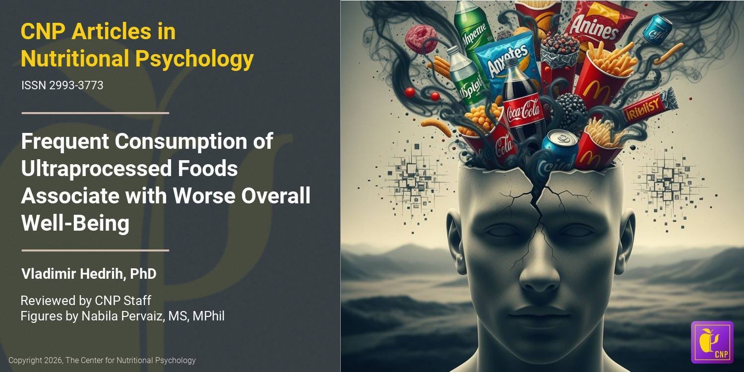
Western high-fat diet consumption during adolescence increases susceptibility to traumatic stress while selectively disrupting hippocampal and ventricular volumes (animal study)
In this 2016 study, adolescent Lewis rats (28 days postnatal) were given unrestricted access to either a Western-like Diet (41.4% kcal from fat) or the control diet (16.5% kcal from fat) for 8 weeks, before being exposed to a cat odor threat (to induce psychological trauma), in order to test if an obesogenic Western-like high-fat diet (WD) pre-disposes rats to post-traumatic stress responsivity. One week into encountering stress, increased anxiety-like behavior was evident in the rats fed on the WD, compared with those on the control diet (indicated by the elevated plus maze and the open field test). The high-fat diet was also associated with significant hippocampal atrophy (20% reduction) and lateral ventricular enlargement (50% increase), observed in the magnetic resonance imaging results. As well as these volumetric disturbances, leptin and FK506-binding protein 51 (FKBP51) levels were elevated, while hippocampal blood vessel density was reduced. Asymmetric structural vulnerabilities to the WD were discovered, particularly in the ventral and left hippocampus and lateral ventricle. This study demonstrates that WD consumption during adolescence influences key substrates implicated in post-traumatic stress disorder. It is important to develop our knowledge on WD’s impact on a person’s course of development since susceptibility to stress translates to greater vulnerability to neuropsychiatric disorders. [NPID: stress, animal, Western-style diet, WS diet, psychological trauma, responsivity, anxiety, hippocampus, hippocampal atrophy, PTSD, neuropsychiatric disorders]
Year: 2016