A study published inNature Communications sought to identify the brain mechanism through which a lack of sleep increases food desire.
They found that a lack of sleep decreases the activity of cortex regions responsible for cognitive processes regarding food intake while increasing the activity of the subcortical amygdala region.
Loss of sleep leads to a heightened desire for high-calorie foods.
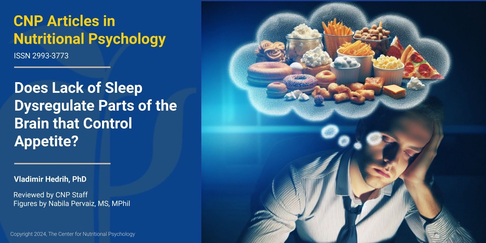
When lacking sleep, some drink coffee, some try to increase their physical activity to stay awake, and some visit the fridge. Most people have experienced this situation when they couldn’t get enough sleep, accompanied by a desire to eat more food than usual. But does lack of sleep really increase our appetite?
Sleep and appetite
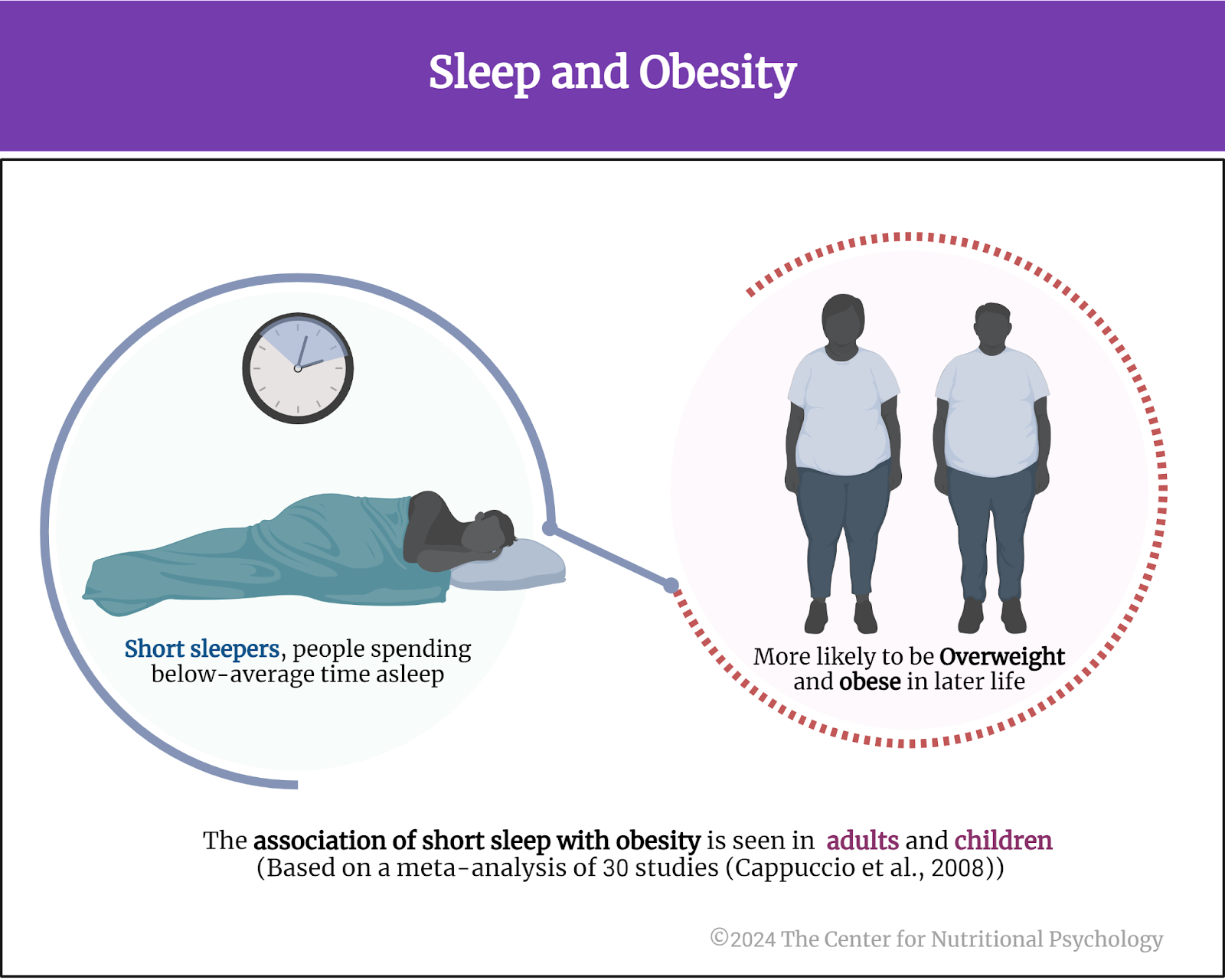
Researchers extensively investigated the link between sleep and the desire to eat. Many found that insufficient sleep is associated with increased food intake. A 2008 meta-analysis of 30 studies found that short sleepers, people spending below-average time asleep, are more likely to be overweight and obese. This association was present in adults and children (Cappuccio et al., 2008). A newer meta-analysis confirmed these findings again, showing that short sleep duration leads to an increased risk of obesity later in life (Bacaro et al., 2020) (see Figure 1).
Figure 1. Sleep and obesity
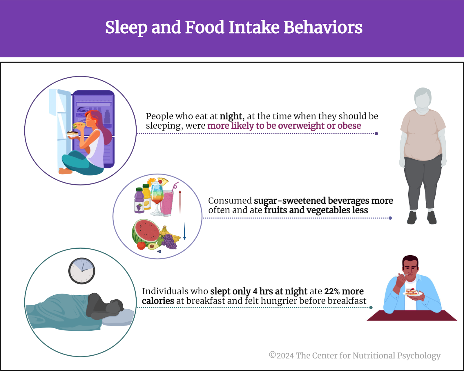
Similarly, newer studies showed that people who eat at night, at the time when they should be sleeping, were more likely to be overweight or obese. They also consumed sugar-sweetened beverages more often and ate fruits and vegetables less often than those not prone to eating at night (Lent et al., 2022). Additionally, individuals suffering from night eating syndrome, a condition in which they tend to eat lots of food at night, have a lower quality of sleep than those without this disorder (Tzischinsky et al., 2021).
One experiment showed that individuals who slept only 4 hours at night ate 22% more calories for breakfast the following day and felt hungrier immediately before breakfast (Brondel et al., 2010)(see Figure 2).
Figure 2. Sleep and food intake behaviors
Despite all the evidence that the desire for food and sleep quality are connected, the neural mechanisms that produce this effect remained more or less unknown (Greer et al., 2013).
The current study
Study author Stephanie Greer and her colleagues wanted to identify the neural mechanism through which a lack of sleep increases the desire for food. They wanted to know what changes in the brain when we lack sleep produce this effect.
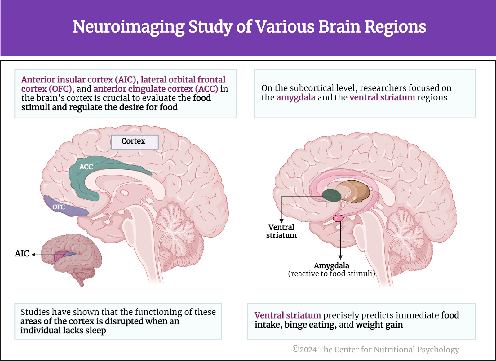
These authors conducted a neuroimaging study in which they focused on a set of cortical and subcortical regions of the brain that researchers consider crucial for evaluating food stimuli and regulating the desire for food. These areas were the anterior insular cortex, lateral orbital frontal cortex, and anterior cingulate cortex in the brain’s cortex. All of these areas have well-established roles in signaling the value of a food stimulus and regulating how we integrate evaluations of various food features to create preferences for food. Studies have also shown that the functioning of these areas of the cortex is disrupted when an individual lacks sleep (Muzur et al., 2002).
On the subcortical level, they focused on the amygdala and the ventral striatum regions. Previous studies showed that the amygdala is very reactive to food stimuli. Activity in the ventral striatum, on the other hand, very precisely predicts immediate food intake, binge eating, and weight gain (Greer et al., 2013) (see Figure 3).
Figure 3.Neuroimaging study of various brain regions
The procedure
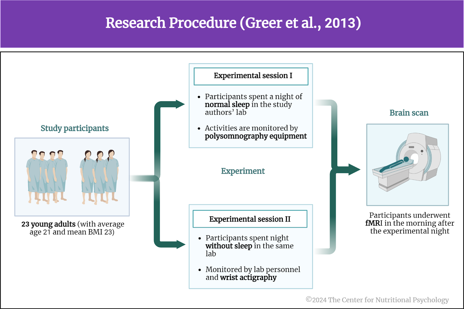
The study participants were 23 young adults, the average age of whom was 21. Of these, 13 were females. Their mean body mass index was 23, meaning that, on average, they were normal weight.
Participants completed two experimental sessions. During one session, they spent a night of normal sleep in the study authors’ lab, monitored by polysomnography equipment. The second session was a night without sleep in the same lab, monitored by lab personnel and wrist actigraphy.
Wrist actigraphs are wearable devices that measure movements. Researchers can use them to determine when the wearer is sleeping and when he/she is awake. Polysomnography equipment consists of a set of devices used to record various physiological parameters during sleep. These can include brain waves (EEG), eye movements, muscle activity, heart rate, and breathing patterns. Polysomnography is used to monitor sleep quality and diagnose sleep disorders.
Study participants underwent functional magnetic resonance imaging (fMRI) the morning after the experimental night (see Figure 4).
Figure 4. Procedure (Greer et al., 2013)
Sleep deprivation reduced activity in the studied cortical regions
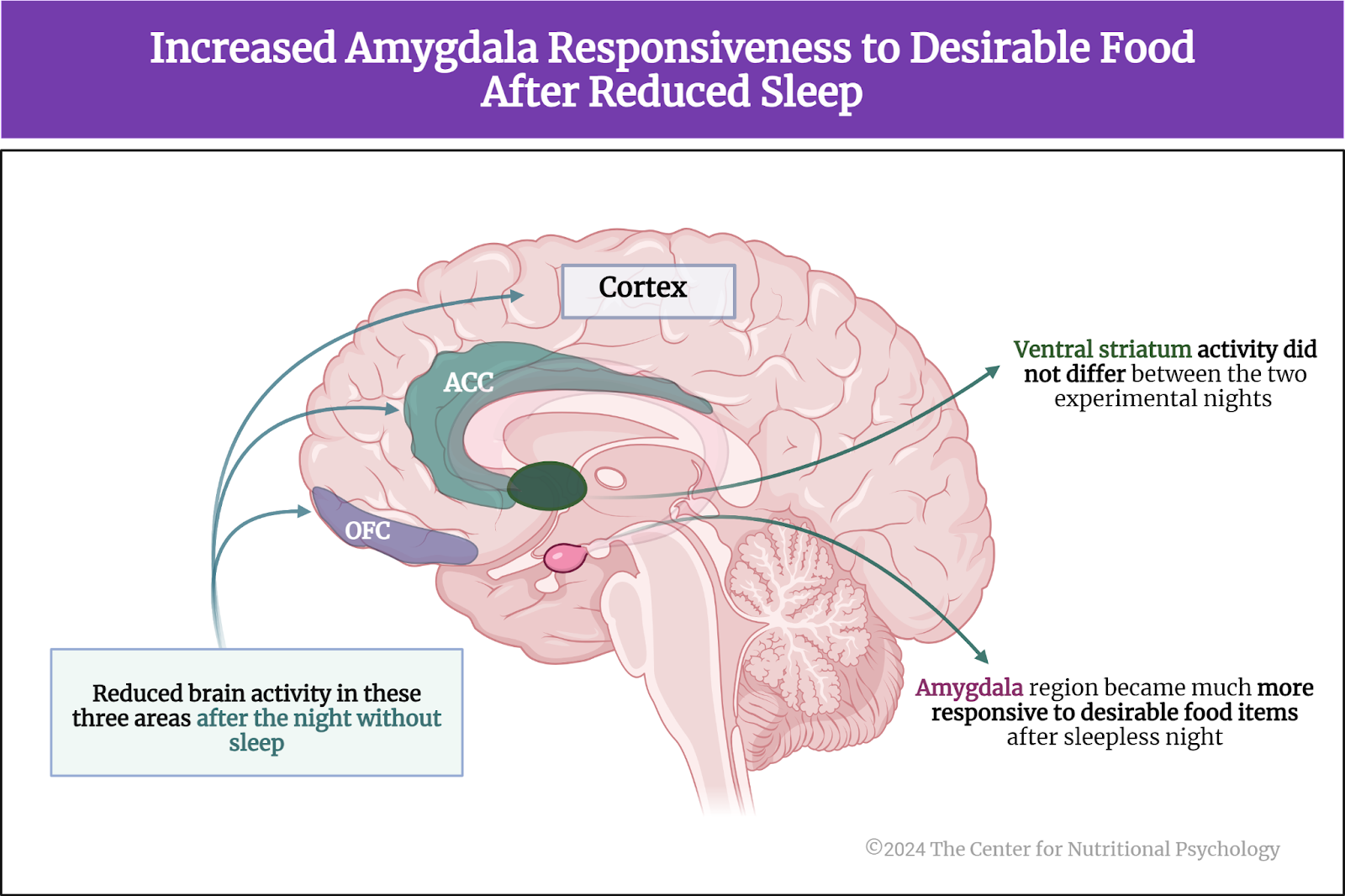
Results showed that brain activity was reduced after the night without sleep in all three studied areas of the cortex – the anterior cingulate cortex, the lateral orbital frontal cortex, and the anterior insular cortex.
In contrast to the cortical regions, the amygdala region became much more responsive to desirable food items after sleepless nights than when participants slept normally. The ventral striatum activity did not differ between the two experimental nights (see Figure 5).
Figure 5. Increased amygdala responsiveness to desirable food after reduced sleep
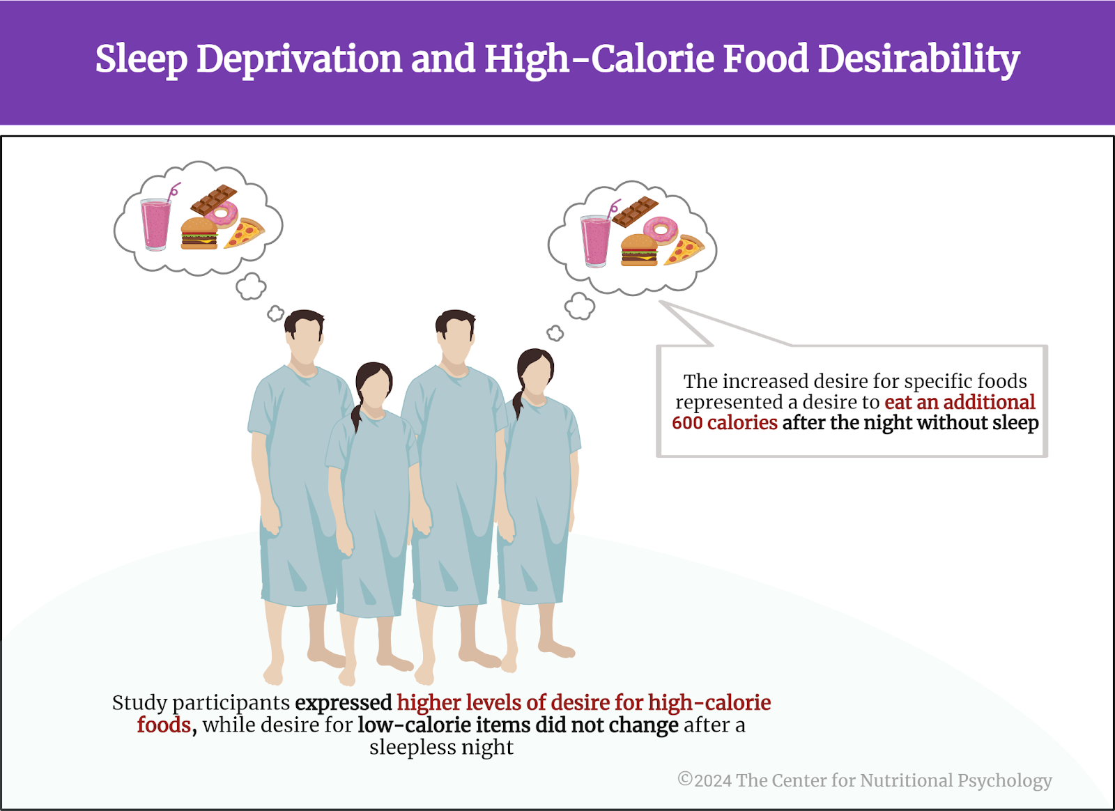
Sleep deprivation increases the desirability of high-calorie foods
Compared to the night when they normally slept, study participants expressed higher levels of desire for high-calorie foods after the night without sleep. There was no difference between the two nights in how much participants desired low-calorie items. Overall, the increase in desire for specific foods represented a desire to eat an additional 600 calories after the night without sleep. The increase in desire for high-calorie foods was so specific that the increase in the desirability of a specific food item after a night without sleep could be predicted based on its calorie content (see Figure 6).
Figure 6. Sleep deprivation and high-calorie food desirability
Conclusion
Overall, the study found that lack of sleep decreases the activity of the cortex areas responsible for cognitive processes regarding food while increasing the activity of the amygdala subcortical region. After a sleepless night, high-calorie food items also became more desirable.
These findings contribute to a better scientific understanding of the neural mechanisms underpinning the link between sleep deprivation and increased desire for food. They very strongly suggest that weight loss and obesity prevention programs should consider sleep quality and include regulating any sleep problems among their goals.
Bacaro, V., Ballesio, A., Cerolini, S., Vacca, M., Poggiogalle, E., Donini, L. M., Lucidi, F., & Lombardo, C. (2020). Sleep duration and obesity in adulthood: An updated systematic review and meta-analysis. Obesity Research & Clinical Practice, 14(4), 301–309. https://doi.org/10.1016/j.orcp.2020.03.004
Brondel, L., Romer, M. A., Nougues, P. M., Touyarou, P., & Davenne, D. (2010). Acute partial sleep deprivation increases food intake in healthy men. The American Journal of Clinical Nutrition, 91(6), 1550–1559. https://doi.org/10.3945/ajcn.2009.28523
Cappuccio, F. P., Taggart, F. M., Kandala, N.-B., Currie, A., ChB, M., Peile, E., & Miller, M. A. (2008). Meta-Analysis of Short Sleep Duration and Obesity in Children and Adults. 31(5).
Greer, S. M., Goldstein, A. N., & Walker, M. P. (2013). The impact of sleep deprivation on food desire in the human brain. Nature Communications, 4. https://doi.org/10.1038/ncomms3259
Lent, M. R., Atwood, M., Bennett, W. L., Woolf, T. B., Martin, L., Zhao, D., Goheer, A. A., Song, S., McTigue, K. M., Lehmann, H. P., Holzhauer, K., & Coughlin, J. W. (2022). Night eating, weight, and health behaviors in adults participating in the Daily24 study. Eating Behaviors, 45. https://doi.org/10.1016/j.eatbeh.2022.101605
Muzur, A., Pace-Schott, E. F., & Hobson, J. A. (2002). The prefrontal cortex in sleep. Trends in Cognitive Sciences, 6(11), 475–481. https://doi.org/10.1016/S1364-6613(02)01992-7
Tzischinsky, O., Latzer, I. T., Alon, S., & Latzer, Y. (2021). Sleep quality and eating disorder-related psychopathologies in patients with night eating syndrome and binge eating disorders. Journal of Clinical Medicine, 10(19). https://doi.org/10.3390/jcm10194613
An enzyme produced in white blood cells regulates some of the psychological symptoms of depression
A study on mice published inNature reported that an enzyme called matrix metalloproteinase 8 (MMP8) regulates the effects of stress on the symptoms of depression.
MMP8 was increased in the blood serum of humans with major depressive disorder and mice exposed to chronic stress.
Injecting mice with an appropriate dose of MMP8 promoted social avoidance.
The results showcase how immune system reactions can affect the brain, leading to profound behavioral changes.
Scientists have studied mental health problems for centuries. However, for most of this time, their focus has solely been on psychological symptoms because the scientific know-how needed to understand the complex biochemical mechanisms underpinning psychology was simply not there until now. Recent advances in biomedical technology allowed modern researchers to start mapping the biochemical mechanisms behind these disorders. One major topic of this type of research is stress-related disorders, such as major depressive disorder.
Major depressive disorder and stress
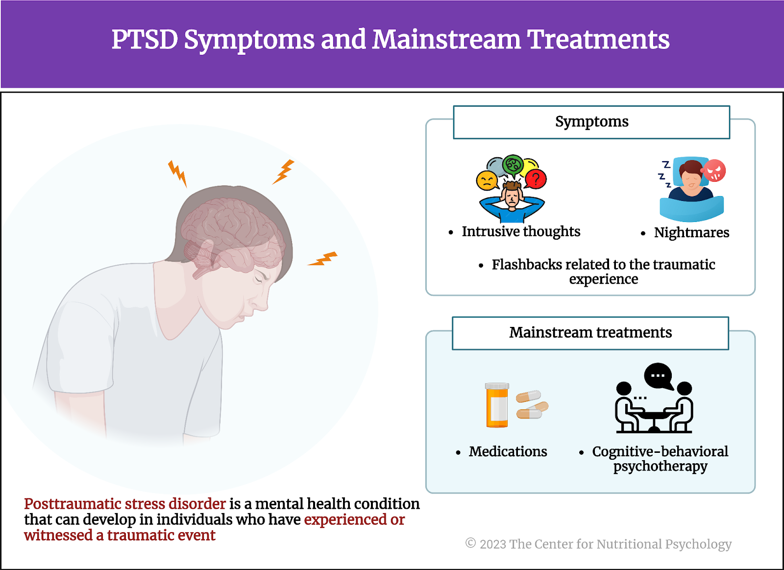
Major depressive disorder, or MDD, is a serious mental health condition characterized by persistent feelings of sadness, hopelessness, and a lack of interest or pleasure in daily activities, often accompanied by physical symptoms such as changes in appetite or sleep patterns. It is one of the most frequent psychiatric disorders worldwide (Steffen et al., 2020; Weinberger et al., 2018).
Stress likely plays a significant role in the development and exacerbation of major depressive disorder
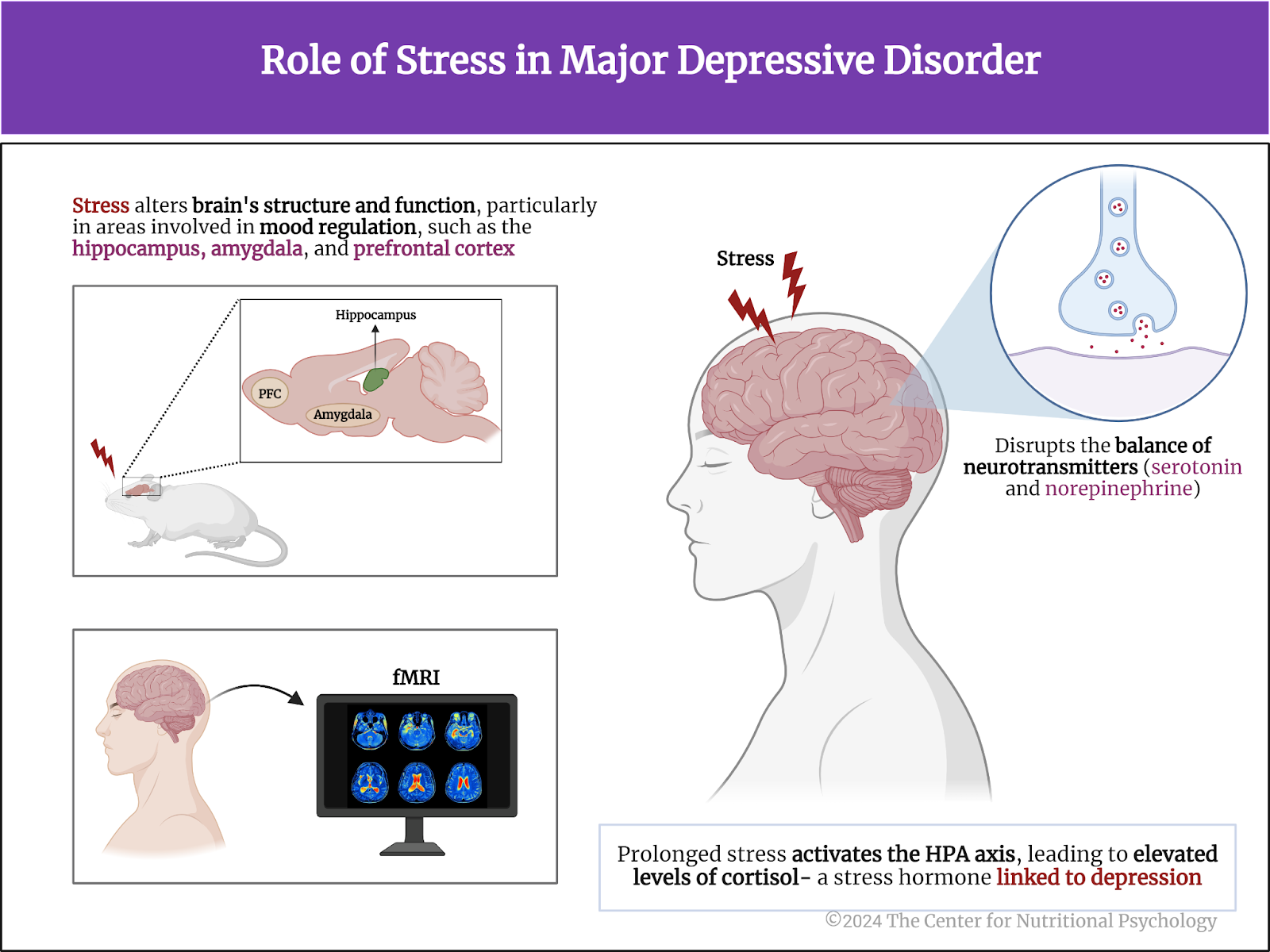
Studies on mice showed that chronic stress can lead to depression-like symptoms and alterations in the brain’s structure and function, particularly in areas involved in mood regulation, such as the hippocampus, amygdala, and prefrontal cortex (PFC) (Khan et al., 2020). Neuroimaging studies have reported alterations to similar brain areas in humans with depression (Zhang et al., 2018)
Stress can also disrupt the balance of neurotransmitters, like serotonin and norepinephrine, which are crucial for maintaining mood stability (Adell et al., 1988). Furthermore, prolonged stress activates the hypothalamic-pituitary-adrenal axis (HPA axis), leading to elevated cortisol levels. This stress hormone has been linked to depression (Tafet & Bernardini, 2003) (see Figure 1).
Figure 1. Role of stress in major depressive disorder
Inflammatory processes and depression
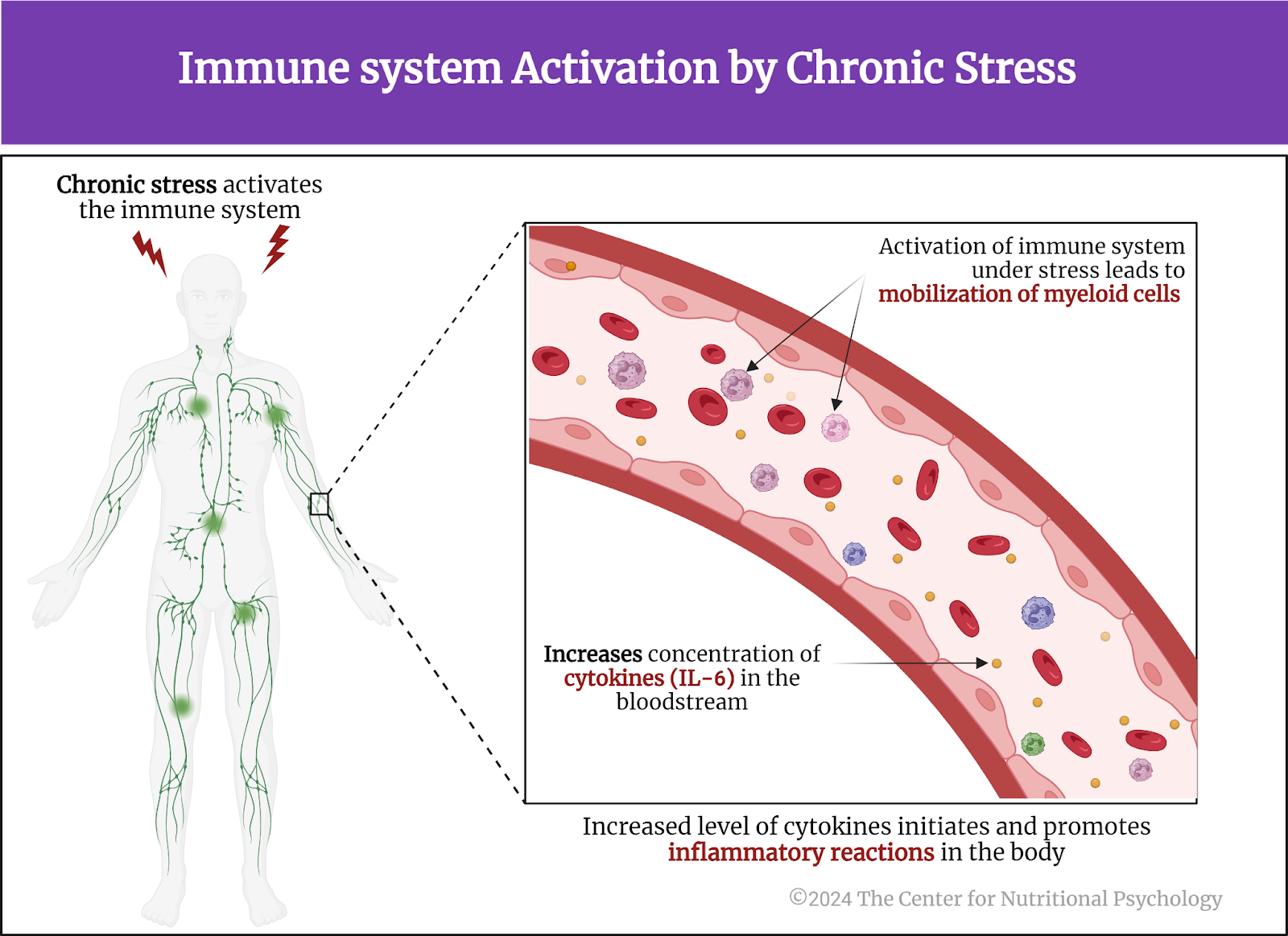
Studies show chronic stress also activates the immune system, the body’s natural defense against bacteria, viruses, and other pathogens. This activation leads to the mobilization of a type of white blood cell called myeloid cells. The organism under stress also increases the concentration of specific signaling proteins called cytokines (such as interleukin-6) in the bloodstream. This initiates and promotes inflammatory reactions in the body (Cathomas et al., 2024) (see Figure 2).
Figure 2. Immune system activation by chronic stress
In line with this, studies on humans show that individuals with stress-related mental health conditions such as major depressive disorder display a state of chronic low-grade inflammation characterized by increased concentrations of these pro-inflammatory proteins in the bloodstream (Dowlati et al., 2010)
The current study
Study author Flurin Cathomas and his colleagues wanted to understand better the role of immune system molecules in the development of psychological symptoms of depression. The initial results they obtained focused their attention on the role of the enzyme matrix metalloproteinase 8 (MMP8).
MMP8 belongs to the metalloproteinase family, a group of enzymes responsible for breaking down extracellular matrix proteins. MMP8 is primarily produced by neutrophils, a type of myeloid white blood cell that plays a crucial role in the immune system’s response to infection and inflammation. They are derived from myeloid progenitor cells in the bone marrow.
These researchers conducted experiments on 4-7-week-old mice purchased externally or bred in the researchers’ laboratory. In the experiments, the study authors exposed these mice to different types of stressors and conducted different biomedical and surgical procedures. The main type of stress-induced in these mice was chronic social defeat. This was done by exposing them for 5 or 10 minutes to a large, aggressive mouse for ten days. After this treatment, some mice started avoiding other mice, developing social avoidance, a pattern of behaviors comparable to that of depression in humans. Other mice did not. Study authors called the first type of mice “susceptible” mice and the later “resistant” mice. They also used mice not exposed to stress as controls (see Figure 3).
Figure 3. Study Procedure (Cathomas, 2024)
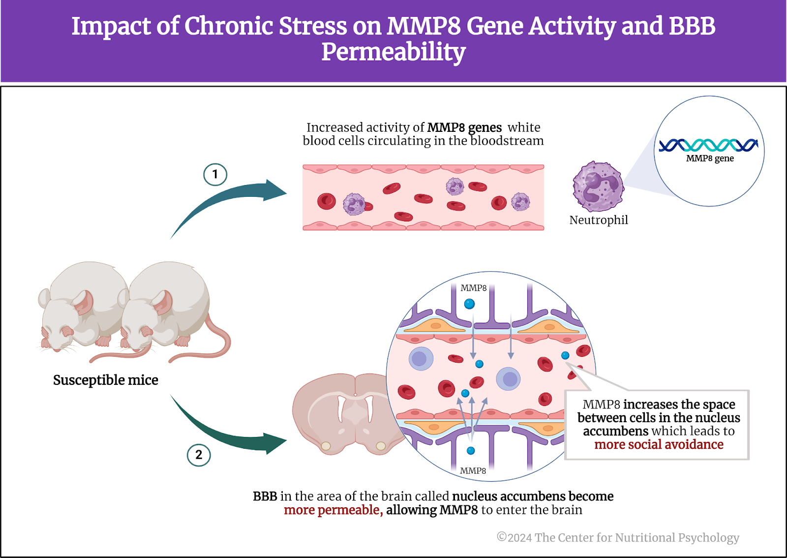
Chronic stress upregulates genes producing MMP8 in the bloodstream of susceptible mice
The study authors examined the activity of white blood cells in mice and grouped them into four categories based on their gene activity patterns. They identified a group of genes that were more active in susceptible mice compared to controls. Many of the genes in this group were proteins known to play a role in the process of inflammation. One of the most differentially active genes in the two groups of mice was the one encoding MMP8.
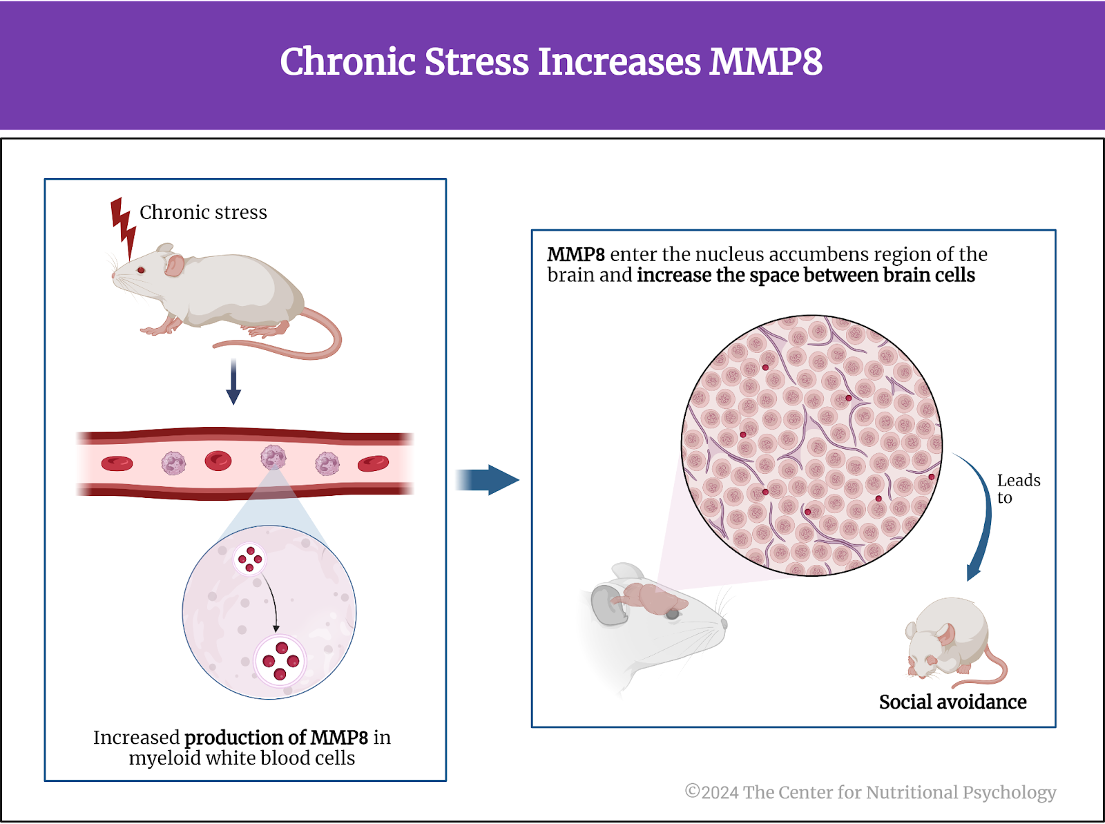
Further analysis showed that this increased activity of MMP8 genes was not found in the brain but only in white blood cells circulating in the bloodstream. Although these MMP8 molecules are produced outside the brain, it turned out that they can still travel to the brain. Chronic stress makes the blood-brain barrier (BBB) in the area of the brain called nucleus accumbens more permeable, allowing MMP8 to enter the brain at that point. Upon entering, they increase the space between cells in this area. A separate experiment showed that increasing the space between cells in the nucleus accumbens leads to more social avoidance in these mice (see Figure 4).
Figure 4. Impact of chronic stress on MMP8 gene activity and BBB permeability
Study authors verified these findings in humans and found that individuals with major depressive disorder also have increased concentrations of MMP8 in their bloodstream.
MMP8 regulates social avoidance
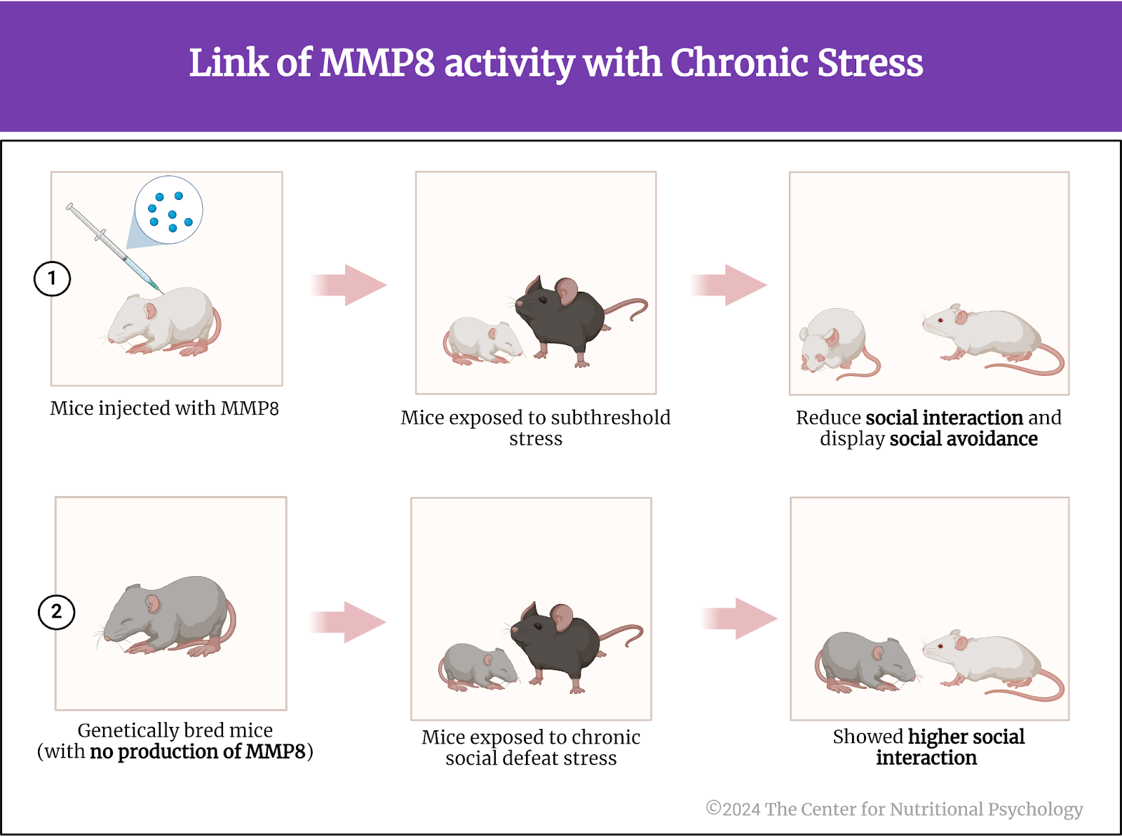
When study authors injected MMP8 into mice that were exposed to subthreshold stress (social defeat stress insufficient to produce behavioral changes), these mice also reduced their social interactions (i.e., started displaying social avoidance). To ensure this is the effect of MMP8 (and not of something else), the study authors bred mice genetically altered not to produce it. Exposing these mice to chronic social defeat stress did not result in social avoidance. They even showed higher social interaction and spent less time in the corner than regular mice. This indicated that MMP8 regulates social avoidance behavior in mice (Figure 5).
Figure 5. Link of MMP8 activity with chronic stress
Conclusion
The study showed that chronic stress increases the enzyme matrix metalloproteinase 8 (MMP8) production in myeloid white blood cells in the bloodstream. These enzymes enter the nucleus accumbens region of the brain and increase the space between brain cells, leading to social avoidance in mice (see Figure 6).
Figure 6. Chronic stress increases MMP8
Although the study was conducted on mice, similar physiological mechanisms also likely exist in humans. Thanks to this similarity, these findings contribute to the scientific understanding of biochemical mechanisms underpinning psychological reactions to chronic stress and symptoms of disorders such as depression. Understanding the biochemical mechanisms of mental disorders will likely lead to the development of much more effective ways to treat them.
The paper “Circulating myeloid-derived MMP8 in stress susceptibility and depression” was authored by Flurin Cathomas, Hsiao-Yun Lin, Kenny L. Chan, Long Li, Lyonna F. Parise, Johana Alvarez, Romain Durand-de Cuttoli, Antonio V. Aubry, Samer Muhareb, Fiona Desland, Yusuke Shimo, Aarthi Ramakrishnan, Molly Estill, Carmen Ferrer-Pérez, Eric M. Parise, C. Matthias Wilk, Manuella P. Kaster, Jun Wang, Allison Sowa, William G. Janssen, Sara Costi, Adeeb Rahman, Nicolas Fernandez, Matthew Campbell, Filip K. Swirski, Eric J. Nestler, Li Shen, Miriam Mera, James W. Murrough, and Scott J. Russo.
References
Adell, A., Garcia-Marquez, C., Armario, A., & Gelpi, E. (1988). Chronic Stress Increases Serotonin and Noradrenaline in Rat Brain and Sensitizes Their Responses to a Further Acute Stress. Journal of Neurochemistry, 50(6), 1678–1681. https://doi.org/10.1111/j.1471-4159.1988.tb02462.x
Cathomas, F., Lin, H.-Y., Chan, K. L., Li, L., Parise, L. F., Alvarez, J., Durand-de Cuttoli, R., Aubry, A. V., Muhareb, S., Desland, F., Shimo, Y., Ramakrishnan, A., Estill, M., Ferrer-Pérez, C., Parise, E. M., Wilk, C. M., Kaster, M. P., Wang, J., Sowa, A., … Russo, S. J. (2024). Circulating myeloid-derived MMP8 in stress susceptibility and depression. Nature, 626(8001), 1108–1115. https://doi.org/10.1038/s41586-023-07015-2
Dowlati, Y., Herrmann, N., Swardfager, W., Liu, H., Sham, L., Reim, E. K., & Lanctôt, K. L. (2010). A Meta-Analysis of Cytokines in Major Depression. Biological Psychiatry, 67(5), 446–457. https://doi.org/10.1016/j.biopsych.2009.09.033
Khan, A. R., Geiger, L., Wiborg, O., & Czéh, B. (2020). Stress-Induced Morphological, Cellular and Molecular Changes in the Brain—Lessons Learned from the Chronic Mild Stress Model of Depression. Cells, 9(4), 1026. https://doi.org/10.3390/cells9041026
Steffen, A., Thom, J., Jacobi, F., Holstiege, J., & Bätzing, J. (2020). Trends in prevalence of depression in Germany between 2009 and 2017 based on nationwide ambulatory claims data. Journal of Affective Disorders, 271, 239–247. https://doi.org/10.1016/J.JAD.2020.03.082
Tafet, G. E., & Bernardini, R. (2003). Psychoneuroendocrinological links between chronic stress and depression. Progress in Neuro-Psychopharmacology and Biological Psychiatry, 27(6), 893–903. https://doi.org/10.1016/S0278-5846(03)00162-3
Weinberger, A. H., Gbedemah, M., Martinez, A. M., Nash, D., Galea, S., & Goodwin, R. D. (2018). Trends in depression prevalence in the USA from 2005 to 2015: Widening disparities in vulnerable groups. Psychological Medicine, 48(8), 1308–1315. https://doi.org/10.1017/S0033291717002781
Zhang, F.-F., Peng, W., Sweeney, J. A., Jia, Z.-Y., & Gong, Q.-Y. (2018). Brain structure alterations in depression: Psychoradiological evidence. CNS Neuroscience & Therapeutics, 24(11), 994–1003. https://doi.org/10.1111/cns.12835
An analysis of the National Health and Nutrition Examination Survey data published inBMC Psychiatry found an association between sugar intake and depression in U.S. adults.
With every 100 grams of additional sugar intake, the prevalence of depression increased by 28%.
Results were similar across subgroups by age, sex, education, and other personal characteristics.
Most people know that there is a link between appetite and mood. When stressed, some of us tend to eat more (Dakanalis et al., 2023). A similar thing happens when we do not have enough sleep (Greer et al., 2013). On the other hand, when we are hungry, we tend to become angry and irritable. That is how the term “hangry” came to be (Hedrih, 2023a; Swami et al., 2022). But is there a link between moods or mental health in general and specific foods? For example, do the dietary choices of individuals suffering from depression differ from the dietary choices of individuals who aren’t suffering from depression?
Do the dietary choices of individuals with depression differ from the dietary choices of individuals without depression?
What is depression?
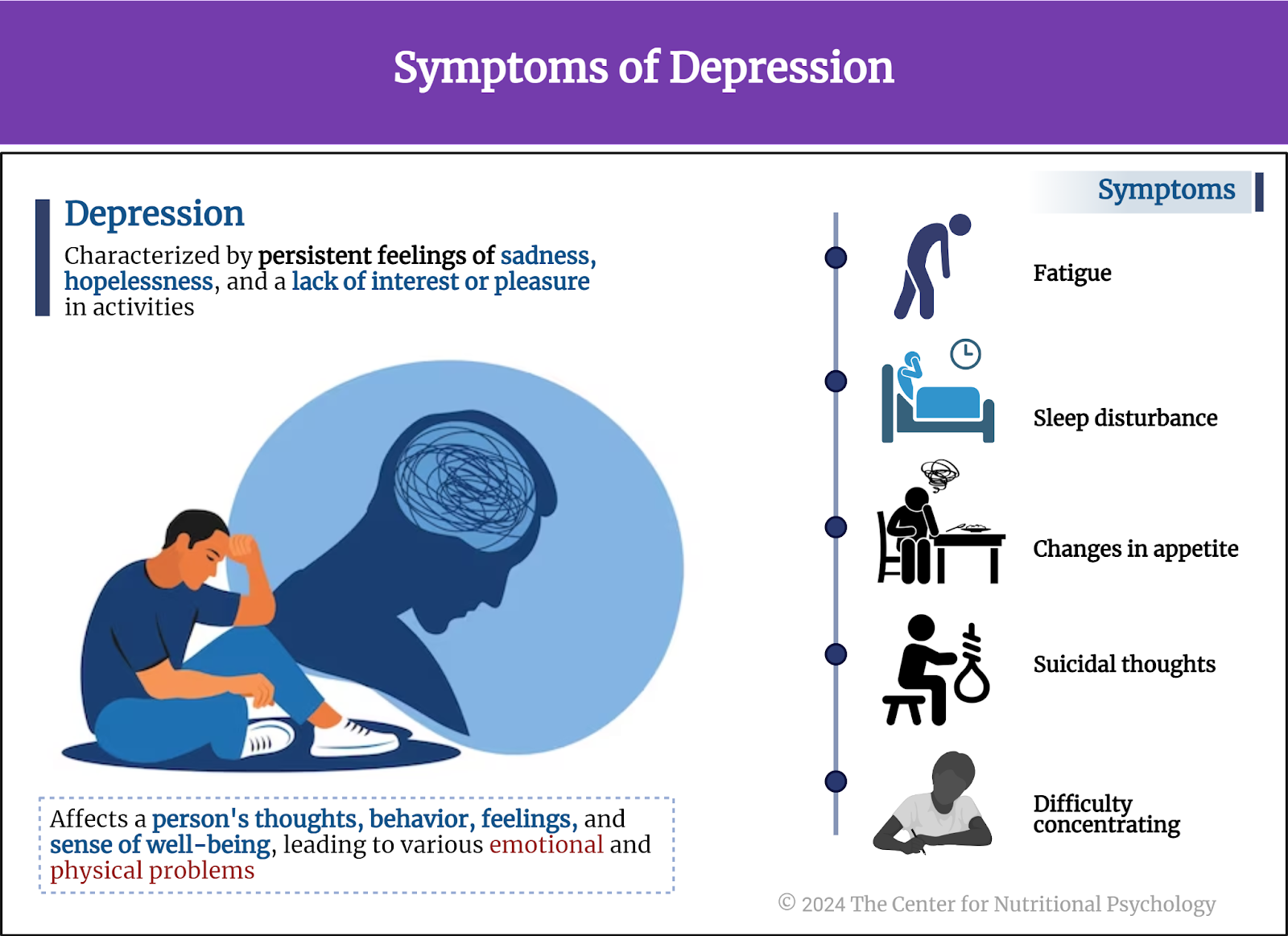
Depression is a common and serious mental health condition characterized by persistent feelings of sadness, hopelessness, and a lack of interest or pleasure in activities. It can affect a person’s thoughts, behavior, feelings, and sense of well-being, leading to various emotional and physical problems. Symptoms can include changes in appetite, sleep disturbances, fatigue, difficulty concentrating, and even thoughts of death or suicide (see Figure 1).
Figure 1. Symptoms of depression
Depression is one of the most prevalent mental health disorders worldwide. The share of depressed individuals has been on the rise in recent decades in many world countries (Steffen et al., 2020; Weinberger et al., 2018). In 2018, the World Health Organization estimated that 4.4% of the global population is affected by it (Zhang et al., 2024). They expect the numbers to rise further in the coming years.
Unfortunately, existing treatments for depression are not very effective. It is estimated that around 30% of individuals suffering from depression do not respond to standard treatments, i.e., these treatments do not result in the withdrawal of depression symptoms (McIntyre et al., 2023). That is why a large number of scientists are working on finding novel ways to diagnose and treat depression. One of the topics that draw interest in this regard is the dietary specificities of people suffering from depression.
Around 30% of individuals suffering from depression do not respond to standard treatments
Dietary choices and depression
In recent decades, many studies have reported links between symptoms of depression and anxiety and various food choices. A large-scale study found that depressed women tend to drink artificially sweetened beverages and eat ultraprocessed food more than the average person (Hedrih, 2023b; Samuthpongtorn et al., 2023). Another study linked the consumption of fried food with symptoms of anxiety and depression. These researchers conducted an experiment showing that acrylamide, a product of frying foods such as potatoes, can induce anxiety-like symptoms in zebrafish (Wang et al., 2023).
Similarly, many studies link increased sugar consumption with adverse health outcomes (Huang et al., 2023). But does this include depression?
Many studies report links between symptoms of depression and anxiety and various food choices (artificially sweetened beverages and fried foods)
The current study
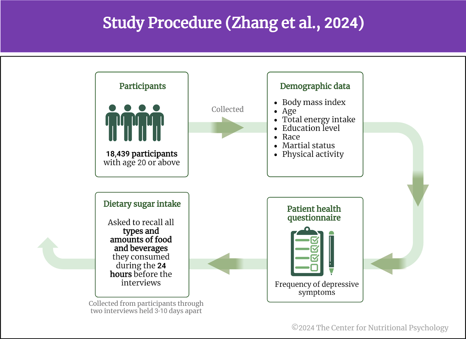
Study author Lu Zhang and her colleagues wanted to explore whether sugar consumption is associated with the severity of depressive symptoms (Zhang et al., 2024). They analyzed data from the National Health and Nutrition Examination Survey database. The National Health and Nutrition Examination Survey is a program of studies conducted by the National Center for Health Statistics (NCHS), part of the Centers for Disease Control and Prevention (CDC), designed to assess the health and nutritional status of adults and children in the United States through interviews and physical examinations.
The study data
Data used in this study came from participants aged 20 or above who provided their survey responses between 2011 and 2018. The study authors used data on the frequency of depressive symptoms (the Patient Health Questionnaire) and dietary sugar intake. Dietary information was collected from participants through two interviews held 3-10 days apart. In these interviews, researchers asked participants to recall all types and amounts of food and beverages they consumed during the 24 hours before the interviews. Dietary sugar intake was calculated from data collected in these interviews.
The study authors also analyzed data on several other participants’ characteristics. These included body mass index, age, total energy intake, education level, race, marital status, physical activity, etc.
This analysis used data from 18,439 participants (see Figure 2).
Figure 2. Study Procedure (Zhang et al., 2024)
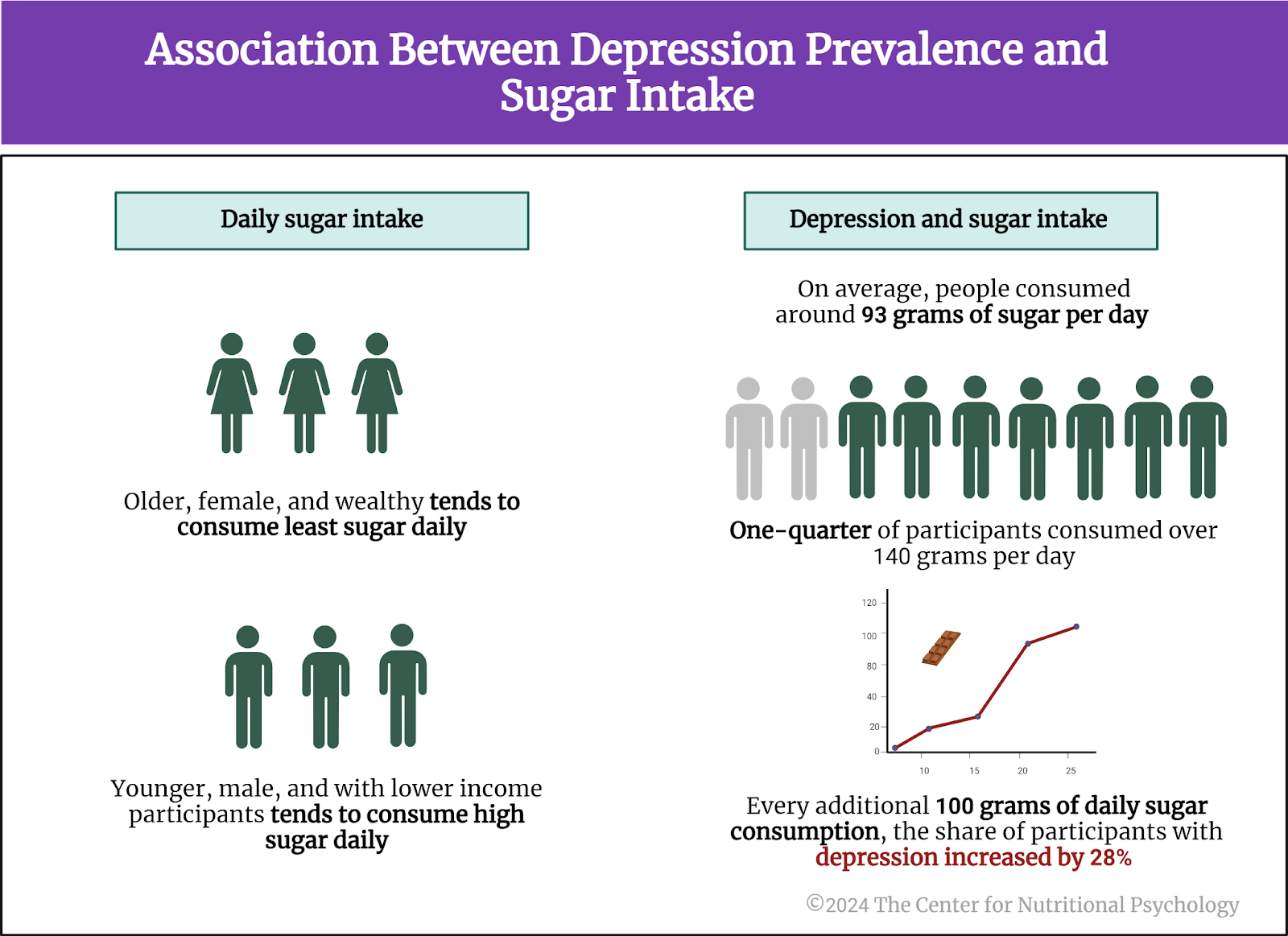
Older, wealthier, more educated, partnered, and female participants tend to consume the least sugar
Results showed that individuals consuming the least sugar daily tended to be older, female, and wealthy. Their overall energy intake was lower; they smoked less and drank more than the average participant. Participants with the highest daily sugar intake tended to be younger, male, and with lower income. They tended to have never smoked and had a higher energy intake than average.
Depressed individuals tended to consume more sugar
There was a clear association between the prevalence of depression and sugar intake. On average, people consumed around 93 grams of sugar per day, but individuals were consuming quite a bit more than that. One-quarter of participants consumed over 140 grams per day. Statistical analyses showed that for every additional 100 grams of daily sugar consumption, the share of participants with depression increased by 28% (see Figure 3).
Figure 3. Association between depression prevalence and sugar intake
Conclusion
The study shows a clear link between dietary sugar consumption and depression. However, the design of the study does not allow us to conclude whether it is the intake of sugar that contributes to depression or whether it is depression that makes individuals consume more sugary drinks and foods. It is also possible that there is some third factor contributing to both depression and the heightened intake of sugar.
Be that as it may, the link between heightened sugar intake and depression is quite clear. However, it is up to future researchers to identify the underlying mechanism and determine whether limiting dietary sugar intake could help improve symptoms in individuals suffering from this disorder.
Dakanalis, A., Mentzelou, M., Papadopoulou, S. K., Papandreou, D., Spanoudaki, M., Vasios, G. K., Pavlidou, E., Mantzorou, M., & Giaginis, C. (2023). The Association of Emotional Eating with Overweight/Obesity, Depression, Anxiety/Stress, and Dietary Patterns: A Review of the Current Clinical Evidence. Nutrients, 15(5), 1173. https://doi.org/10.3390/nu15051173
Greer, S. M., Goldstein, A. N., & Walker, M. P. (2013). The impact of sleep deprivation on food desire in the human brain. Nature Communications, 4. https://doi.org/10.1038/ncomms3259
Hedrih, V. (2023a). Food and Mood: Is the Concept of ‘Hangry’ Real? CNP Articles in Nutritional Psychology. https://www.nutritional-psychology.org/food-and-mood-is-the-concept-of-hangry-real/
Hedrih, V. (2023b). Women Consuming Lots of Artificially Sweetened Beverages Might Have a Higher Risk of Depression, Study Finds. CNP Articles in Nutritional Psychology. https://www.nutritional-psychology.org/women-consuming-lots-of-artificially-sweetened-beverages-might-have-a-higher-risk-of-depression-study-finds/
Huang, Y., Chen, Z., Chen, B., Li, J., Yuan, X., Li, J., Wang, W., Dai, T., Chen, H., Wang, Y., Wang, R., Wang, P., Guo, J., Dong, Q., Liu, C., Wei, Q., Cao, D., & Liu, L. (2023). Dietary sugar consumption and health: Umbrella review. BMJ (Clinical Research Ed.), 381, e071609. https://doi.org/10.1136/bmj-2022-071609
McIntyre, R. S., Alsuwaidan, M., Baune, B. T., Berk, M., Demyttenaere, K., Goldberg, J. F., Gorwood, P., Ho, R., Kasper, S., Kennedy, S. H., Ly-Uson, J., Mansur, R. B., McAllister-Williams, R. H., Murrough, J. W., Nemeroff, C. B., Nierenberg, A. A., Rosenblat, J. D., Sanacora, G., Schatzberg, A. F., … Maj, M. (2023). Treatment-resistant depression: Definition, prevalence, detection, management, and investigational interventions. World Psychiatry, 22(3), 394–412. https://doi.org/10.1002/wps.21120
Samuthpongtorn, C., Nguyen, L. H., Okereke, O. I., Wang, D. D., Song, M., Chan, A. T., & Mehta, R. S. (2023). Consumption of Ultraprocessed Food and Risk of Depression. JAMA Network Open, 6(9), e2334770. https://doi.org/10.1001/jamanetworkopen.2023.34770
Steffen, A., Thom, J., Jacobi, F., Holstiege, J., & Bätzing, J. (2020). Trends in prevalence of depression in Germany between 2009 and 2017 based on nationwide ambulatory claims data. Journal of Affective Disorders, 271, 239–247. https://doi.org/10.1016/J.JAD.2020.03.082
Swami, V., Hochstöger, S., Kargl, E., & Stieger, S. (2022). Hangry in the field: An experience sampling study on the impact of hunger on anger, irritability, and affect. PLOS ONE, 17(7), e0269629. https://doi.org/10.1371/JOURNAL.PONE.0269629
Wang, A., Wan, X., Zhuang, P., Jia, W., Ao, Y., Liu, X., Tian, Y., Zhu, L., Huang, Y., Yao, J., Wang, B., Wu, Y., Xu, Z., Wang, J., Yao, W., Jiao, J., & Zhang, Y. (2023). High-fried food consumption impacts anxiety and depression due to lipid metabolism disturbance and neuroinflammation. Proceedings of the National Academy of Sciences of the United States of America, 120(118). https://doi.org/10.1073/pnas.2221097120
Weinberger, A. H., Gbedemah, M., Martinez, A. M., Nash, D., Galea, S., & Goodwin, R. D. (2018). Trends in depression prevalence in the USA from 2005 to 2015: Widening disparities in vulnerable groups. Psychological Medicine, 48(8), 1308–1315. https://doi.org/10.1017/S0033291717002781
Zhang, L., Sun, H., Liu, Z., Yang, J., & Liu, Y. (2024). Association between dietary sugar intake and depression in US adults: A cross-sectional study using data from the National Health and Nutrition Examination Survey 2011–2018. BMC Psychiatry, 24(110), 1–10. https://doi.org/10.1186/s12888-024-05531-7
In an online experiment published in Appetite, participants rated various characteristics of foods, including energy density, level of processing, and carbohydrate-to-fat ratio.
Results showed that carbohydrate-to-fat ratio and taste were the primary determinants of food liking and the desire to eat.
Contrary to findings from previous studies, the level of processing was not linked to either the desire to eat the food or its taste ratings.
Have you ever wondered what makes us like and want to eat certain foods while disliking others? Most people will say that they like tasty food when asked this question. But what makes some foods tastier than others? Many scientific studies explored this question.
What makes some foods tastier than others?
Ultraprocessed foods and obesity
Scientists first studied this question in the context of the obesity pandemic currently plaguing the world (Wong et al., 2022). Their findings showed that many overweight people tend to eat lots of ultraprocessed foods. Ultraprocessed foods have undergone extensive processing and often contain additives to improve their taste and appeal (Monteiro et al., 2019). Researchers found that some components of ultraprocessed foods trigger neural processes similar to those found in various addictions, creating intense desires for those foods (Gearhardt et al., 2023; Hedrih, 2023).
Some components of ultraprocessed foods trigger neural processes similar to those found in various addictions, creating intense desires for those foods
Fat, carbohydrates, and food reward processing in the brain
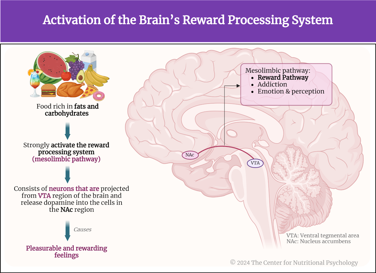
Other studies indicate that foods rich in fat and carbohydratescreate the strongest activation of the reward processing system in the brain, the system of neural circuits that produces the pleasurable and rewarding feelings we experience when we eat something tasty (see Figure 1).
Figure 1. Activation of the brain’s reward processing system
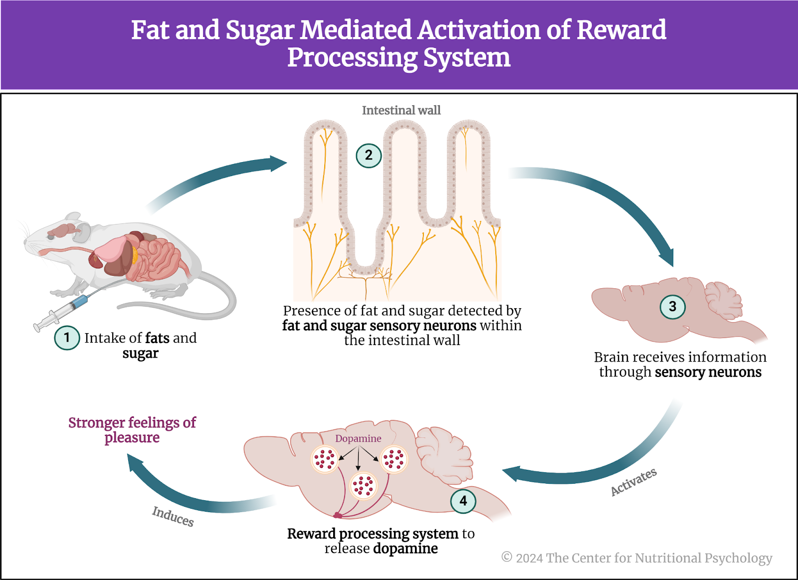
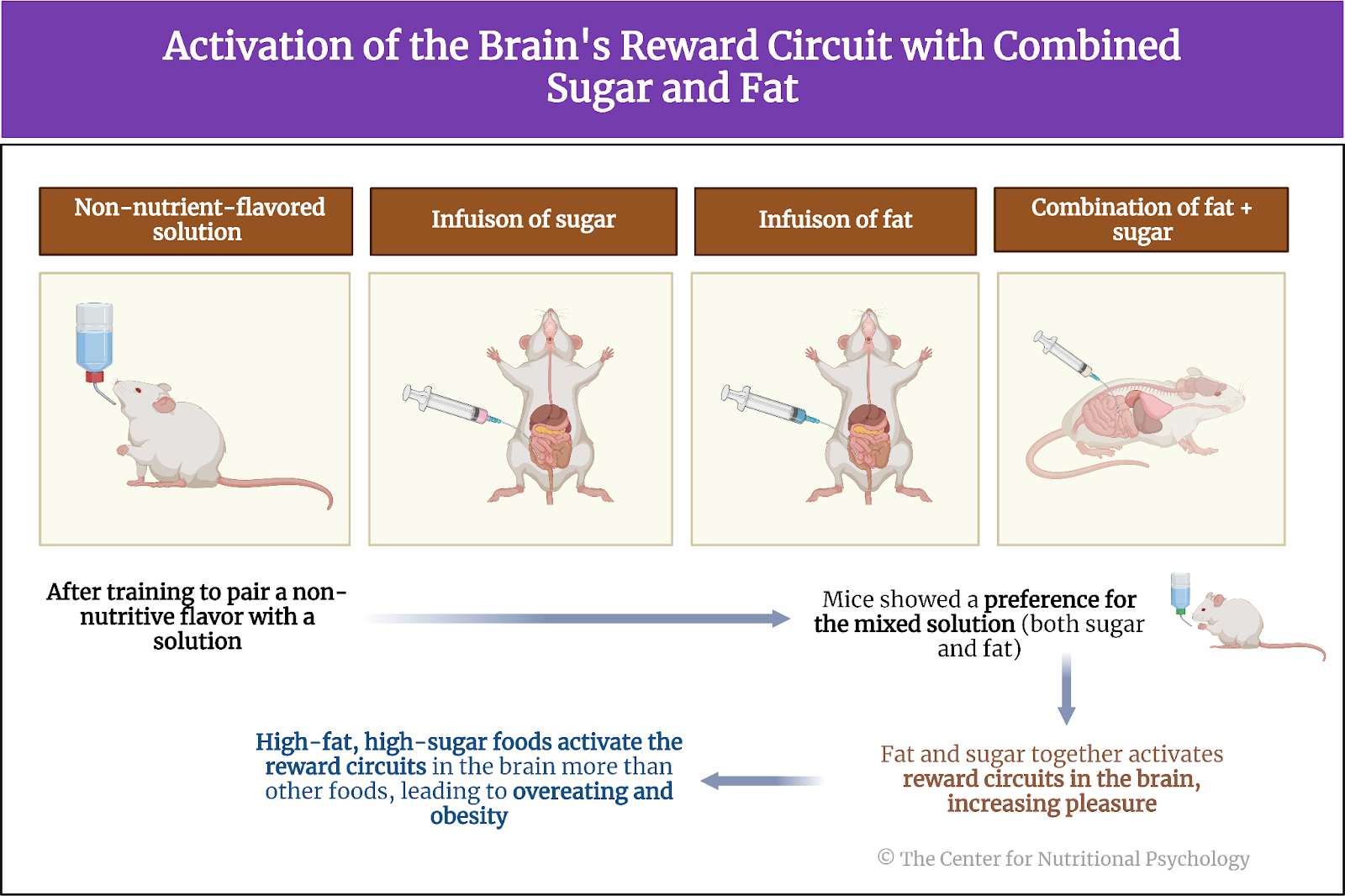
A recent study on mice showed that our brain has separate neural reward circuits for sugars and fats. Due to this, foods that are both fatty and sweet trigger both of these circuits simultaneously resulting in stronger feelings of pleasure than what foods that are just sweet or just fatty could produce (Hedrih, 2024; McDougle et al., 2024). Research has shown for quite some time that feeding mice a fat-rich diet (mouse food already contains carbohydrates) will cause them to overeat and become obese (Ikemoto et al., 1996) (see Figure 2).
Figure 2. Fat and Sugar Mediated Activation of Reward Processing System
The current study
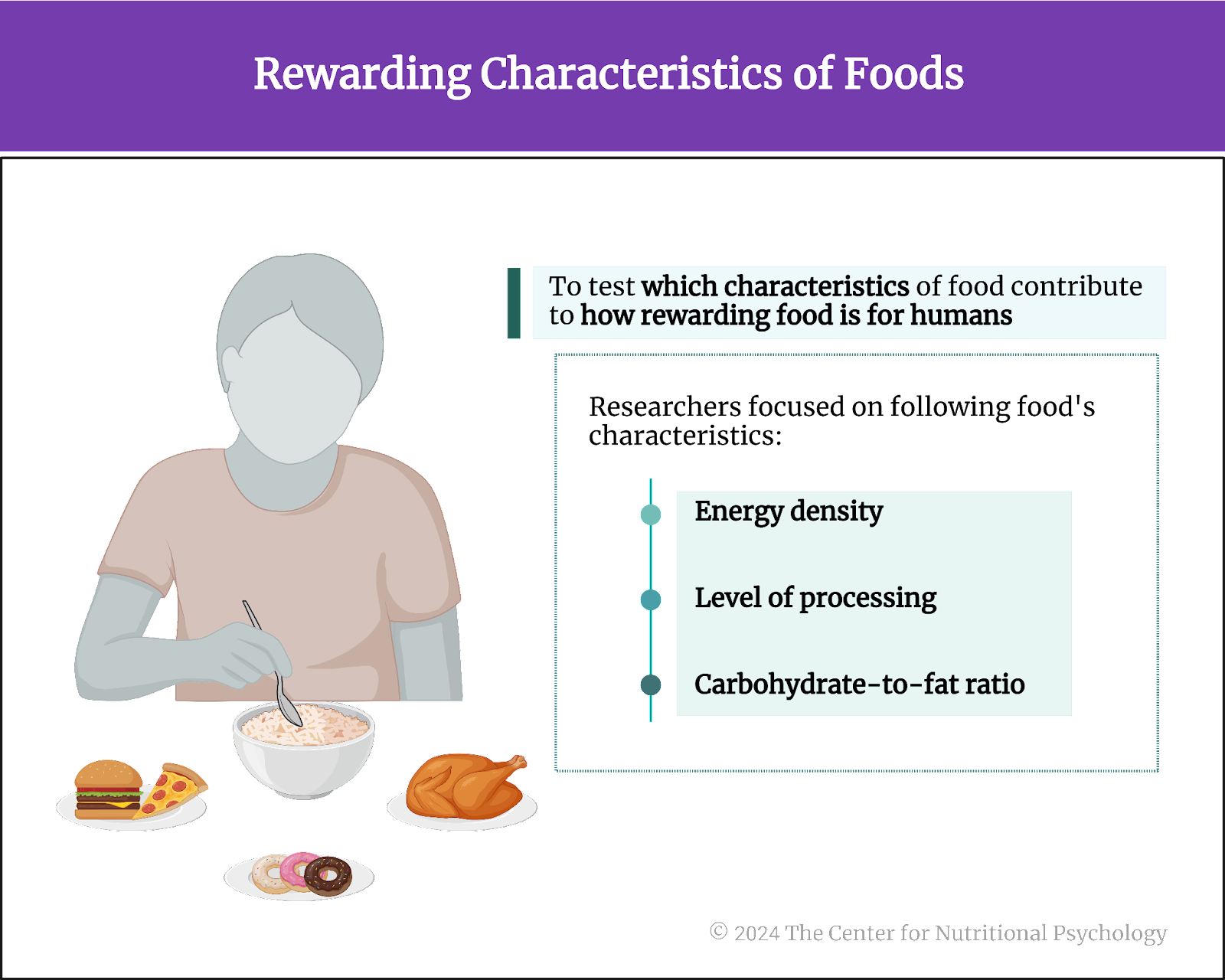
Study author Peter J. Rogers and his colleagues wanted to test which characteristics of food contribute to how rewarding the food is for humans(Rogers et al., 2024). They focused on 1) the energy density of food, 2) its level ofprocessing, and 3) the carbohydrate-to-fat ratio(see Figure 3). The energy density of food refers to the amount of energy (calories) in a given weight of food, typically expressed in calories per gram.
Figure 3. Rewarding characteristics of foods
The procedure
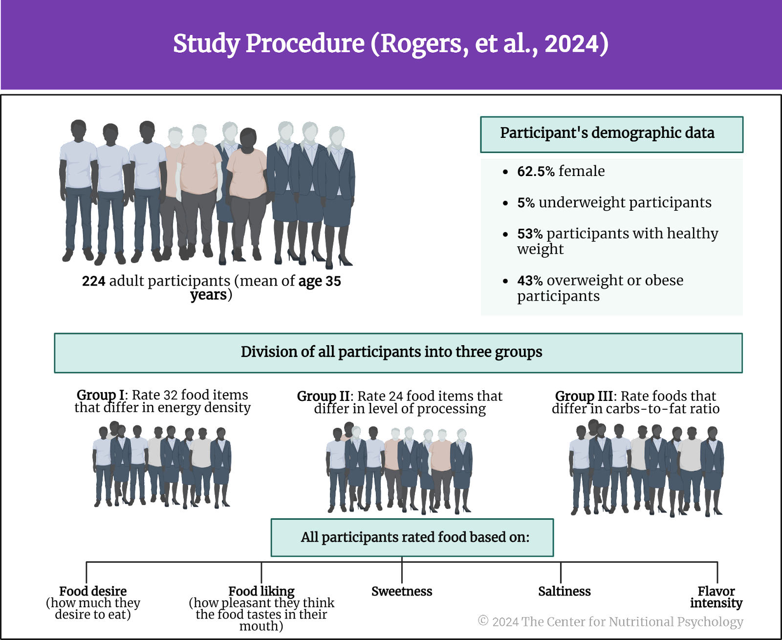
The authors recruited 224 adult participants through social media. Participants were required not to be vegetarians or vegans and to be familiar with the food they would be rating. The mean age of participants was 35 years. 62.5% of them were female. 5% of participants were underweight, 53% had a healthy weight, and 43% were overweight or obese.
The study authors divided these participants into three groups. One group would rate a set of food items differing in energy density (32 foods), and the second group would do that for foods differing in the level of processing (24 foods). In contrast, the third group rated foods differing in their carbohydrate-to-fat ratio (see Figure 4).
Figure 4. Study Procedure (Rogers et al., 2024)
Participants rated their assigned food set on food reward (how much they desire to eat), liking (how pleasant they think the food tastes in their mouth), sweetness, saltiness, and flavor intensity.
Food items were presented as pictures of a 50-gram portion on a light-beige colored plate. To ensure that participants were somewhat hungry at the time of the study, the authors instructed them not to eat anything for 2 hours before the study. Participants were supposed to imagine taking a bite of the food shown in the picture and tasting it. They would then provide the rating.
Foods presented in the energy density group included (in increasing order of energy density) fresh apple slices (the lowest energy density), a boiled egg, Edam cheese slices, crisps, and original Pringles (the highest energy density). Foods in the level of processing set included avocado (no processing), Parma ham, and wine gums (a type of gummy candy, the highest level of processing). The carbohydrate-to-fat ratio group included dried apple slices (high share of carbohydrates, little fat), Cornish dairy ice cream (moderate amounts of both fats and carbohydrates), and Frankfurter sausage (high in fats, little carbohydrates).
Participants liked very energy-dense foods and medium-processed foods the most
Results showed that participants liked and wanted to eat energy-dense foods the most. However, the least liked foods were not those with the lowest energy density but foods with medium energy density levels. The situation with food rewards was the same. Taste intensity tended to increase with energy density. Participants rated the least energy-dense foods as having the least intensive taste.
In the set of foods that differed by the level of processing, participants found processed, but not ultraprocessed foods, to be the most to their liking, the most rewarding, and with the highest taste intensity. Minimally processed foods received the lowest ratings. However, participants found unprocessed foods even more rewarding (i.e., they wanted to eat them more) than medium-processed foods.
Participants found processed, but not ultraprocessed foods, to be the most to their liking, the most rewarding, and with the highest taste intensity
Foods containing both fats and carbohydrates received the best ratings
In the carbohydrate-to-fat ratio food group, participants found foods containing roughly equal amounts of carbohydrates and fats to be the most to their liking and the most rewarding. Foods consisting solely of carbohydrates or solely of fats tended to receive the worst ratings. Differences in food intensity in this group of food items were less pronounced.
The study authors developed a statistical model to predict food reward and liking and included various food constituents, intensity of taste, and carbohydrate-to-fat ratio. Statistical tests showed that participants liked foods containing roughly equal amounts of fats and carbohydrates, with the most intense taste and low fiber content. They also found such foods the most rewarding (see Figure 5).
Figure 5. Research findings
Conclusion
The study showed that people find foods with intensive taste containing roughly equal amounts of fats and carbohydrates the most to their liking and the most rewarding. Contrary to previous studies, participants did not find ultraprocessed foods the most rewarding or like them the most. This shows that in this study, it was not the level of processing that determined how liked and rewarding a food item would be.
These findings can help researchers and health practitioners better understand the psychological values of different food items. This might allow for better diets and improved obesity prevention programs.
Gearhardt, A. N., Bueno, N. B., DiFeliceantonio, A. G., Roberto, C. A., Jiménez-Murcia, S., & Fernandez-Aranda, F. (2023). Social, clinical, and policy implications of ultra-processed food addiction. BMJ, e075354. https://doi.org/10.1136/bmj-2023-075354
Hedrih, V. (2023). Scientists Propose that Ultra-Processed Foods be Classified as Addictive Substances. CNP Articles in Nutritional Psychology. https://www.nutritional-psychology.org/scientists-propose-that-ultra-processed-foods-be-classified-as-addictive-substances/
Hedrih, V. (2024, February 19). Consuming Fat and Sugar (At The Same Time) Promotes Overeating, Study Finds. CNP Articles in Nutritional Psychology. https://www.nutritional-psychology.org/16563-2/
Ikemoto, S., Takahashi, M., Tsunoda, N., Maruyama, K., Itakura, H., & Ezaki, O. (1996). High-fat diet-induced hyperglycemia and obesity in mice: Differential effects of dietary oils. Metabolism, 45(12), 1539–1546. https://doi.org/10.1016/S0026-0495(96)90185-7
McDougle, M., de Araujo, A., Singh, A., Yang, M., Braga, I., Paille, V., Mendez-Hernandez, R., Vergara, M., Woodie, L. N., Gour, A., Sharma, A., Urs, N., Warren, B., & de Lartigue, G. (2024). Separate gut-brain circuits for fat and sugar reinforcement combine to promote overeating. Cell Metabolism. https://doi.org/10.1016/j.cmet.2023.12.014
Monteiro, C. A., Cannon, G., Levy, R. B., Moubarac, J. C., Louzada, M. L. C., Rauber, F., Khandpur, N., Cediel, G., Neri, D., Martinez-Steele, E., Baraldi, L. G., & Jaime, P. C. (2019). Ultra-processed foods: What they are and how to identify them. Public Health Nutrition, 22(5), 936–941. https://doi.org/10.1017/S1368980018003762
Rogers, P. J., Vural, Y., Berridge-Burley, N., Butcher, C., Cawley, E., Gao, Z., Sutcliffe, A., Tinker, L., Zeng, X., Flynn, A. N., Brunstrom, J. M., & Brand-Miller, J. C. (2024). Evidence that carbohydrate-to-fat ratio and taste, but not energy density or NOVA level of processing, are determinants of food liking and food reward. Appetite, 193, 107124. https://doi.org/10.1016/j.appet.2023.107124
Wong, M. C., Mccarthy, C., Fearnbach, N., Yang, S., Shepherd, J., & Heymsfield, S. B. (2022). Emergence of the obesity epidemic: 6-decade visualization with humanoid avatars. The American Journal of Clinical Nutrition, 115(4), 1189–1193. https://doi.org/10.1093/AJCN/NQAC005
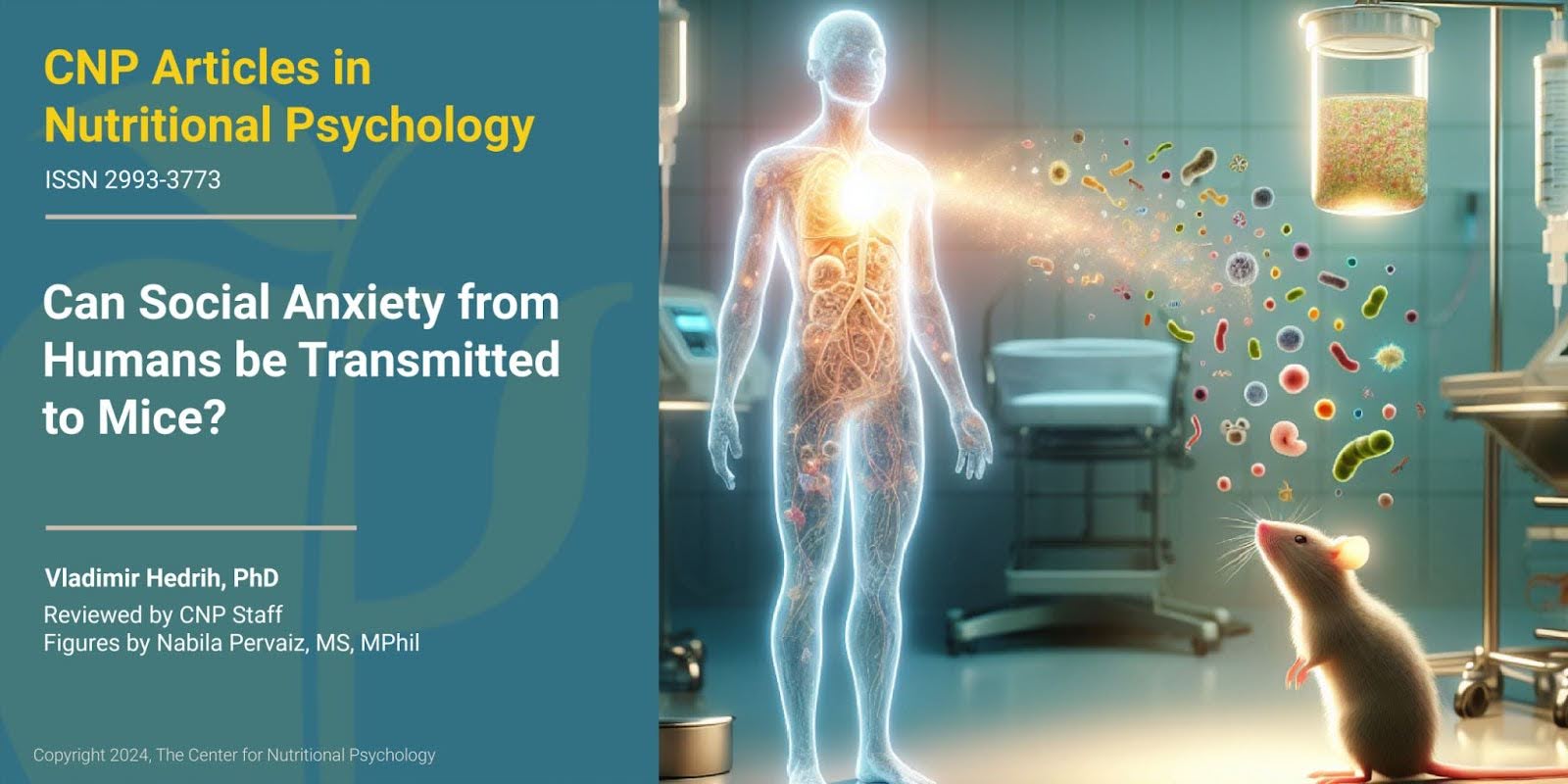
A study published inPNAS: Neuroscience explored whether social anxiety can be transmitted from humans to mice via gut microbiota
Mice receiving gut microbiota from human participants with social anxiety disorder became more sensitive to social fear
While the nonsocial behaviors of these mice remained normal, biochemical changes linked to higher levels of social fear were also detected
What is social anxiety?
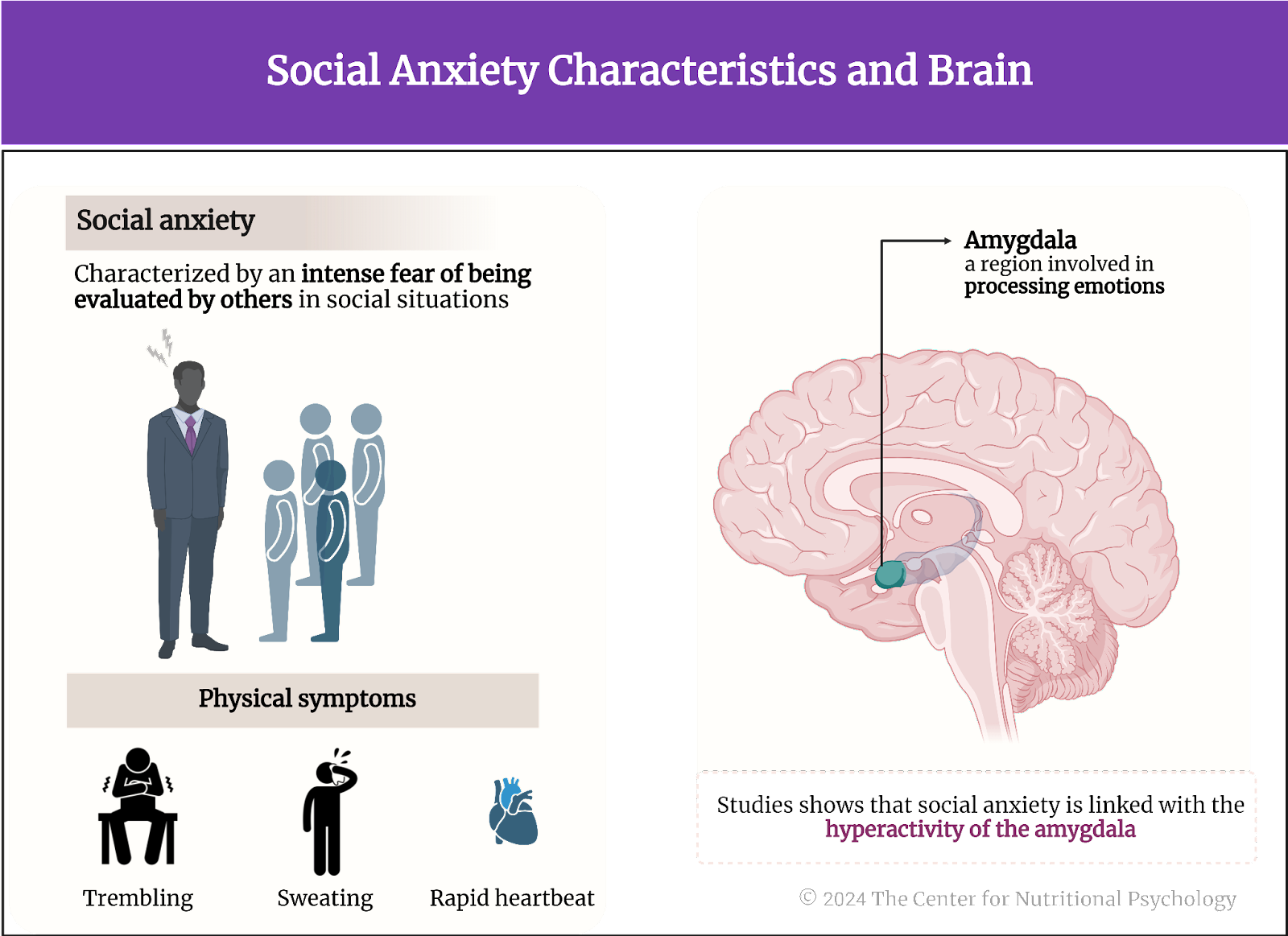
Social anxiety is a common human condition characterized by an intense fear of being evaluated by others in social situations (Morrison & Heimberg, 2013). People with social anxiety constantly worry about acting in a way that will be embarrassing or humiliating. They may even avoid social situations to prevent embarrassment. This fear can interfere with daily activities, work, and relationships. It can also lead to physical symptoms such as sweating, trembling, or a rapid heartbeat. If social anxiety reaches the level of severity where it impairs everyday functioning, it becomes a social anxiety disorder or social phobia.
Studies of brain activity link social anxiety with the hyperactivity of the amygdala region of the brain (Phan et al., 2006), a region involved in processing emotions, and with abnormalities in neural networks that use neurotransmitters serotonin and dopamine (e.g., Wee et al., 2008). However, the causes of these changes remain unknown (see Figure 1).
Figure 1. Social anxiety Characteristics and brain
The microbiota-gut-brain axis and social anxiety
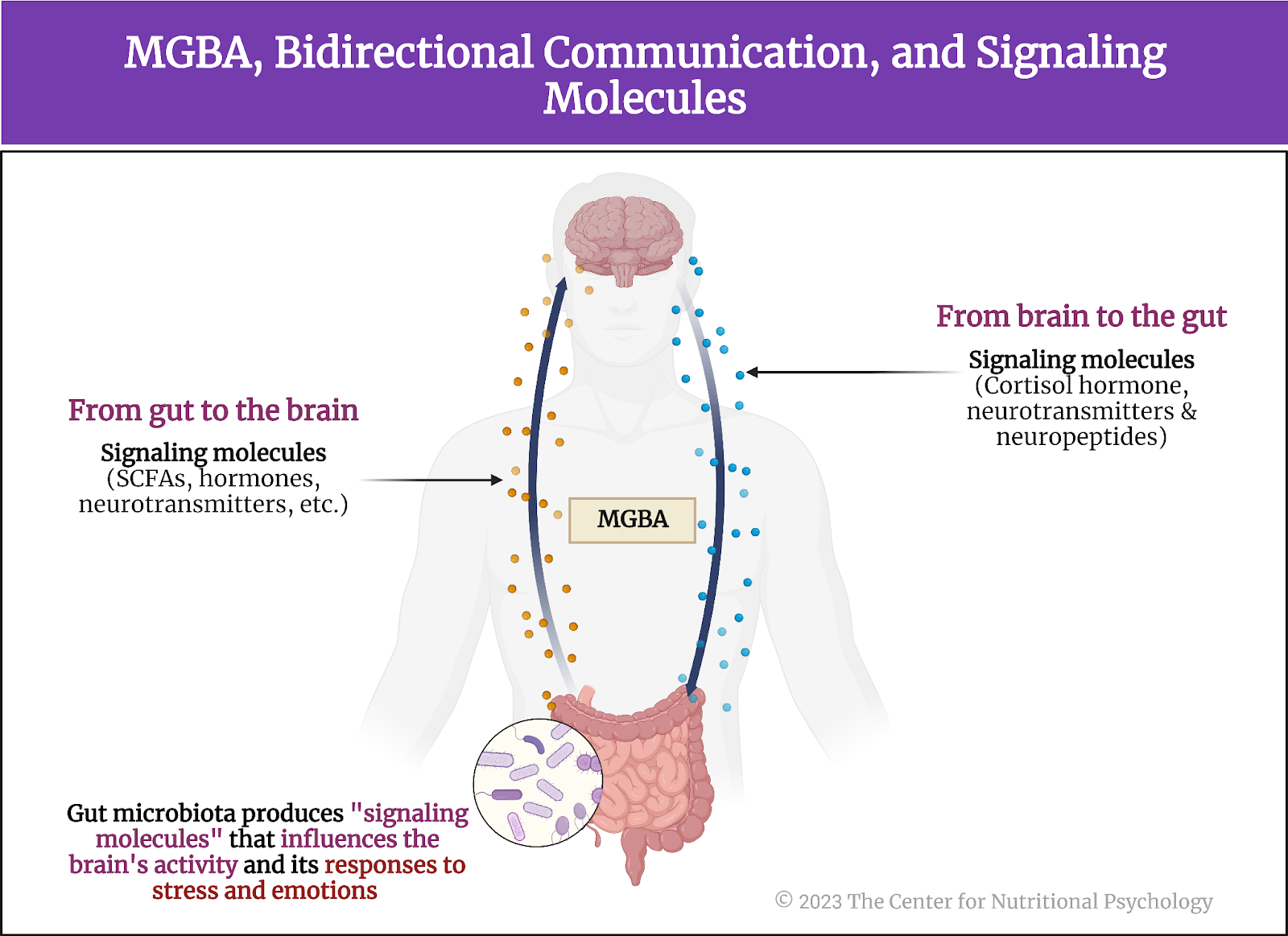
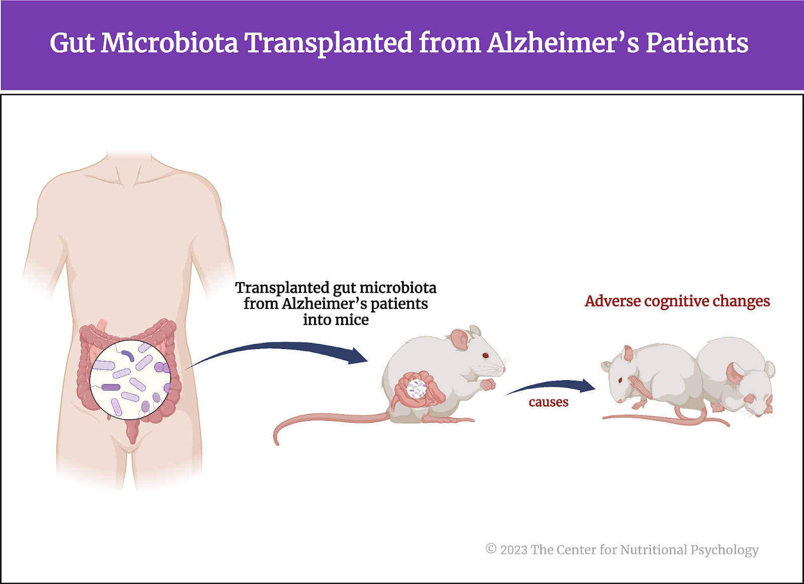
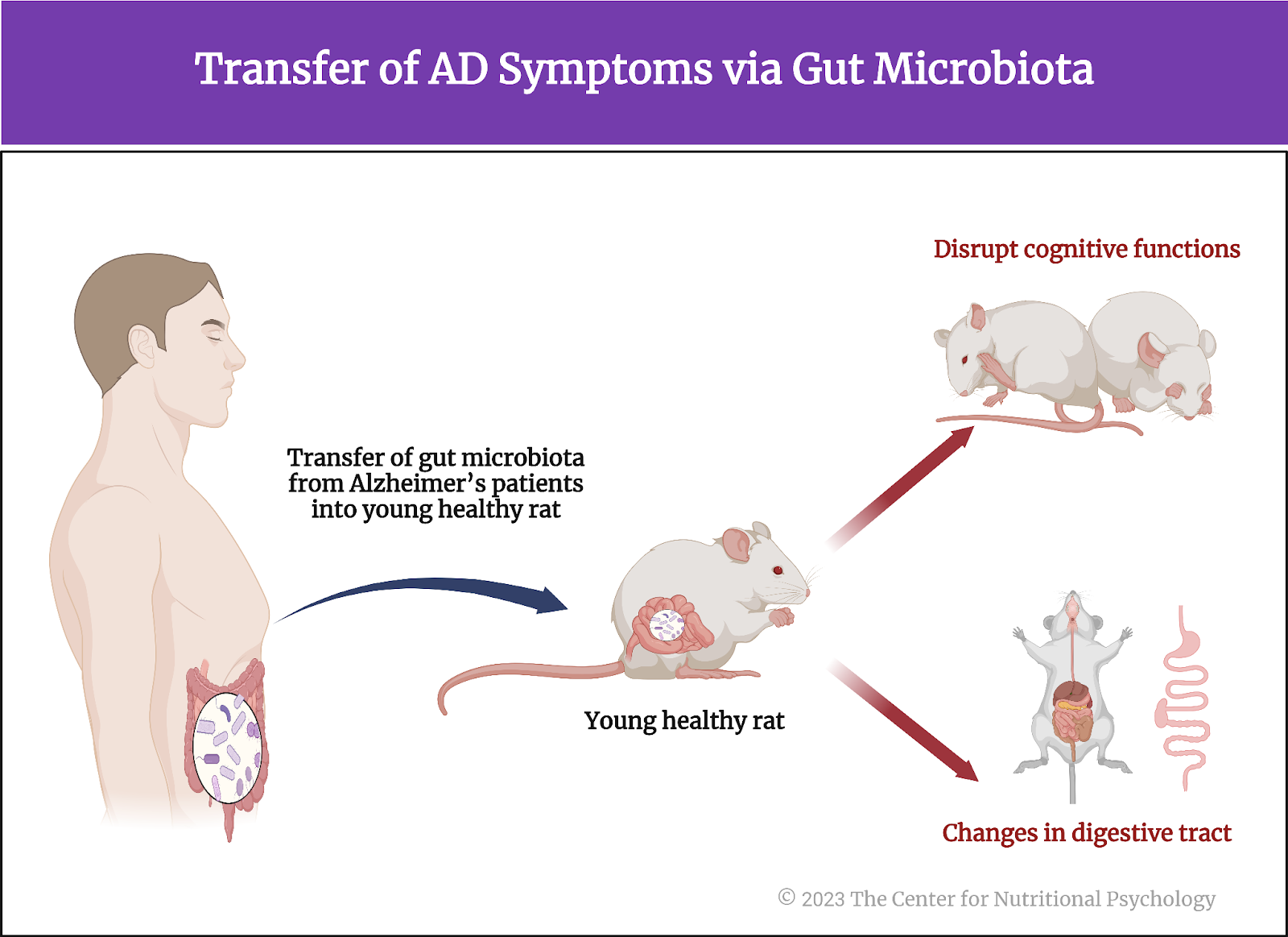
The recent discovery of the microbiota-gut-brain axis (MGBA), a bidirectional pathway through which microorganisms residing in our gut can affect processes in the brain and vice versa, showed that microbiota changes can affect different biochemical processes in the brain. Study after study on the microbiota-gut-brain axis shows that gut microbiota composition is linked to various mental health symptoms, that it can regulate reward processing in the brain, craving, certain aspects of social cognition, and binge drinking (Carbia et al., 2023; García-Cabrerizo et al., 2021; V. Hedrih, 2023). Transplanting microbiota from patients with Alzheimer’s disease into rats makes rats develop cognitive impairments (Grabrucker et al., 2023; Hedrih, 2024) (see Figure 2).
Figure 2. Microbiota composition linked to various mental health symptoms
Transplanting microbiota from humans with Alzheimer’s disease into rats makes rats develop cognitive impairments
The current study
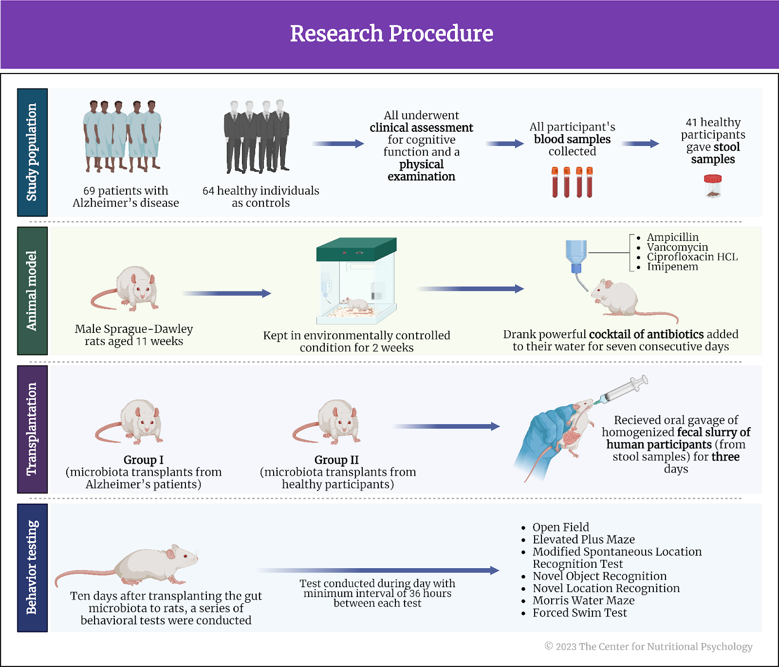
Recent research findings also show that the gut microbiota composition of individuals with social anxiety disorder differs from that of healthy humans. This made the author of this study, Nathaniel L. Ritz, and his colleagues wonder whether gut microbiota might have a causal role in the development of social anxiety. To test this, they conducted a study in which they transplanted the gut microbiota of individuals with social anxiety disorder and healthy individuals into the guts of mice (Ritz et al., 2024).
Study procedure
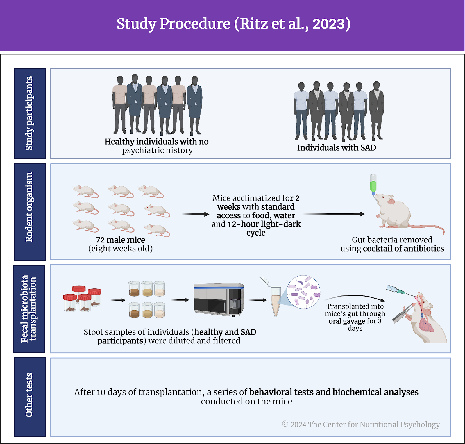
The study involved 12 human participants: 6 diagnosed with social anxiety disorder (SAD) and six healthy individuals without psychiatric history. The study authors selected the social anxiety group from participants of a previous study on the links between social anxiety and gut microbiota composition. Healthy participants came from University College Cork in Ireland. Both groups provided stool samples needed for transplantation into mice.
The study authors transplanted gut microbiota from the feces of human participants into the guts of 72 male mice. The mice were eight weeks old at the start of the study. They were acclimatized for two weeks and kept in a 12-hour light-dark cycle with free access to standard mouse food and water. Researchers then depleted mice’s gut microbiota using a strong cocktail of antibiotics (ampicillin, vancomycin, imipenem) to prepare them for human microbiota. Each mouse received microbiota from a randomly chosen human participant.
Stool samples from human participants were diluted, filtered, and inserted into the mice’s guts via oral gavage for three consecutive days. Oral gavage is a procedure where researchers use a syringe with a feeding tube to directly insert contents into the guts of mice through their mouths. Half of the mice received microbiota from individuals with social anxiety disorder, while the other half received it from healthy participants.
The study authors waited ten days after the transplantation procedure and then conducted a series of behavioral tests on the mice. They also collected stool samples of mice and conducted different biochemical analyses (see Figure 3).
Figure 3. Study procedure (Ritz et al., 2023)
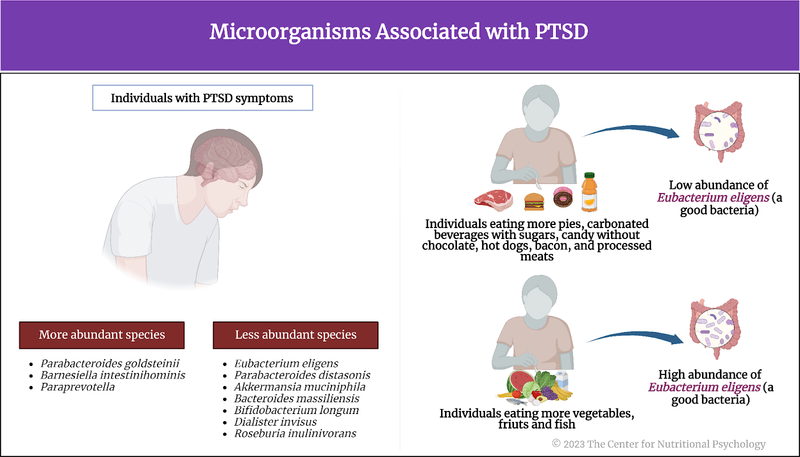
Gut microbiota from individuals with social anxiety differed in composition
Results showed differences in the gut microbiota composition between mice transplanted with microbiota from healthy participants and those from participants with social anxiety disorder. They differed in the abundance of three bacterial species – Bacteroides nordii, Bacteroides cellulosiyticus, and Phocaeicola massiliensi.
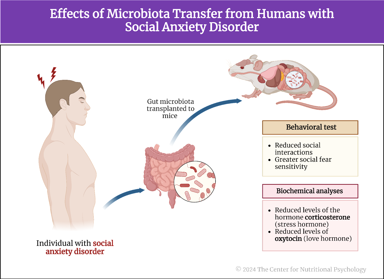
Gut microbiota from humans with social anxiety disorder induced social fear sensitivity in mice
Mice with microbiota from social anxiety disorder participants showed reduced social interactions in behavioral tests. However, their non-social exploratory behaviors did not differ from those of mice that received microbiota from healthy human participants. Based on this, the study authors concluded that transplantation of gut microbiota from humans with social anxiety disorder induced greater social fear sensitivity in these mice.
Biochemical changes also indicated heightened susceptibility to social fear
Mice that received gut microbiota from humans with social anxiety disorder had reduced levels of the hormone corticosterone. Corticosterone is a steroid hormone the adrenal cortex produces, primarily regulating stress responses, energy metabolism, immune reactions, and electrolyte balance.
They also showed reduced levels of oxytocin in a specific region of the brain (the bed nucleus of the stria terminalis) and lower activity of genes related to this neurotransmitter in other brain regions (the medial amygdala and the prefrontal cortex) (see Figure 4). Oxytocin is a hormone and neurotransmitter often called the “love hormone” due to its role in social bonding, sexual reproduction, childbirth, and maternal behaviors. Thus, its reduced levels and production make a mouse more susceptible to social fear and less motivated for social interactions.
Figure 4. Effects of microbiota transfer from humans with social anxiety disorder
Conclusion
The study showed that it is possible to induce social fear sensitivity in mice by transplanting gut microbiota from humans with social anxiety disorder (i.e., highly sensitive to social fear). These results confirm that gut microorganisms can play a causal role in the development of social fear. If future studies show that changes to gut microbiota can also reduce social anxiety (instead of increasing it), it may open the possibility of developing ways to treat social anxiety using tailor-made probiotics.
It’s possible to induce social fear sensitivity in mice by transplanting gut microbiota from humans with social anxiety disorder
The paper “Social anxiety disorder-associated gut microbiota increases social fear” was authored by Nathaniel L. Ritz, Marta Brocka, Mary I. Butler, Caitlin S. M. Cowan, Camila Barrera-Bugueño, Christopher J. R. Turkingtona, Lorraine A. Draper, Thomaz F. S. Bastiaanssen, Valentine Turpin, Lorena Morales, David Campos, Cassandra E. Gheorghe, Anna Ratsika, Virat Sharma, Anna V. Golubeva, Maria R. Aburto, Andrey N. Shkoporov, Gerard M. Moloney, Colin Hill, Gerard Clarke, David A. Slattery, Timothy G. Dinan, and John F. Cryan.
References
Carbia, C., Bastiaanssen, T. F. S., Iannone, F., García-cabrerizo, R., Boscaini, S., Berding, K., Strain, C. R., Clarke, G., Stanton, C., Dinan, T. G., & Cryan, J. F. (2023). The Microbiome-Gut-Brain axis regulates social cognition & craving in young binge drinkers. eBioMedicine, 89, 104442. https://doi.org/10.1016/j.ebiom.2023.104442
García-Cabrerizo, R., Carbia, C., O´Riordan, K. J., Schellekens, H., & Cryan, J. F. (2021). Microbiota-gut-brain axis as a regulator of reward processes. Journal of Neurochemistry, 157(5), 1495–1524. https://doi.org/10.1111/JNC.15284
Grabrucker, S., Marizzoni, M., Silajdžić, E., Lopizzo, N., Mombelli, E., Nicolas, S., Dohm-Hansen, S., Scassellati, C., Moretti, D. V., Rosa, M., Hoffmann, K.,
Cryan, J. F., O’Leary, O. F., English, J. A., Lavelle, A., O’Neill, C., Thuret, S., Cattaneo, A., & Nolan, Y. M. (2023). Microbiota from Alzheimer’s patients induce deficits in cognition and hippocampal neurogenesis. Brain. https://doi.org/10.1093/brain/awad303
Hedrih, V. (2023, September 2). Gut Microbiota’s Role in Mental Health: The Positives and Negatives. CNP Articles in Nutritional Psychology. https://www.nutritional-psychology.org/how-your-gut-microbiota-is-linked-to-both-positive-and-negative-aspects-of-mental-health/
Hedrih, V. (2024, January 29). Can Symptoms of Alzheimer’s be Transferred to Rats via the Gut Microbiota of Alzheimer’s Patients? CNP Articles in Nutritional Psychology. https://www.nutritional-psychology.org/can-symptoms-of-alzheimers-be-transferred-to-rats-via-the-gut-microbiota-of-alzheimers-patients/
Morrison, A., & Heimberg, R. (2013). Social Anxiety and Social Anxiety Disorder. Annual Review of Clinical Psychology, 9, 249–274. https://doi.org/10.1146/annurev-clinpsy-050212-185631
Phan, K. L., Fitzgerald, D., Nathan, P., & Tancer, M. (2006). Association between Amygdala Hyperactivity to Harsh Faces and Severity of Social Anxiety in Generalized Social Phobia. Biological Psychiatry, 59, 424–429. https://doi.org/10.1016/j.biopsych.2005.08.012
Ritz, N. L., Brocka, M., Butler, M. I., Cowan, C. S. M., Barrera-Bugueño, C., Turkington, C. J. R., Draper, L. A., Bastiaanssen, T. F. S., Turpin, V., Morales, L., Campos, D., Gheorghe, C. E., Ratsika, A., Sharma, V., Golubeva, A. V., Aburto, M. R., Shkoporov, A. N., Moloney, G. M., Hill, C., … Cryan, J. F. (2024). Social anxiety disorder-associated gut microbiota increases social fear. Proceedings of the National Academy of Sciences, 121(1), e2308706120. https://doi.org/10.1073/pnas.2308706120
Wee, N. J. van der, Veen, J. F. van, Stevens, H., Vliet, I. M. van, Rijk, P. P. van, & Westenberg, H. G. (2008). Increased Serotonin and Dopamine Transporter Binding in Psychotropic Medication–Naïve Patients with Generalized Social Anxiety Disorder Shown by 123I-β-(4-Iodophenyl)-Tropane SPECT. Journal of Nuclear Medicine, 49(5), 757–763. https://doi.org/10.2967/jnumed.107.045518
A series of experiments on mice published inCell Reports found that hunger reduces inflammatory responses after an injury.
The anti-inflammatory effects of hunger were more robustthan those of non-steroidal anti-inflammatory drugs.
AgRP neurons projecting to the paraventricular nucleus of the brain’s hypothalamus region mediate the majority of hunger’s anti-inflammatory effects.
People need food to survive. Severe malnutrition causes the body to break down its own tissues to meet energy needs. The body first utilizes the stored fat, but if starvation continues, it eventually uses muscle mass to fulfill its nutritional needs. Prolonged starvation impairs vital functions and leads to organ failure, weakened immune response, and severe hormonal imbalances (Sidiropoulos, 2007). Cognitive functions also deteriorate. But what happens when people restrict their food intake only for limited periods?
Restricted food intake While the consequences of a chronic lack of food are dire, cultures worldwide have long believed that restricted food intake for limited periods of time can produce beneficial psychological and physiological consequences.
The practice of fasting, or abstaining from all or some kinds of food or drink for a specific period of time, is an important part of almost all major religions. Religions practice fasting as a means of spiritual discipline, self-reflection, and expressing devotion or penitence. It is often seen as a way to cleanse the body and mind, allowing for deeper religious or spiritual contemplation and connection with the divine or higher power.
Cultures worldwide have long believed that restricted food intake for limited periods of time can produce beneficial psychological and physiological consequences
Research studies show that restricted food intake for a limited period leads to complex physiological changes in the central and peripheral nervous systems. These changes can inhibit inflammation by preventing the activation of inflammasomes, multi-protein complexes that activate an inflammatory response. Additionally, restricted food intake reduces the production and release of pro-inflammatory cytokines, a group of signaling proteins secreted by immune cells that promote inflammation (Klima et al., 2023). On the psychological side, however, restrained eating can increase the feeling of hunger and food craving (Dicker-Oren et al., 2022).
Restricted food intake for a limited period can inhibit inflammation by preventing the activation of inflammasomes, multi-protein complexes that activate an inflammatory response, and reduce the production and release of pro-inflammatory cytokines
While inflammation is an adaptive response, helping the body fight off infection or facilitating the repair of damaged tissue, long-term inflammation can become maladaptive and impair basic actions necessary for survival. It is also the cause or a contributing factor to many diseases and adverse medical conditions.
On the psychological side, however, restrained eating can increase the feeling of hunger and food craving (Dicker-Oren et al., 2022)
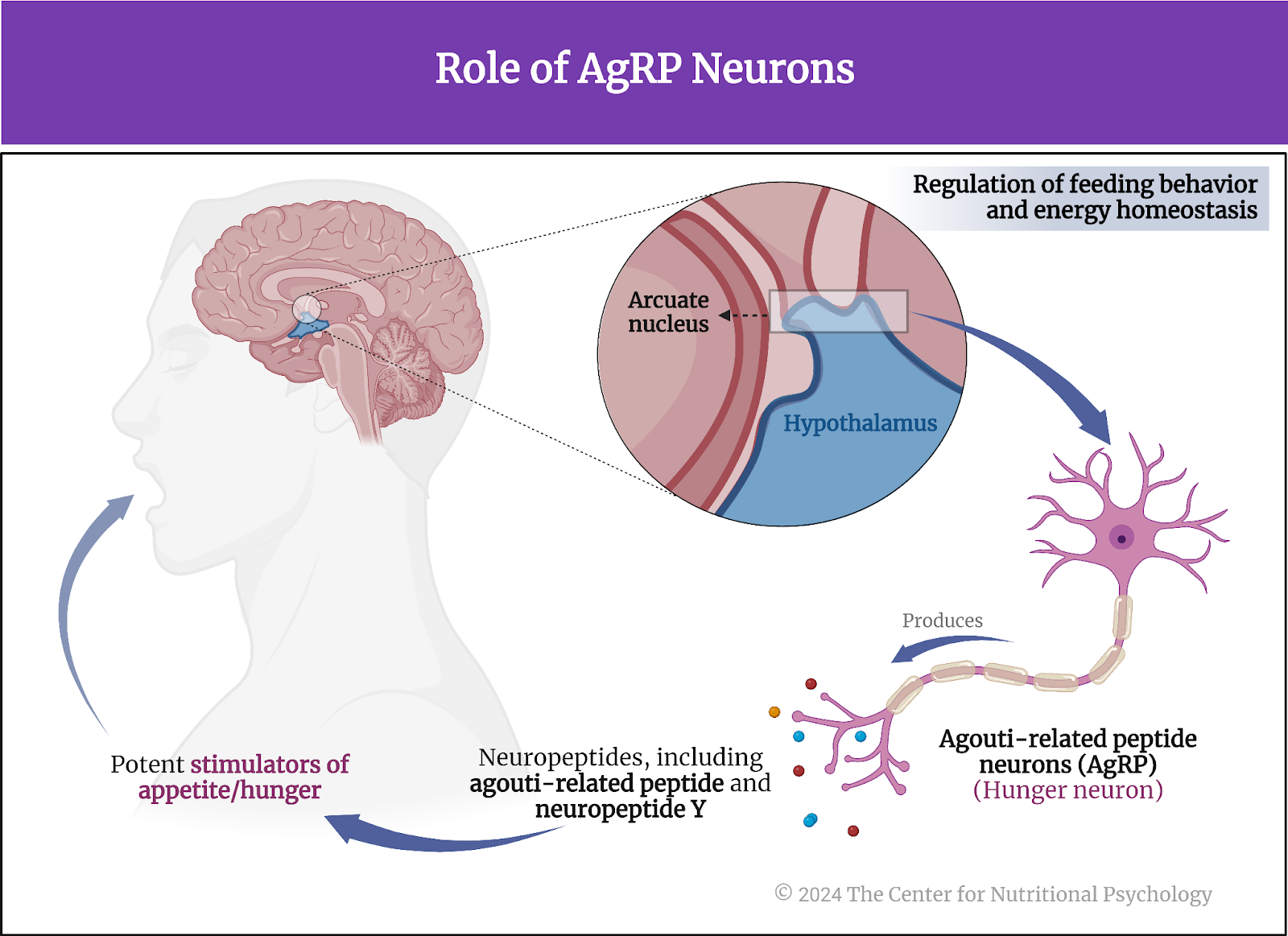
Agouti-related peptide (AgRP) neurons – hunger neurons An important part of the neural circuits that react to restricted food intake are agouti-related peptide neurons, or AgRP neuronsfor short. Often referred to as “hunger neurons,” they are a specific type of neuron located in the brain’s arcuate nucleus of the hypothalamus region. These neurons play a critical role in regulating feeding behavior and energy homeostasis. AgRP neurons produce neuropeptides, including agouti-related peptide (AgRP) and neuropeptide Y (NPY), which are potent stimulators of appetite (Sternson & Atasoy, 2014) (see Figure 1).
Figure 1. Role of AgRP neurons
The activity of AgRP neurons increases food intake and reduces energy expenditure, leading to weight gain. They are part of a complex neural network that responds to various signals related to energy status, such as hormones indicating hunger (ghrelin) or satiety (leptin and insulin). In conditions of energy deficit, such as fasting, AgRP neurons are stimulated, promoting hunger and encouraging food-seeking behavior (Atasoy et al., 2012; Chen et al., 2016).
“Hunger neurons” (AgRP) are a specific type of neuron located in the brain’s arcuate nucleus of the hypothalamus region and play a critical role in regulating feeding behavior and energy homeostasis
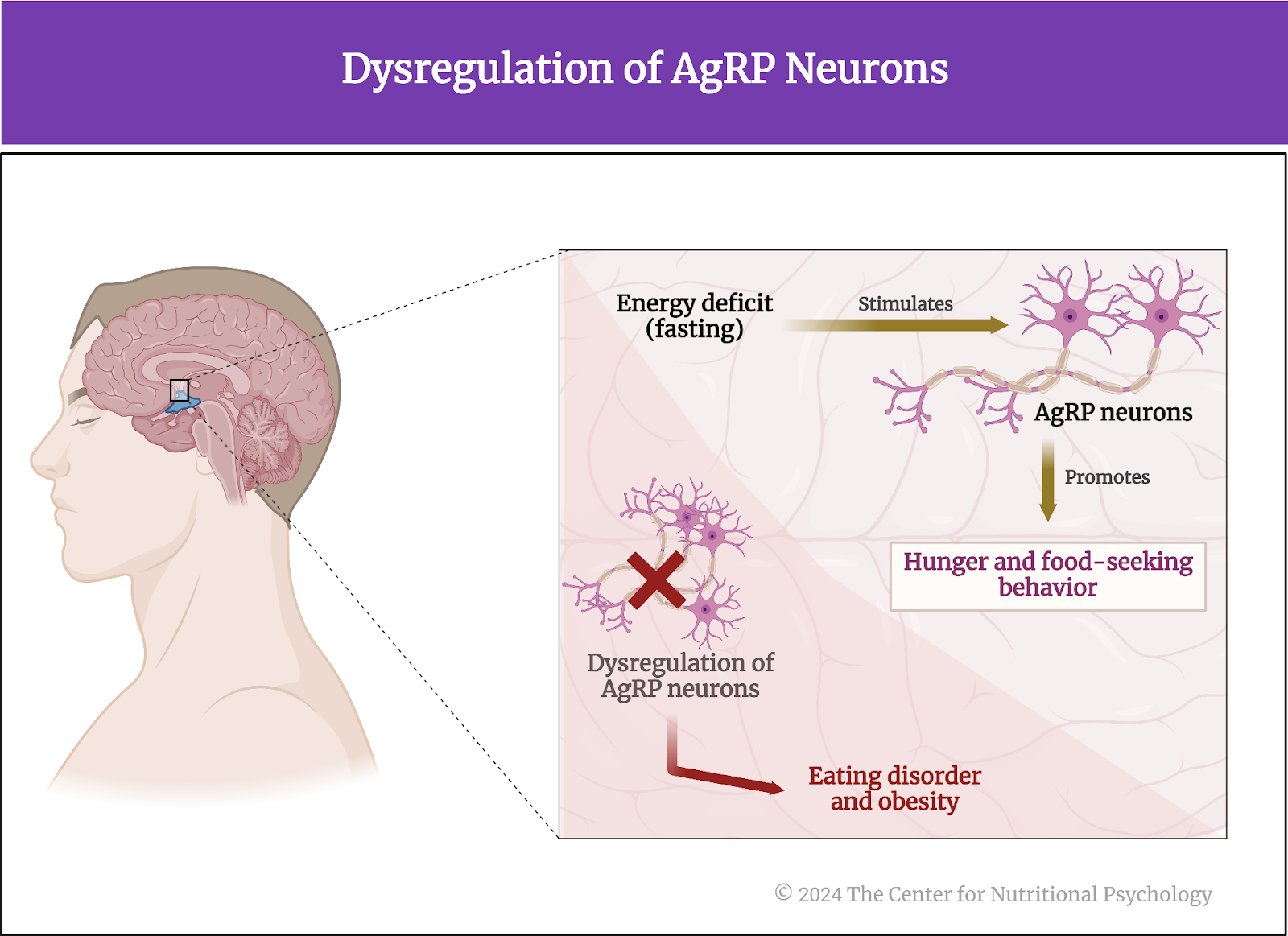
This system is crucial for survival, ensuring that energy intake is increased when energy stores are low. However, dysregulation of AgRP neurons and the pathways they influence can contribute to eating disorders and obesity (see Figure 2).
Figure 2. Dysregulation of AgRP neurons
The current study Study author Michelle L. Klima and her colleagues wanted to map the neural pathways from the brain to the periphery that convey the anti-inflammatory effects of food deprivation. They note that it is likely that hunger circuits (AgRP neurons) in the central nervous system play a role in this because previous studies have shown that manipulations of gene expressions in these neurons can influence immune responses (Klima et al., 2023).
They conducted a series of experiments on mice. The mice used in this study were male and female, kept in group housing, and at least eight weeks old.
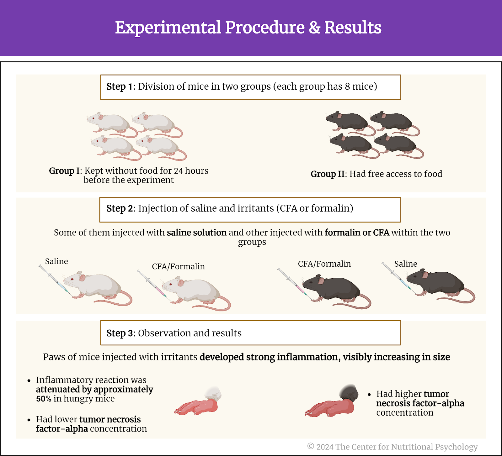
Hunger reduces inflammation in response to irritants In the first experiment, study authors injected either formalin, complete Freund’s adjuvant (CFA), or a saline solution into the paws of these mice. Formalin and CFA are strong irritants that cause an inflammatory reaction and swelling of the paw. The saline solution injection was a control procedure. It is not expected to produce an inflammatory response, so the researchers use it as a reference for comparison with the reactions caused by the injections of the other two substances.
In the experiment, the study authors had two groups of mice – 8 mice that were kept without food for 24 hours before the experiment, i.e., hungry, and another eight that had free access to food. Some mice were injected with irritants and others with a saline solution within the two groups.
Results showed that the paws of mice injected with irritants developed strong inflammation, visibly increasing in size. However, this inflammatory reaction was attenuated by approximately 50% in hungry mice compared to mice fed normally. Female mice had stronger paw inflammation after the CFA injection than male mice, but the inflammation reduction effects of hunger were similar in both sexes.
Concentrations of inflammatory cytokine tumor necrosis factor-alpha, an indicator of inflammation, were much lower in hungry mice, confirming that hunger had a strong anti-inflammatory effect. Researchers also compared the anti-inflammatory effects of hunger with the effects of non-steroidal anti-inflammatory drugs ketoprofen and ketorolac. They found that hunger reduced paw inflammation 20% more than these two drugs (see Figure 3).
Figure 3. Experimental procedure and results
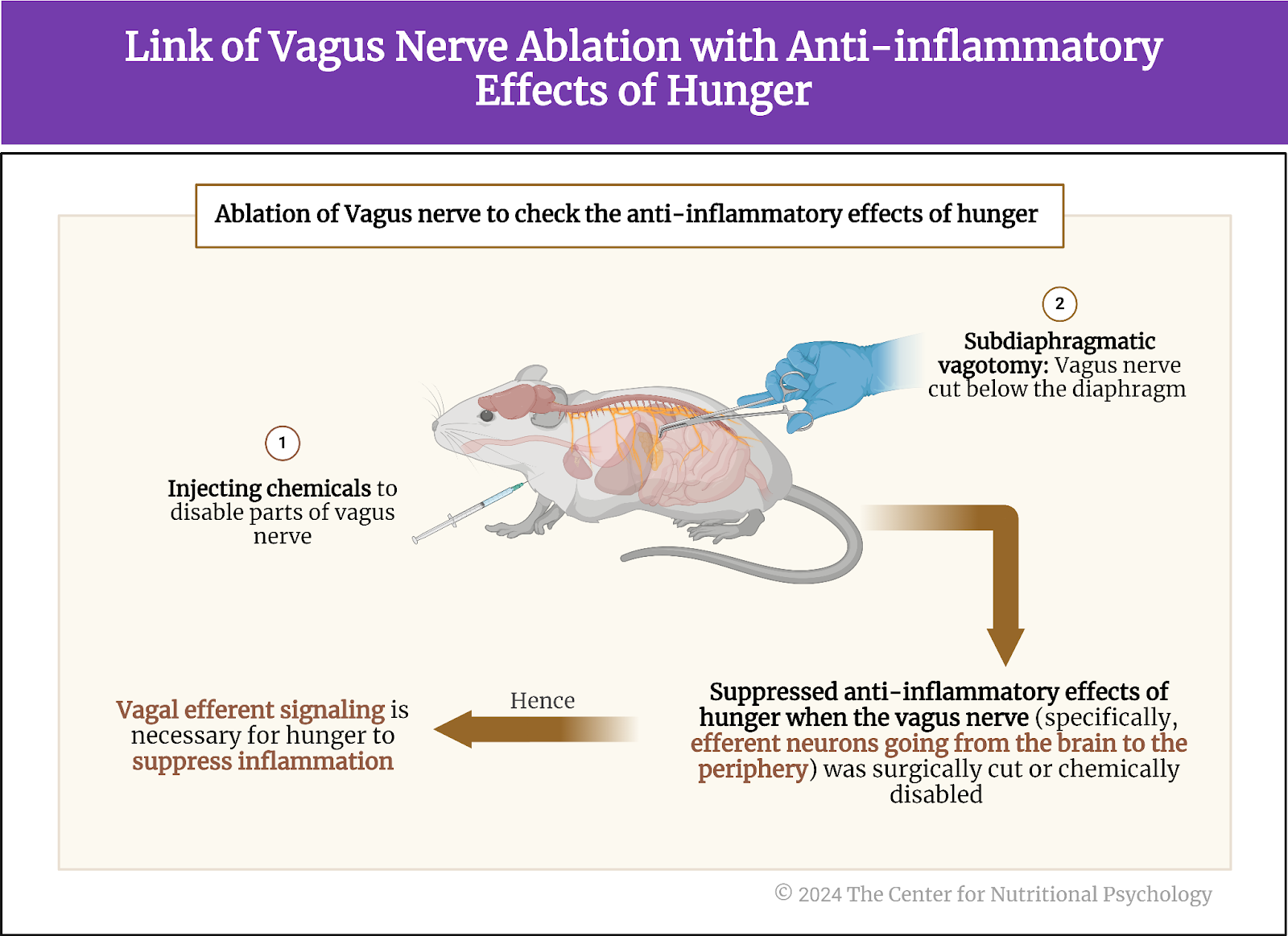
Anti-inflammatory effects of hunger go from the brain through the vagus nerve In their next experiment, study authors disabled the vagus nerve of the mice either by cutting it below the diaphragm or by injecting chemicals to disable parts of this nerve.
The vagus nerve extends from the brainstem to the abdomen, innervating various organs. It is crucial in the parasympathetic nervous system, responsible for the body’s rest and digest functions. The vagus nerve helps regulate heart rate, digestion, and respiratory rate but is also involved in inflammatory responses. It also conveys sensory information from the internal organs to the brain.
Results showed that the anti-inflammatory effects of hunger were suppressed when the vagus nerve was disabled in this way. This happened both when the nerve was physically cut and when chemicals were used.
Further experimentation showed that the anti-inflammatory effect is suppressed only when neurons going from the brain to the periphery (efferent neurons) are destroyed or disabled. Destroying or disabling neurons that carry information from the periphery to the brain (afferent neurons) did not affect the anti-inflammatory effects of hunger. This told researchers that the anti-inflammatory effects of hunger go from the brain through the vagus nerve to other areas of the body (see Figure 4).
Figure 4. Link of vagus nerve ablation with anti-inflammatory effects of hunger
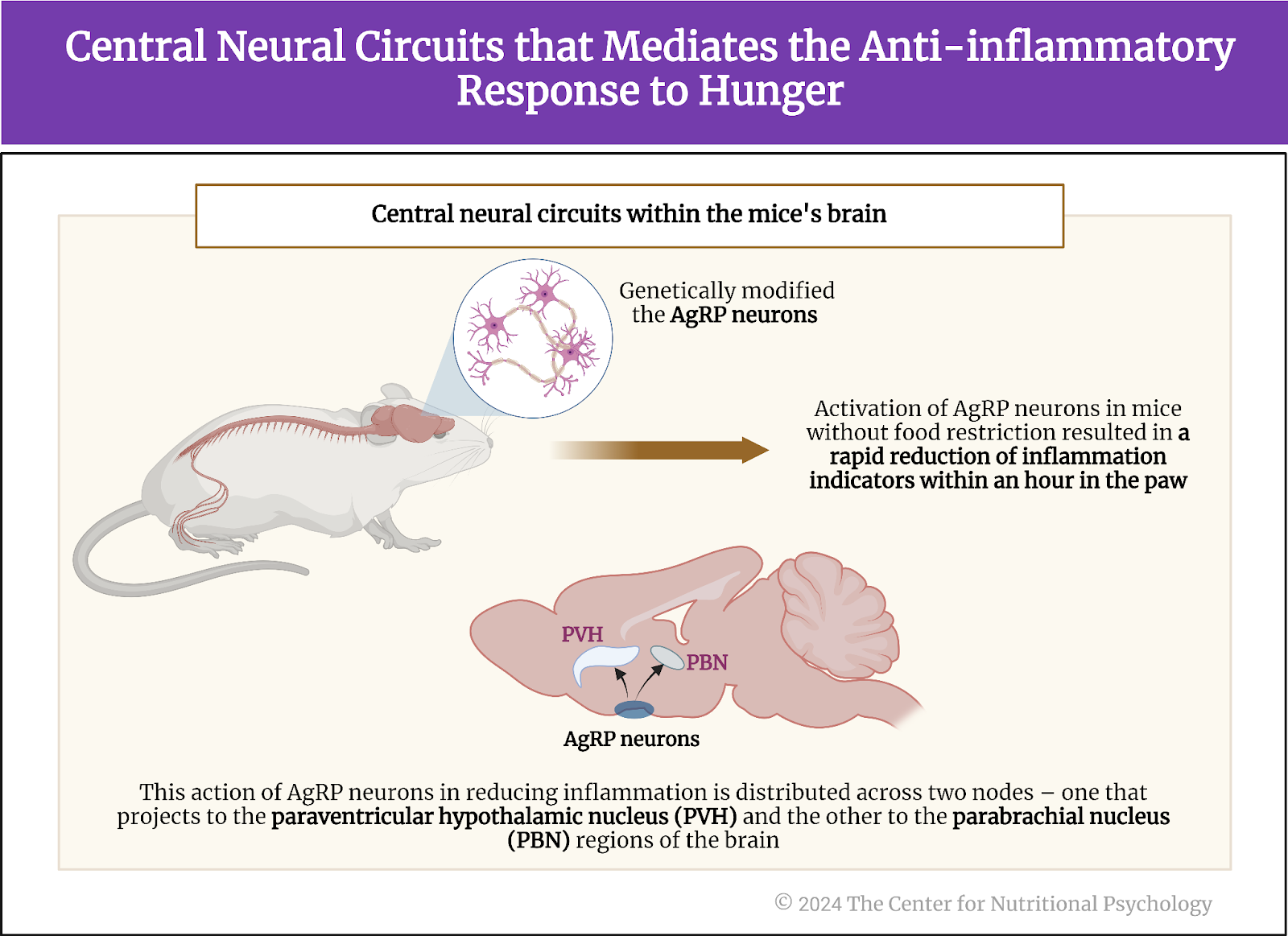
Activation of AgRP neurons reduces inflammation Next, the study authors looked for neural circuits in the brain that trigger the anti-inflammatory response to hunger. Knowing that AgRP neurons are activated by hunger, they first looked at them. These researchers genetically modified the AgRP neurons in a group of mice to be activated by specific chemicals or light.
Results showed an anti-inflammatory response followed when they artificially activated these neurons without restricting food to mice. Paws of mice in which study authors artificially activated AgRP neurons showed a much less intense inflammatory reaction after an irritant (CFA) injection than mice from the control group that ate normally and whose AgRp neurons were not artificially activated. The anti-inflammatory reaction caused by the activation of AgRP neurons was rapid, taking less than 1 hour for the reduction in multiple indicators of inflammation in the paw to become visible (see Figure 5).
Figure 5. Central neural circuits that mediate the anti-inflammatory response to hunger
The study authors further sought to identify which AgRP neurons specifically activated the anti-inflammatory response. To do this, they activated specific groups of AgRP neurons one by one artificially in normally fed mice. They found that the anti-inflammatory action of AgRP neurons is distributed over two nodes – one projecting to the paraventricular hypothalamic nucleus and the other to the parabrachial nucleus regions of the brain. The magnitude of the anti-inflammatory effect of the first one was larger, mediating the bulk of the anti-inflammatory effects of these neurons.
Conclusions Through a series of experiments on mice, this study showed that hunger produces an anti-inflammatory effect stronger than the effects of non-steroid anti-inflammatory medications such as ketoprofen and ketorolac. It showed that the bulk of the anti-inflammatory effect is mediated by hunger neurons, i.e., Agouti-related peptide (AgRP) neurons in the arcuate nucleus in the brain’s hypothalamus region. Even artificially triggering the activity of these neurons when a mouse is not hungry causes a rapid anti-inflammatory response.
These findings improve scientific understanding of the neural pathways of inflammation and can help healthcare researchers develop better ways to treat inflammation. Many diseases, from autoimmune diseases to skin disorders, are caused or aggravated by maladaptive immune responses of the body. Finding ways to better modulate or control the body’s immune response may be key to developing more effective treatments for them.
Atasoy, D., Betley, J. N., Su, H. H., & Sternson, S. M. (2012). Deconstruction of a neural circuit for hunger. Nature, 488(7410), 172–177. https://doi.org/10.1038/nature11270
Chen, Y., Lin, Y.-C., Zimmerman, C. A., Essner, R. A., & Knight, Z. A. (2016). Hunger neurons drive feeding through a sustained, positive reinforcement signal. ELife, 5, e18640. https://doi.org/10.7554/eLife.18640.001
Dicker-Oren, S. D., Gelkopf, M., & Greene, T. (2022). The dynamic network associations of food craving, restrained eating, hunger and negative emotions. Appetite, 175(March), 106019. https://doi.org/10.1016/j.appet.2022.106019
Klima, M. L., Kruger, K. A., Goldstein, N., Pulido, S., Low, A. Y. T., Assenmacher, C. A., Alhadeff, A. L., & Betley, J. N. (2023). Anti-inflammatory effects of hunger are transmitted to the periphery via projection-specific AgRP circuits. Cell Reports, 42(11). https://doi.org/10.1016/j.celrep.2023.113338
Sidiropoulos, M. (2007). Anorexia Nervosa: The physiological consequences of starvation and the need for primary prevention efforts. CASE PRESENTATION. McGill Journal of Medicine, 10(1), 20–25.
Sternson, S. M., & Atasoy, D. (2014). Agouti-related protein neuron circuits that regulate appetite. Neuroendocrinology, 100, 95–102. https://doi.org/10.1159/000369072
A study of UK Biobank data published inPNAS reported that frequent consumption of fried food increased the risk of anxiety by 12% and the risk of depression by 7%.
Fried potato consumption was specifically associated with a somewhat increased risk of depression and anxiety symptoms.
An experiment on zebrafish confirmed that chronic exposure to acrylamide, a substance created when food is fried, creates anxiety-like symptoms in these fish.
The consolidation of research in the diet-mental health relationship within nutritional psychology has shown that our food choices and patterns can influence our moods and state of mind, and our moods and state of mind can influence our food choices and patterns (NPRL). People tend to eat when stressed or bored (Dicker-Oren et al., 2022; Stevenson et al., 2023). They can also feel angry when hungry (called hanger), in which one experiences anger due to hunger (Hedrih, 2023a).
Food choices and patterns can influence our moods and state of mind, which can influence our food choices and patterns
But could our dietary choices be associated with serious mental health disorders? Disorders like anxiety or depression?
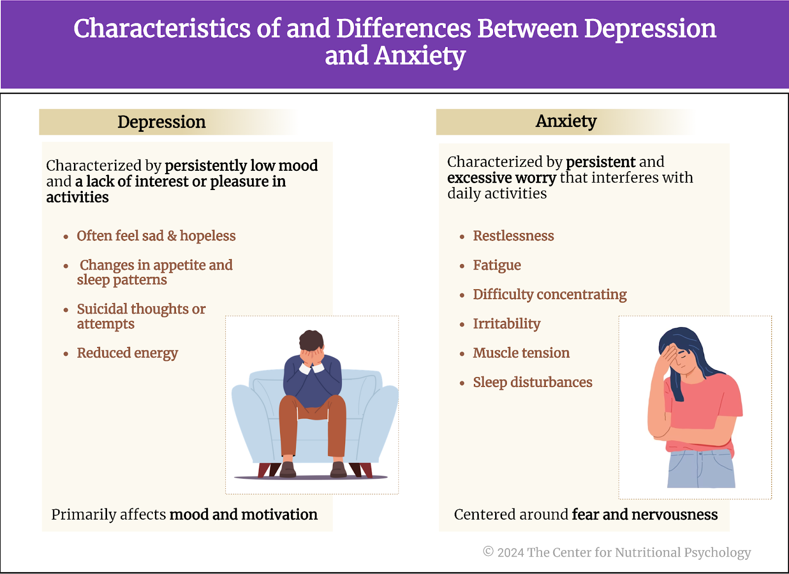
Depression and anxiety Depression and anxiety are two of the most frequent mental health disorders. Depression is characterized by persistently low mood and a lack of interest or pleasure in activities. A depressed individual will often feel sad, hopeless, and with little energy. This can lead to adverse changes in appetite and sleep patterns, but sometimes also thoughts of suicide.
Anxiety, on the other hand, is a condition marked by persistent and excessive worry that interferes with daily activities. It is typically accompanied by physical symptoms like restlessness, fatigue, difficulty concentrating, irritability, muscle tension, and sleep disturbances. While both conditions involve emotional distress,depression primarily affects mood and motivation, whereas anxiety is centered around fear and nervousness (see Figure 1).
Figure 1. Characteristics of and differences between depression and anxiety
Studies indicate that the share of individuals suffering from depression has been increasing both in the U.S. and globally in the past decades (Steffen et al., 2020; Weinberger et al., 2018). Anxiety and depression often occur together. Estimates state that the number of people suffering from anxiety increased after the global COVID-19 pandemic by 26% (Wang et al., 2023).
The number of people suffering from anxiety increased after the global COVID-19 pandemic by 26% (Wang et al., 2023)
Diet and mental health While the precise causes of anxiety and depression are unknown, a great number of genetic, biological, environmental, and psychological factors have been found to play a role in their development. Recent studies have also linked certain dietary habits, such as the consumption of ultra-processed foods or sweetened beverages, to increased depression risks (Hedrih, 2023b; Samuthpongtorn et al., 2023).
Other researchers bring into focus the so-called Western diet, a diet based on fried and processed foods, refined grains, sugary products, and beer, as a dietary pattern associated with an increased risk of depression and anxiety (Wang et al., 2023). Within this diet, researchers single out fried food as particularly contributing to these adverse links with mental health.
The Western diet is a dietary pattern associated with an increased risk of depression and anxiety
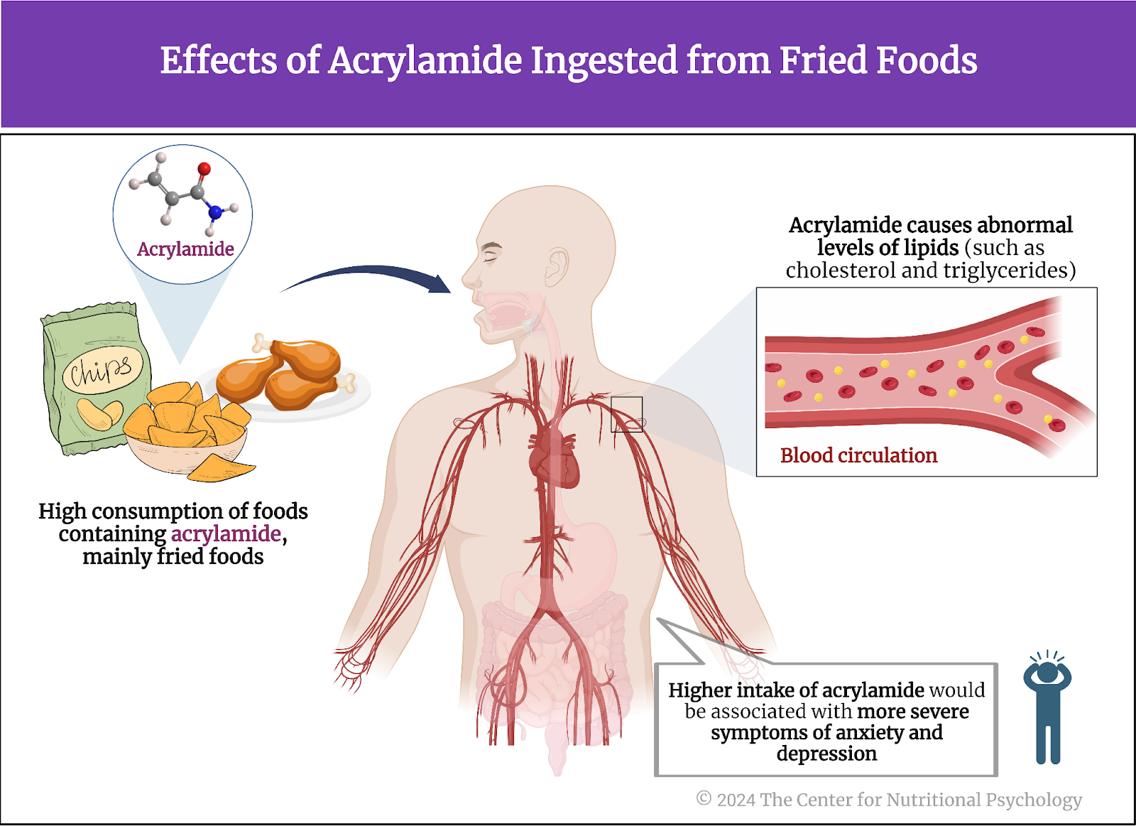
Fried food and health Frying is a method of food preparation that people widely use at home and in restaurants. However, the process of frying changes the nutrient composition of the food and can produce various hazardous compounds. One such substance is acrylamide. Acrylamide is created during the process of frying foods that are rich in carbohydrates, such as potatoes. Acrylamide is known to be toxic for neurons in higher concentrations. Studies have linked its prolonged intake to an increased risk of cardiovascular disease, neurological disorders, obesity, metabolic syndrome, and depression.
The process of frying changes the food’s nutrient composition and can produce various hazardous compounds
The current study Study author Anli Wang and her colleagues wanted to examine the association between the intake of a substance found in fried food called acrylamide and depression and anxiety in a large sample from the general population. Acrylamide is a substance found in fried food.
These authors expected that high consumption of fried food and, consequently, higher intake of acrylamide would be associated with more severe symptoms of anxiety and depression. Ingested acrylamide would lead to abnormal levels of lipids, such as cholesterol and triglycerides, in the blood and inflammation. This would, in turn, impact the likelihood that depression or anxiety would develop (see Figure 2).
Figure 2. Effects of acrylamide ingested from fried foods
In this study, the authors analyzed human data from the UK Biobank and conducted an experiment on zebrafish.
The UK Biobank TheUK Biobank is a large-scale biomedical database and research resource containing in-depth genetic and health information from half a million UK participants. The project, which began in 2006 and is ongoing, aims to improve the prevention, diagnosis, and treatment of a wide range of serious and life-threatening illnesses.
UK Biobank participants provided blood, urine, and saliva samples and detailed health and lifestyle information, which researchers worldwide can access to conduct health-related research. Many valuable scientific discoveries and insights have come from this database, and new studies using UK Biobank data are continuously being published.
For this study, researchers analyzed data from 140,728 individuals from the UK Biobank. Of these, during the 11.3 years of follow-up, 8,294 had symptoms of anxiety, and 12,735 had depression symptoms (see Figure 3).
Figure 3. Study procedure
The Zebrafish experimentI n addition to analyzing the UK Biobank data, the researchers conducted an experiment on zebrafish. This experiment exposed zebrafish groups to different acrylamide concentrations for 180 days. After the exposure period, they conducted a series of tests to evaluate anxiety-like symptoms in these fish. At the end of the experiment, they killed the fish and harvested their brains to analyze tissue changes.
Zebrafish are a small, freshwater fish species extensively used in biomedical research due to their genetic similarity to humans, transparent embryos that allow easy observation of developmental processes, and rapid reproduction.
These researchers looked for specific symptoms in zebrafish exposed to acrylamide –specifically thigmotaxis and scototaxis. Scototaxis is a tendency of fish to prefer darker areas over lighter ones in their environment. Thigmotaxis is a preference for staying close to physical objects or walls in their aquarium. In research, both of these tendencies are indications that the fish is experiencing anxiety-like conditions and that it is under stress. The more pronounced they are, the higher the anxiety-like conditions in the fish are.
UK Biobank People consuming fried food more often had somewhat higher risks of depression and anxiety
Results from the UK Biobank showed that individuals consuming at least one fried meal per day were more likely to be younger, male, and active smokers than those consuming fried food less often. After taking into account the age and sex of participants, statistical analyses showed that individuals consuming fried food more often were somewhat more likely to have anxiety and depression symptoms.
Notably, individuals consuming fried food were 12% more likely to have anxiety symptoms and 7% more likely to have symptoms of depression. Results of looking at specific food types highlighted fried potatoes and fried white meat. Consumption of each of these foods was associated with a 4% higher risk of anxiety and a 7% higher risk of depression. However, after adjusting for various other factors, the link with fried white meat consumption disappeared, but the one with fried potato consumption remained (see Figure 4).
Figure 4. Fried food consumption and depression and anxiety symptoms
Individuals consuming fried food were 12% more likely to have anxiety symptoms and 7% more likely to have symptoms of depression
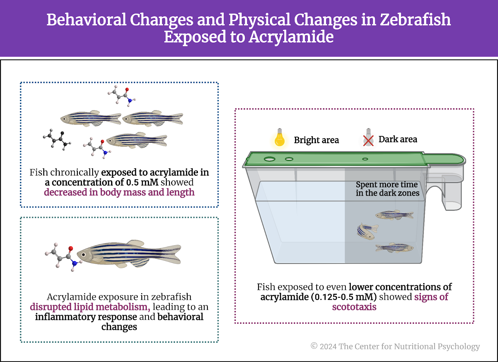
Zebrafish exposed to acrylamide developed behaviors indicative of stress and anxiety The experiment on zebrafish showed that fish chronically exposed to acrylamide in a concentration of 0.5 mM decreased in body mass and length. Fish exposed to even lower concentrations of acrylamide (0.125-0.5 mM) showed signs of scototaxis. They spent more time in the dark zones than zebrafish without this exposure. The time the fish spent in the dark zones was associated with the concentration of acrylamide used.
Other behavioral comparisons between zebrafish exposed to acrylamide and those in the control group also confirmed the finding about anxiety-like symptoms in fish exposed to this substance. Analysis of tissues of zebrafish showed that exposure to acrylamide disturbed their lipid metabolism and initiated an inflammatory response, which, in turn, likely led to the observed behavioral changes (see Figure 5).
Figure 5. Behavioral changes and physical changes in Zebrafish exposed to acrylamide
Conclusion An analysis of UK Biobank data showed that individuals frequently consuming fried food have a slightly higher risk of having depression or anxiety symptoms. An experiment on zebrafish confirmed that chronic exposure to acrylamide, a substance created when food rich in carbohydrates is fried, might cause this link. Exposure to acrylamide can disturb the metabolism of lipids and initiate an inflammatory response that may, in turn, contribute to the development of anxiety and depression symptoms.
Chronic exposure to acrylamide, a substance created when food rich in carbohydrates is fried, might cause this link
These findings contribute to a better scientific understanding of the diet-mental health relationship (DMHR). They may help individuals and mental health practitioners prevent or better treat anxiety and depression conditions by modifying their diet. The insights brought by this study may also allow for the creation of better menus and meal plans in the hospitality industry and various institutional food services.
Dicker-Oren, S. D., Gelkopf, M., & Greene, T. (2022). The dynamic network associations of food craving, restrained eating, hunger and negative emotions. Appetite, 175(March), 106019. https://doi.org/10.1016/j.appet.2022.106019
Hedrih, V. (2023a). Food and Mood: Is the Concept of ‘Hangry’ Real? CNP Articles in Nutritional Psychology. https://www.nutritional-psychology.org/food-and-mood-is-the-concept-of-hangry-real/
Hedrih, V. (2023b). Women Consuming Lots of Artificially Sweetened Beverages Might Have a Higher Risk of Depression, Study Finds. CNP Articles in Nutritional Psychology. https://www.nutritional-psychology.org/women-consuming-lots-of-artificially-sweetened-beverages-might-have-a-higher-risk-of-depression-study-finds/
Samuthpongtorn, C., Nguyen, L. H., Okereke, O. I., Wang, D. D., Song, M., Chan, A. T., & Mehta, R. S. (2023). Consumption of Ultraprocessed Food and Risk of Depression. JAMA Network Open, 6(9), e2334770. https://doi.org/10.1001/jamanetworkopen.2023.34770
Steffen, A., Thom, J., Jacobi, F., Holstiege, J., & Bätzing, J. (2020). Trends in prevalence of depression in Germany between 2009 and 2017 based on nationwide ambulatory claims data. Journal of Affective Disorders, 271, 239–247. https://doi.org/10.1016/J.JAD.2020.03.082
Stevenson, R. J., Bartlett, J., Wright, M., Hughes, A., Hill, B. J., Saluja, S., & Francis, H. M. (2023). The development of interoceptive hunger signals. Developmental Psychobiology, 65(2), 1–11. https://doi.org/10.1002/dev.22374
Wang, A., Wan, X., Zhuang, P., Jia, W., Ao, Y., Liu, X., Tian, Y., Zhu, L., Huang, Y., Yao, J., Wang, B., Wu, Y., Xu, Z., Wang, J., Yao, W., Jiao, J., & Zhang, Y. (2023). High-fried food consumption impacts anxiety and depression due to lipid metabolism disturbance and neuroinflammation. Proceedings of the National Academy of Sciences of the United States of America, 120(118). https://doi.org/10.1073/pnas.2221097120
Weinberger, A. H., Gbedemah, M., Martinez, A. M., Nash, D., Galea, S., & Goodwin, R. D. (2018). Trends in depression prevalence in the USA from 2005 to 2015: widening disparities in vulnerable groups. Psychological Medicine, 48(8), 1308–1315. https://doi.org/10.1017/S0033291717002781
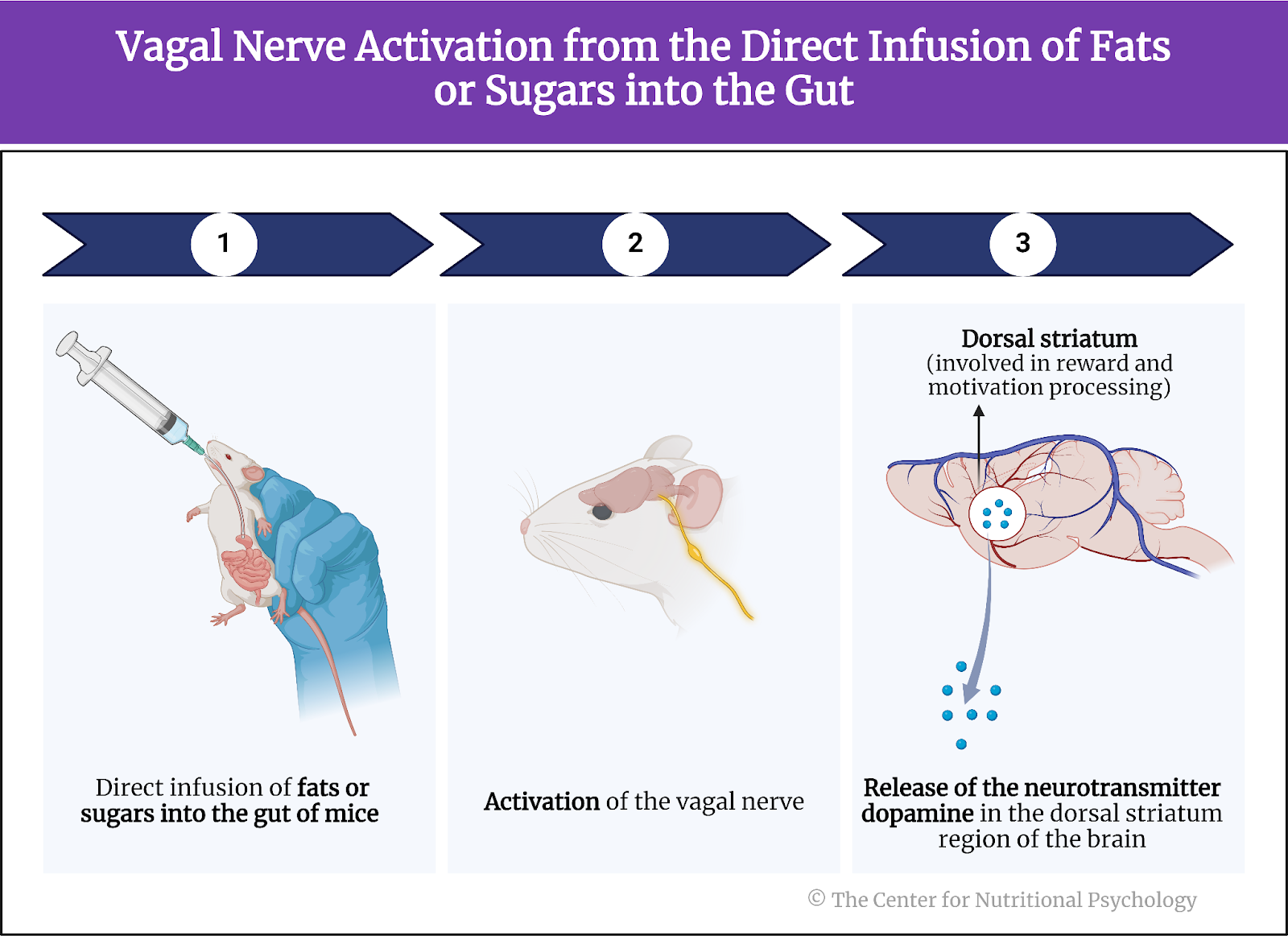
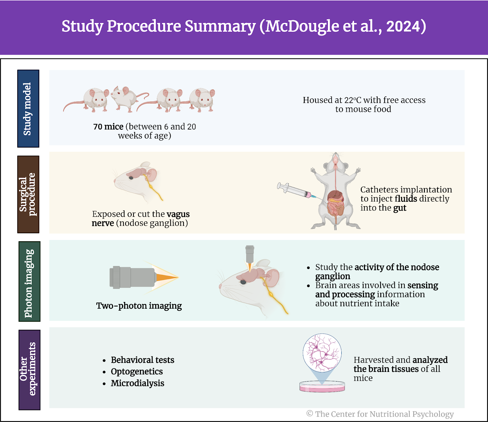
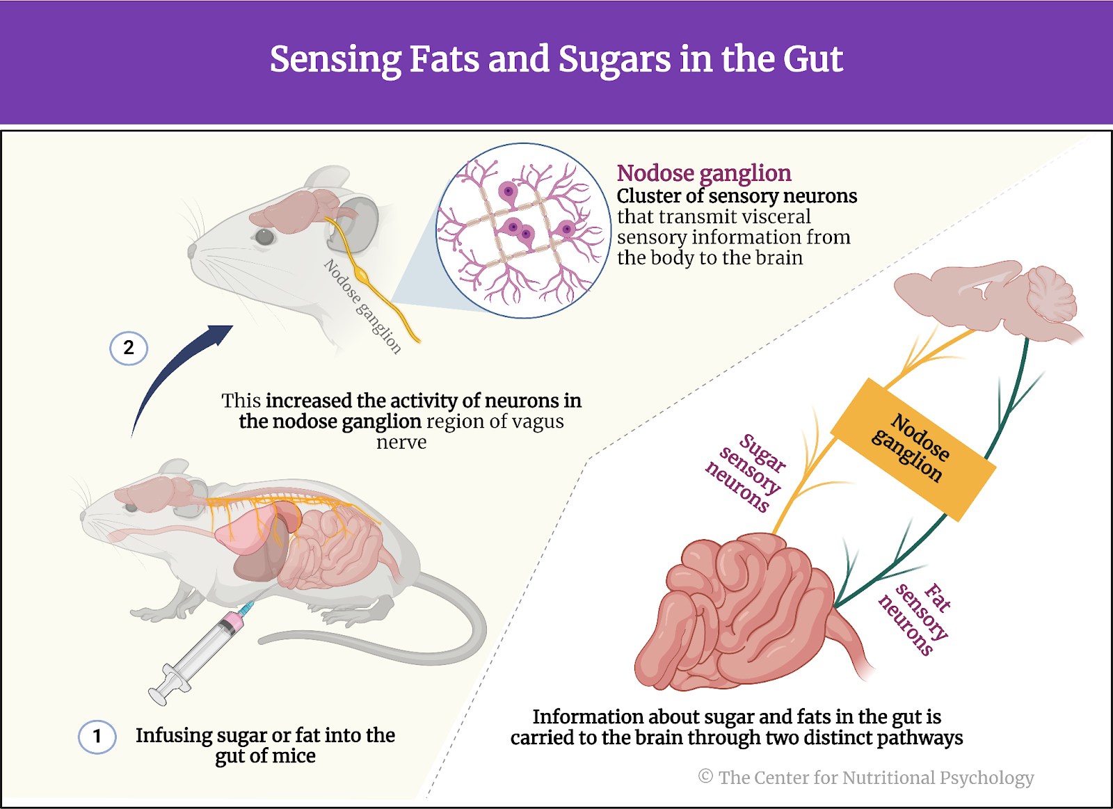
A study on mice published inCell Metabolism found that gut-brain neural circuits conveying and processing information about the presence of fat and sugar in the gut are separate
Foods that contain both sugar and fats activate both circuits, releasing more dopamine compared to foods of equal caloric value containing only fat
The large amounts of dopamine released in this way create feelings of pleasure that promote overeating on the types of food that caused the release
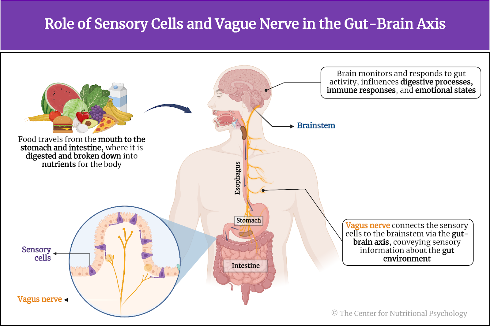
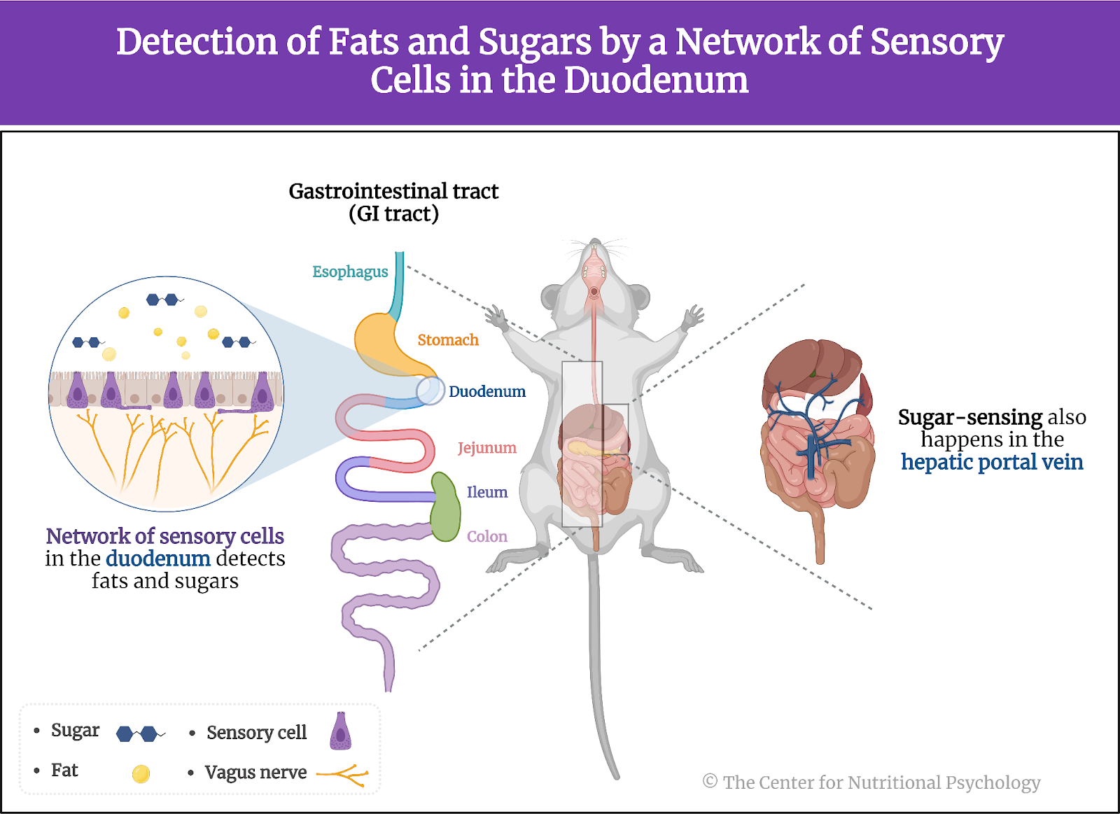
When we want to find out what a piece of food is like, we can taste or smell it. When we smell something, sensory cells located in a small patch of tissue (olfactory epithelium) at the top of our nasal cavity react to the odorant molecules coming from that piece of food, and the olfactory nerve to carries the information to our brain, allowing us to experience smell. Similarly, when we taste a piece of food, taste buds in our mouth react to it, and specific nerves carry the information to our brain. While we eat, this evaluation of the qualities of food using our sensory organs happens continuously. However, not all of our food sensations come from our external senses. Our body also has sensory cells in the gut that inform the brain about what we eat.
The vagus nerve After we ingest food through our mouth, it enters the esophagus. It continues towards the stomach and, after that, into the intestine. Food is further digested and broken down into components absorbed into the body as nutrients in these parts of the gastrointestinal system.
There are sensory cells located throughout the gastrointestinal system. The vagus nerve, one of the longest nerves in the body, connects these cells to the brain through a communication pathway called the gut-brain axis. In this way, it conveys sensory information about the state of the intestinal environment, including nutrient levels, gut microbiota activity, and intestinal wall integrity from the gut to the brain. These signals are then integrated into the brainstem, allowing the brain to monitor and respond to gut activity, influencing digestive processes, immune responses, and emotional states. (Bonaz et al., 2018) (see Figure 1).
Figure 1. Role of sensory cells and vagus nerve in the Gut-Brain Axis.
Among other things, the vagus nerve carries information to the brain about the nutritional value of our food. However, scientists so far have not fully understood the intricate details of how this system functions (McDougle et al., 2024).
The obesity pandemic Recent decades have seen a continued increase in the share of obese individuals throughout the developed world. Many call this the obesity pandemic (Wong et al., 2022). This has motivated many studies into the causes of this pandemic. Results point to an intricate interplay between specific types of nutrients and other substances in the food and the properties of the human central nervous system as important contributing causes of obesity (Wilding, 2001).
Notably, while human brains (and brains of many other species) have a food intake regulation mechanism that determines when we will feel a desire to eat or when we will feel satiated, studies indicate that parts of this mechanism might be malfunctioning in obese individuals(Pujol et al., 2021; Seabrook et al., 2023). Studies indicate that feeding rodents high-fat diets instead of their regular chow can, in time, dysregulate their food intake regulation mechanisms, leading them to eat more calories than they need, resulting in obesity (Ikemoto et al., 1996). In humans, studies have linked the dysregulation of this mechanism with the consumption of foods that are both fatty and sweet (a property rarely found in natural foods) but also to various additives that create addiction-like reactions to foods that contain them (Hedrih, 2023).
The current study Study author Molly McDougle and her colleagues wanted to explore the neural pathways through which the brain detects the intake of fats and sugars (i.e., fatty and sweet foods). They wanted to know whether the intake of these two types of nutrients triggers the same sensory neurons and whether they engage overlapping or separate neural circuits.
What was known before this study was that a direct infusion of fats or sugars into the gut (of mice) activates the vagus nerve. This, in turn, leads to the release of the neurotransmitter dopamine in the dorsal striatum region of the brain. The dorsal striatum is involved in reward and motivation processing, and dopamine is a key neurotransmitter that signals pleasure and reward (see Figure 2).
Figure 2. Vagal nerve activation from the direct infusion of fats or sugars into the gut
Study procedure The study was conducted on 70 mice. The mice used in this study were between 6 and 20 weeks of age. Thirty-five mice were male, and 35 were female. They were housed at 22 degrees C and had free access to mouse food.
The study authors performed a series of experiments on these mice. The experiments involved surgical procedures that exposed or cut the vagus nerve and/or exposed the nodose ganglion (a part of the vagus nerve) for further activities. These surgeries also involved the implantation of catheters that allowed researchers to inject fluids directly into the gut of these mice, bypassing the mouth and, thus, taste receptors.
These researchers used 2-photon imaging to study the activity of the nodose ganglion in the mice (while the mice were alive) and of brain areas involved in sensing and processing information about nutrient intake. Two-photon imaging is a fluorescence microscopy technique that allows for deep-tissue, high-resolution imaging of live cells and tissues. It uses two photons of lower energy to excite a fluorophore simultaneously. Fluorophores are special molecules that emit a visible glow when excited by light of a specific wavelength, thus allowing researchers to track and observe different biological processes or structures.
Other experimental procedures included behavioral tests, techniques using light to control neurons that have been genetically modified to express ion channels sensitive to light (optogenetics), sampling extracellular fluid from the dorsal striatum region of the brain to measure the quantities of dopamine (microdialysis) and others. In the end, the study authors harvested and analyzed the brain tissues of these mice (see Figure 3).
Figure 3. Study procedure summary (McDougle et al., 2024).
Fats and sugars are sensed by separate neurons of the vagal nerve Results showed that fats and sugars increased the activity of neurons in the vagus nerve, but mostly not of the same ones. Neurons comprising the nodose ganglion mostly reacted to the presence of fat or sugars. Very few neurons reacted to both. These neurons reacting to nutrients in the intestine comprised around 17% of the neurons in the nodose ganglion. The nodose ganglion is a cluster of sensory neurons located on the vagus nerve, transmitting visceral sensory information from the body to the brain.
When researchers infused sugar or fat into the guts of mice, the neurons of the nodose ganglion they previously marked increased their frequencies of nerve impulses. Analysis of the neural circuits activated by fats and sugars showed that they differ. They were largely separate. Information about sugar and fats in the gut is carried to the brain through two distinct pathways. These pathways convey information about the type of nutrient in the gut, but they also likely convey information about its concentration (see Figure 4).
Figure 4. Sensing fats and sugars in the gut
The duodenum is the key sensing site for both sugar and fat Tracing the locations of sensory cells, these neurons led these researchers to discover an extensive network of sensory cells in the duodenum. Almost all sensory cells detecting fats and many of those detecting sugars were located there. The duodenum is the first section of the small intestine, connecting the stomach to the jejunum. It plays a crucial role in the initial phase of digestion by receiving partially digested food from the stomach and digestive enzymes from the pancreas and liver. On the other hand, study authors found that sugar-sensing also happens in the hepatic portal vein, a blood vessel that carries nutrient-rich blood from the gastrointestinal tract and spleen to the liver (see Figure 5).
Figure 5. Detection of fats and sugars by a network of sensory cells in the duodenum
Reinforcement learning based on sugar and fat happens through separate circuits Experiments where researchers destroyed fat-sensing neurons but left the sugar-sensing ones intact or vice versa indicated that reinforcement learning, a type of learning where a mouse can associate a specific object or sensation with a specific nutrient, can be conducted using only the neurons sensing that specific nutrient. Mice without fat-sensing neurons but with intact sugar-sensing neurons were able to form a preference for a flavor associated with sugar intake.
Mice without fat-sensing neurons but with intact sugar-sensing neurons were able to form a preference for a flavor associated with sugar intake
In the nigrostriatal region of the brain, both information about fat and sugar in the gut led to the release of dopamine (thought to produce the feeling of pleasure and a rewarding experience). However, these nutrients activated parallel and largely separate neuronal populations at each node of the reward circuit in this part of the brain. This means that distinguishable neural circuits are responsible for reinforcement learning for fat and sugar, i.e., for learning to associate stimuli with pleasure caused by the ingestion of sugar and pleasure caused by the ingestion of fat (see Figure 6).
Figure 6. Release of dopamine in response to fat and sugar
Consuming fat and sugar at the same time promotes overeating for pleasure In another group of experiments, study authors found that when they gave mice a choice between taking solutions of fat, sugar (sucrose), or a mixture of the two nutrients, mice consumed equal amounts of fat or sugar solutions. However, the number of licks of the solution containing both sugar and fats nearly doubled compared to just sugar or just fat alone.
Consuming fat and sugar at the same time promotes overeating for pleasure
Researchers then offered mice a non-nutrient-flavored solution to lick, injecting sugar, fat, or a combination of the two directly into their guts (using the catheters they installed). After training to pair a non-nutritive flavor with a solution, mice again showed a preference for the mixed solution (both sugar and fat), indicating that the preference for this solution is independent of taste. This means that the preference for the sugar and fat combination has to do with sensing those nutrients in the gut and not with their taste in the mouth.
The preference for the sugar and fat combination has to do with sensing those nutrients in the gut and not with their taste in the mouth
Study authors conclude that this is because this mixed solution activates both the brain’s rewarding circuits for fat and sugar, increasing the overall feeling of pleasure. Analysis of the quantity of released dopamine in the dorsal striatum region of the brain found that it is much higher after the injection of the mixed sugar-fat solution than after the injection of a solution with equal caloric value but containing only fat.
This suggests that foods rich in fat and sugar produce higher activation of reward circuits for the same amount of calories than foods containing just one of these two nutrients. This would translate to higher feelings of pleasure after consumption, motivating the individual to consume more food of that type, which could lead to obesity (see Figure 7).
Figure 7. Activation of the brain’s rewarding circuits with combined sugar and fat
Conclusion The study showed that sensing the presence of sugars and fats in the intestines of mice is achieved through different sets of neurons. The study also found that these two types of nutrients activate parallel but largely separate reward circuits in the brain. Due to this, consuming foods that contain both sugars and fats activates reward circuits in the brain for both of these substances, resulting in a higher release of dopamine and, thus, higher feelings of pleasure, compared to consuming foods of equal caloric value but containing only fats or only sugars. These peculiarities of neural pathways make mice, and possibly humans, more likely to overeat foods rich in fats and sugars driven by feelings of pleasure the simultaneous activation of these two neural pathways creates.
The discovery of these separate mechanisms and understanding of the ways they work can help scientists. Still, people working in nutrition better understand how the properties of certain food items lead to obesity. This can, in sequence, help better plan individual diets and develop policies to help stop the ongoing obesity epidemic.
The paper “Separate gut-brain circuits for fat and sugar reinforcement combine to promote overeating” was authored by Molly McDougle, Alan de Araujo, Arashdeep Singh, Mingxin Yang, Isadora Braga, Vincent Paille, Rebeca Mendez-Hernandez, Macarena Vergara, Lauren N. Woodie, Abhishek Gour, Abhisheak Sharma, Nikhil Urs, Brandon Warren, and Guillaume de Lartigue.
References
Bonaz, B., Bazin, T., & Pellissier, S. (2018). The vagus nerve at the interface of the microbiota-gut-brain axis. In Frontiers in Neuroscience (Vol. 12, Issue FEB). Frontiers Media S.A. https://doi.org/10.3389/fnins.2018.00049
Hedrih, V. (2023). Scientists Propose that Ultra-Processed Foods be Classified as Addictive Substances. CNP Articles in Nutritional Psychology. https://www.nutritional-psychology.org/scientists-propose-that-ultra-processed-foods-be-classified-as-addictive-substances/
Ikemoto, S., Takahashi, M., Tsunoda, N., Maruyama, K., Itakura, H., & Ezaki, O. (1996). High-fat diet-induced hyperglycemia and obesity in mice: Differential effects of dietary oils. Metabolism, 45(12), 1539–1546. https://doi.org/10.1016/S0026-0495(96)90185-7
McDougle, M., de Araujo, A., Singh, A., Yang, M., Braga, I., Paille, V., Mendez-Hernandez, R., Vergara, M., Woodie, L. N., Gour, A., Sharma, A., Urs, N., Warren, B., & de Lartigue, G. (2024). Separate gut-brain circuits for fat and sugar reinforcement combine to promote overeating. Cell Metabolism. https://doi.org/10.1016/j.cmet.2023.12.014
Pujol, J., Blanco-Hinojo, L., Martínez-Vilavella, G., Deus, J., Pérez-Sola, V., & Sunyer, J. (2021). Dysfunctional Brain Reward System in Child Obesity. Cerebral Cortex, 31, 4376–4385. https://doi.org/10.1093/cercor/bhab092
Seabrook, L. T., Naef, L., Baimel, C., Judge, A. K., Kenney, T., Ellis, M., Tayyab, T., Armstrong, M., Qiao, M., Floresco, S. B., & Borgland, S. L. (2023). Disinhibition of the orbitofrontal cortex biases decision-making in obesity. Nature Neuroscience, 26(1), 92–106. https://doi.org/10.1038/s41593-022-01210-6
Wilding, J. P. H. (2001). Causes of obesity. Practical Diabetes International, 18(8), 288–292. https://doi.org/10.1002/PDI.277
Wong, M. C., Mccarthy, C., Fearnbach, N., Yang, S., Shepherd, J., & Heymsfield, S. B. (2022). Emergence of the obesity epidemic: 6-decade visualization with humanoid avatars. The American Journal of Clinical Nutrition, 115(4), 1189–1193. https://doi.org/10.1093/AJCN/NQAC005
A study published inNature Mental Healthexamined the links between post-traumatic stress disorder (PTSD), dietary patterns, and gut microbiome in U.S. nurses
Results showed that nurses with higher PTSD symptom levels tended to eat less plant-based food and more red/processed meat
Microbial processes related to the production of pantothenate and coenzyme A can potentially be protective against PTSD
When we are exposed to distressing events or conditions that overwhelm our ability to cope, our body can produce a severe emotional response, and we experience psychological trauma. We feel that we have lost control of events around us. Our ability to integrate these emotional experiences into the story of our lives is reduced. After experiencing psychological trauma, people often start dividing the subjective timeline of their lives into the time before and the time after the traumatic event. Long-lasting psychological and health consequences may often follow (Hamburger et al., 2021). Among other things, the experience of psychological trauma can lead to the development of a serious mental health disorder called post-traumatic stress disorder or PTSD.
After experiencing psychological trauma, people often start dividing the subjective timeline of their lives into the time before and after the traumatic event
What is post-traumatic stress disorder? Post-traumatic stress disorder is a mental health condition that can develop in individuals who have experienced or witnessed a traumatic event. Symptoms of PTSD include intrusive thoughts, nightmares, and flashbacks related to the traumatic experience, causing significant distress. Individuals with PTSD may actively avoid reminders of the trauma, experience heightened arousal, and have negative changes in mood and cognition.
Mainstream treatments for PTSD include psychotherapy, particularly cognitive-behavioral psychotherapy, and medications. Studies indicate that these treatments can be effective in reducing PTSD symptoms (see Figure 1). In some individuals, they are very effective (Watts et al., 2013). However, around 20% of patients suffering from PTSD drop out of treatment before symptoms have withdrawn, while up to 60% of individuals undergoing treatment do not respond to it, i.e., experience no reduction of symptoms as a result (Wittmann et al., 2021).
Figure 1. PTSD symptoms and mainstream treatments
For these reasons, researchers are intensely working on new treatment options. They are trying alternative ways to achieve a reduction of symptoms, such as acupuncture (Watts et al., 2013) or psychedelic drugs (Barone et al., 2019). Researchers are also looking at chemicals that could potentially prevent the disorder from forming in the first place if taken immediately after the traumatic event, such as hydrocortisone (Hennessy et al., 2022).
Health impact of PTSD Individuals suffering from PTSD often suffer from other psychiatric disorders as well. Epidemiological surveys indicate that the vast majority of individuals with PTSD meet the criteria for at least one other disorder, while a substantial percentage have three or more other psychiatric disorders. These most commonly include major depressive disorder, substance use disorder, and anxiety disorders (Brady et al., 2000).
The majority of individuals with PTSD meet the criteria for at least one other disorder, while a substantial percentage have three or more other psychiatric disorders
Individuals with PTSD also have a higher likelihood of various somatic diseases, particularly chronic ones. These include asthma, cardiovascular disease, chronic pain and inflammation, obesity, type 2 diabetes, gastrointestinal disorders, and cognitive decline (Ke et al., 2023).
However, researchers are still looking for mechanisms through which this association between PTSD and somatic disorders is achieved. One promising avenue of research is the study of the gut microbiome, the community of organisms living in the human digestive system. The recent discovery of the microbiota-gut-brain axis, a bidirectional communication pathway through which gut microorganisms affect processes in the brain and vice versa (Carbia et al., 2023; García-Cabrerizo et al., 2021), has made it even more likely that at least a part of the link between PTSD and somatic disorder includes the gut microbiome.
Researchers are looking for mechanisms connecting PTSD and somatic disorders -with one promising avenue of research being the gut microbiome
The current study Study author Shanlin Ke and his colleagues hypothesized that gut microbiome might play a role in PTSD. They note that previous studies have already linked the gut microbiome with various other mental health disorders through the microbiota-gut-brain axis (Hedrih, 2023; Leclercq et al., 2020; Valles-Colomer et al., 2019). Additionally, studies show that the gut microbiome influences the brain region involved in learned fear. Fear is a key feature of PTSD. Gut microbiota depends on food intake, while individuals with PTSD tend to be more prone to eating unhealthy foods (Ke et al., 2023).
Previous studies have already linked the gut microbiome with various other mental health disorders through the microbiota-gut-brain axis (MGBA)
With this in mind, these researchers analyzed data from two studies of female registered nurses in the U.S. in order to systematically examine the associations of trauma exposure and PTSD status with dietary and microbiome data.
The study procedure Data came from 191 nurses who were part of the NHS-II study. NHS-II is a large longitudinal study of U.S. women with over 100,000 registered nurses that started in 1989. Nurses whose data were analyzed here participated in two substudies – the 2008 PTSD substudy that collected data about trauma exposure and PTSD symptoms and the 2013 mind-body study that, among other things, collected up to four stool samples. Stool samples were collected 48-72 hours apart. This was followed by a second set of collections six months later. Stool samples were analyzed to make inferences about the composition of the gut microbiome.
Of the nurses included in this study, 44 had probable PTSD, 119 were exposed to trauma but did not develop PTSD, and 28 participants had no trauma exposure (see Figure 2).
Figure 2. Study Procedure
No association between PTSD and overall microbiome structure Statistical analyses showed no associations between overall gut microbiota composition and PTSD status. Gut microbiota diversity was also not associated with PTSD.
However, there were associations between various other factors and the overall microbiome structure. Body mass index, depression, and the use of antidepressants were all associated with the overall composition of gut microbiome species.
Childhood trauma experiences and antidepressant use were associated with the relative abundance of species forming different metabolic pathways. The gut microbiome metabolic pathways refer to types of activities within the gastrointestinal tract that specific species of microorganisms perform.
Individuals with PTSD eat less plant-based food and more red/processed meat Examination of associations between dietary habits and PTSD revealed that individuals with more pronounced PTSD symptoms tend to eat less plant-based foods and more red/processed meats compared to individuals with lower PTSD symptom levels. The diets of these individuals were less healthy overall.
Individuals with more pronounced PTSD symptoms tend to eat less plant-based foods and more red/processed meats compared to individuals with less PTSD symptoms
Individuals with PTSD symptoms had lower abundances of Eubacterium eligens After statistical analyses showed no associations between the overall gut microbiome composition and PTSD, these authors examined associations between PTSD symptoms and the abundance of individual species of bacteria. They looked for species of microorganisms that differed in abundance in individuals with different levels of PTSD symptoms. Three species were clearly more abundant in individuals with more PTSD symptoms – Parabacteroides goldsteinii, Barnesiella intestinihominis, and Paraprevotella unclassified. Seven species with the highest negative association with PTSD symptoms, i.e., species less abundant in individuals with more PTSD symptoms, were also identified (Eubacterium eligens, Parabacteroides distasonis, Akkermansia muciniphila, Bacteroides massiliensis, Bifidobacterium longum, Dialister invisus, and Roseburia inulinivorans). These species play diverse roles in the human digestive system, contributing to functions ranging from fermentation of dietary fibers to protection of the gut lining.
Eubacterium eligens (a good bacteria) was less abundant in individuals eating more pies, carbonated beverages with sugars, candy without chocolate, hot dogs, bacon, and processed meats. It tended to be more abundant in individuals eating more vegetables (for example, raw carrot, spinach/collard greens cooked, and yellow squash), fruits (for example, orange and banana), and fish. The abundance of this bacteria was the most strongly associated with PTSD symptoms of all analyzed microorganisms. Eubacterium eligens in the gut synthesize pantothenate and Coenzyme A (see Figure 3).
Figure 3. Microorganisms associated with PTSD
Researchers tested a number of statistical models proposing that specific dietary habits mediate the impact of PTSD symptoms on the abundance of specific species of gut bacteria. These analyses revealed that it is possible that the impact of PTSD symptoms on the abundance of Eubacterium eligens is mediated by the consumption of raw carrots. Similarly, it is possible that Adlercreutzia equolifaciens mediates the link between PTSD symptoms and the intake of dairy-cottage/ricotta cheese.
Conclusion In summary, the study showed that PTSD symptoms in U.S. nurses are not associated with the overall structure of the gut microbiome, but the abundance of several bacterial species was associated with the severity of PTSD symptoms in spite of this. Additionally, it turned out that participants with PTSD tend to eat less plant-based food and more red/processed meat. Their overall dietary habits tended to be less healthy (see Figure 4).
Figure 4. Research findings (Ke et al., 2023)
The study also established statistical links between PTSD symptoms, dietary habits, and bacteria. Future studies could explore the nature of these links in more detail. Potentially, these could lead to the discovery of ways to affect PTSD symptoms through dietary interventions and modifying the abundance of certain species of gut bacteria. However, the feasibility of such an approach is yet to be established.
Visit the research category within the Nutritional Psychology Research Library on “Diet, Trauma and PTSD” to learn more about the connection between diet and trauma. To access evidence-based continuing education on the connection between the microbiome and mental health, see the CNP Education page.
References
Barone, W., Beck, J., Mitsunaga-Whitten, M., & Perl, P. (2019). Perceived Benefits of MDMA-Assisted Psychotherapy beyond Symptom Reduction: Qualitative Follow-Up Study of a Clinical Trial for Individuals with Treatment-Resistant PTSD. Journal of Psychoactive Drugs, 51(2), 199–208. https://doi.org/10.1080/02791072.2019.1580805
Brady, K., Killeen, T., Brewerton, T., & Lucerini, S. (2000). Comorbidity of Psychiatric Disorders and Posttraumatic Stress Disorder. Journal of Clinical Psychiatry, 61(suppl 7), 22–32.
Carbia, C., Bastiaanssen, T. F. S., Iannone, F., García-cabrerizo, R., Boscaini, S., Berding, K., Strain, C. R., Clarke, G., Stanton, C., Dinan, T. G., & Cryan, J. F. (2023). The Microbiome-Gut-Brain axis regulates social cognition & craving in young binge drinkers. EBioMedicine, 89, 104442. https://doi.org/10.1016/j.ebiom.2023.104442
García-Cabrerizo, R., Carbia, C., O´Riordan, K. J., Schellekens, H., & Cryan, J. F. (2021). Microbiota-gut-brain axis as a regulator of reward processes. Journal of Neurochemistry, 157(5), 1495–1524. https://doi.org/10.1111/JNC.15284
Hamburger, A., Hancheva, C., & Volkan, V. (Eds.). (2021). Social Trauma – An Interdisciplinary Textbook. Springer Nature Switzerland AG. https://doi.org/10.1007/978-3-030-47817-9
Hedrih, V. (2023). Women Consuming Lots of Artificially Sweetened Beverages Might Have a Higher Risk of Depression, Study Finds. CNP Articles in Nutritional Psychology. https://www.nutritional-psychology.org/women-consuming-lots-of-artificially-sweetened-beverages-might-have-a-higher-risk-of-depression-study-finds/
Hennessy, V. E., Troebinger, L., Iskandar, G., Das, R. K., & Kamboj, S. K. (2022). Accelerated forgetting of a trauma-like event in healthy men and women after a single dose of hydrocortisone. Translational Psychiatry, 12(1). https://doi.org/10.1038/s41398-022-02126-2
Ke, S., Wang, X.-W., Ratanatharathorn, A., Huang, T., Roberts, A. L., Grodstein, F., Kubzansky, L. D., Koenen, K. C., & Liu, Y.-Y. (2023). Association of probable post-traumatic stress disorder with dietary pattern and gut microbiome in a cohort of women. Nature Mental Health, 1(11), 900–913. https://doi.org/10.1038/s44220-023-00145-6
Leclercq, S., Le Roy, T., Furgiuele, S., Coste, V., Bindels, L. B., Leyrolle, Q., Neyrinck, A. M., Quoilin, C., Amadieu, C., Petit, G., Dricot, L., Tagliatti, V., Cani, P. D., Verbeke, K., Colet, J. M., Stärkel, P., de Timary, P., & Delzenne, N. M. (2020). Gut Microbiota-Induced Changes in β-Hydroxybutyrate Metabolism Are Linked to Altered Sociability and Depression in Alcohol Use Disorder. Cell Reports, 33(2). https://doi.org/10.1016/J.CELREP.2020.108238
Valles-Colomer, M., Falony, G., Darzi, Y., Tigchelaar, E. F., Wang, J., Tito, R. Y., Schiweck, C., Kurilshikov, A., Joossens, M., Wijmenga, C., Claes, S., Van Oudenhove, L., Zhernakova, A., Vieira-Silva, S., & Raes, J. (2019). The neuroactive potential of the human gut microbiota in quality of life and depression. Nature Microbiology, 4(4), 623–632. https://doi.org/10.1038/s41564-018-0337-x
Watts, B. V., Schnurr, P. P., Mayo, L., Young-Xu, Y., Weeks, W. B., & Friedman, M. J. (2013). Meta-analysis of the efficacy of treatments for posttraumatic stress disorder. Journal of Clinical Psychiatry, 74(6). https://doi.org/10.4088/JCP.12r08225
Wittmann, L., Muller, J., Morina, N., Maercker, A., & Schnyder, U. (2021). Predicting Treatment Response in Psychotherapy for Postraumatic Stress Disorder: a Pilot Study. Psihologija, 54(1), 1–14. https://doi.org/10.2298/PSI190905007W
A study published inBrain showed that symptoms of Alzheimer’s disease can be transferred to young rats via the gut microbiota of Alzheimer’s patients
Transplantation of these microorganisms into the guts of healthy rats induced cognitive deficits
The deficits resulted from the disruption of adult hippocampal neurogenesis, the capacity of the hippocampus to produce new neurons
Our gut is home to trillions of different microorganisms. These microorganisms sustain themselves using the food we eat and help our digestion process. For example, many food items contain substances called resistant starches. Our digestive system cannot digest these resistant starches, but some bacteria in our gut can. They ferment those types of starches, creating substances called short-chain fatty acids (SCFA) that our bodies can use (Li et al., 2023) (see Figure 1).
Figure 1. Digestion of resistant starch by gut bacteria
Similarly, these bacteria help digest substances like dietary fiber, other complex polysaccharides, lignans, and many others, converting them into substances our body can use as nutrients or that convey various health benefits. However, when pathogenic, unhealthy microorganisms infect our digestive tract, we experience an upset stomach. This includes symptoms such as nausea, vomiting, diarrhea, abdominal pain, and others.
The microbiota-gut-brain axis (MGBA) This community of microorganisms living in our gut is called the gut microbiota. However, its effects on us go far beyond helping digestion. Gut microbiota can also influence our central nervous system –andbe influenced by it. The mechanism through which this bidirectional communication link between gut microbiota and the brain is achieved is called the microbiota-gut-brain axis (MGBA). It is crucial in regulating various physiological and psychological processes (Carbia et al., 2023; García-Cabrerizo et al., 2021).
The Microbiota-Gut-Brain Axis is crucial in regulating various physiological and psychological processes
This bidirectional communication mechanism occurs via several biomolecules, including the hormone cortisol, short-chain fatty acids (SCFAs), and tryptophan. Emerging studies reveal that the gut microbiota produces substances (called ‘signaling molecules’) that can influence the brain’s activity and responses to stress and emotions (more about this can be found in NP 120 Parts I & II). The MGBA is closely tied to the immune system, influencing the body’s inflammatory responses and potentially contributing to neuroinflammation (Zhu et al., 2023) (see Figure 2).
Figure 2. MGBA, bidirectional communication, and signaling molecules
What is Alzheimer’s disease? Alzheimer’s disease is a progressive neurodegenerative disorder characterized by a decline in cognitive function, memory loss, and changes in behavior. It is the most common form of dementia, affecting primarily older adults. The exact cause of Alzheimer’s disease is not fully understood. Still, it is associated with the accumulation of abnormal protein deposits in the brain, called beta-amyloid plaques and tau tangles. These deposits disrupt communication between nerve cells and eventually lead to their degeneration and death (Grabrucker et al., 2023) (see Figure 3).
Figure 3. Alzheimer’s Disease (AD)
Alzheimer’s disease and adult hippocampal neurogenesis The hippocampus is a brain area that is particularly vulnerable to Alzheimer’s disease. It plays a crucial role in learning, memory, and emotional regulation. The hippocampus hosts a population of neural stem cells, special undifferentiated cells that can produce new neurons throughout their lifespan. This process of creating new neural cells is called adult hippocampal neurogenesis. It is crucial for cognitive processes like spatial learning, distinguishing between similar events and environments, and emotion regulation (see Figure 4).
Figure 4. Adult hippocampal neurogenesis
Interestingly, hippocampal neurogenesis is impaired in Alzheimer’s even before abnormal protein deposits can be detected in the brain, including in the hippocampus. This indicates that dysfunction of this system is an early indicator that Alzheimer’s disease is developing (Grabrucker et al., 2023).
The hippocampus is a brain area that is particularly vulnerable to Alzheimer’s disease. It plays a crucial role in learning, memory, and emotional regulation
Causes of Alzheimer’s Genetic factors, such as specific gene mutations, are linked to early-onset Alzheimer’s, while other risk factors include age, family history, and certain lifestyle factors. Despite ongoing research, there is currently no cure for Alzheimer’s disease, and treatment focuses on managing symptoms and improving the quality of life for affected individuals.
However, the recent discovery of the MGBA has opened a new avenue of research demonstrating that this communication pathway is a significant mediator of behavior throughout the lifespan. Studies indicate clear links between gut microbiota composition and behavior (e.g., Leclercq et al., 2020; Valles-Colomer et al., 2019).
Studies indicate clear links between gut microbiota composition and behavior
Recently, links between the MGBA and Alzheimer’s have begun to appear. Studies on mice indicate that transplanting gut microbiota from Alzheimer’s patients into mice can cause adverse cognitive changes in these mice (Kim et al., 2021; Wang et al., 2022) (see Figure 5). But could it also disrupt the adult hippocampal neurogenesis? And would these changes correlate with the level of cognitive impairment of the person the transplanted microbiota came from?
Figure 5. Gut microbiota transplanted from Alzheimer’s patients
The current study Current studies clearly implicate the gut microbiota in the pathological features of Alzheimer’s disease, but until this particular study, it has remained unclear whether cognitive symptoms in human Alzheimer’s patients and underlying cellular changes (such as the disruption of adult hippocampal neurogenesis) could be transmitted to a healthy organism via the gut microbiota. Study author Stefanie Grabrucker and her colleagues wanted to find out. They also wanted to uncover the mechanism through which this happens.
These authors conducted a study in which they took fecal samples containing gut microbiota from humans suffering from Alzheimer’s disease and transplanted them into the guts of young adult male rats.
Current studies implicate gut microbiota in the pathological features of AD, but until now, it has remained unclear whether cognitive symptoms and underlying cellular changes in human Alzheimer’s patients could be transmitted to a healthy organism via the gut microbiota
The study procedure Study participants were 69 patients with Alzheimer’s disease, and 64 healthy individuals were included as controls. They were recruited at the IRCCS Centro San Giovanni di Dio Fatebenefratelli, Brescia, Italy. They all underwent clinical assessment for cognitive function and a physical examination. All participants gave blood samples for further analysis. Fifty-four participants with Alzheimer’s disease and 41 healthy participants gave stool samples in a sterile cup at their homes. Researchers processed these samples for transplanting gut microbiota found in feces into rats.
Animals used in the study were male Sprague-Dawley rats aged 11 weeks. They were kept in environmentally controlled conditions at a temperature of 21oC and under a 12-hour light-dark cycle (12 hours in light and 12 hours in the dark).
Transplanting gut microbiota from stool samples into rats Researchers kept rats for two weeks after arrival at the laboratory without any treatment to let them acclimatize. After this period, they added a powerful cocktail of antibiotics to their water for seven consecutive days. This combination of antibiotics consisted of ampicillin (1 g/l), vancomycin (500 mg/l), ciprofloxacin HCL (200 mg/l), and imipenem (250 mg/l). This treatment aimed to deplete rats’ own microbiota so that their digestive system could readily accept the microorganisms to be transplanted from human participants.
After the antibiotic treatment, the study authors randomly allocated the rats into two groups. one was to receive microbiota transplants from Alzheimer’s patients, while the other would receive it from healthy control participants.
They then applied oral gavage of homogenized fecal slurry of human participants (from stool samples) on them for three days. Gavage is a force-feeding method involving inserting a tube into the animal’s esophagus and delivering a measured amount of food directly into the stomach. In this way, study authors transplanted the gut microbiota of human study participants into the guts of these rats (see Figure 6).
Behavior testing and other analyses Ten days after transplanting the gut microbiota to rats, these researchers conducted a series of behavioral tests on the rats. They conducted the tests during the day (i.e., light cycle), between 9:00 and 19:00. There was a minimum interval of 36 hours between behavioral tests. The tests used were Open Field, Elevated Plus Maze, Modified Spontaneous Location Recognition Test, Novel Object Recognition, Novel Location Recognition, Morris Water Maze, and Forced Swim Test. Their goal was to examine the rats’ cognitive capacity.
Study authors also collected and analyzed fecal samples of the rats during the study. They took blood from their tails ten days after microbiota transplantation for analysis. At the end of the study, the rats were killed, and additional analyses were done on their brain tissue and blood from the trunk (see Figure 6).
Figure 6. Research procedure
Microbiota composition differed between Alzheimer’s patients and controls Researchers used metagenomics (bacterial 16S rRNA gene sequencing) on participants’ stool samples to estimate the composition of participants’ gut microbiota. Results revealed no differences in microbiota diversity between participants with and without Alzheimer’s.
However, there were differences in the abundances of specific groups of bacterial species. Alzheimer’s patients had a higher abundance of Bacteroides (particularly of various species associated with inflammatory processes) and Desulfovibrio genera of bacteria. They had lower abundances of Clostridium sensu stricto 1 and the short-chain fatty acid butyrate-producing genera Coprococcus.
Further analyses revealed that participants with Alzheimer’s who had better cognitive function and higher mental clarity (assessed using the Mini-Mental State Examination) tended to have higher abundances of Coproccocus bacteria. These individuals tended to have lower abundances of Desulfovibrio and Dialister species of bacteria.
Gut microbiota from Alzheimer’s patients induced cognitive deficits in rats Analyses of rat stool samples indicated that gut microbiota transplantation was successful. Comparing rats with microbiota from healthy human participants and those from Alzheimer’s patients, study authors noted that the fecal matter of rats with microbiota from Alzheimer’s patients had higher water content. These rats also increased their water intake, and their colon length decreased. There were other structural changes in the colons of these rats as well.
Behavioral tests showed no changes in rats that received microbiota transplants from healthy human participants. On the other hand, rats with microbiota from Alzheimer’s patients showed impaired ability to recognize familiar locations. They also showed impairment in tasks that relied on different types of memory. Study authors note that all of these cognitive functions depend on the normal functioning of hippocampal neurogenesis.
Rats with microbiota from Alzheimer’s patients showed an impaired ability to recognize familiar locations
Hippocampal neurogenesis was reduced in rats with microbiota from Alzheimer’s patients Direct assessment of hippocampal neurogenesis in rats that received gut microbiota from Alzheimer’s patients showed that hippocampal neurogenesis was indeed disrupted, confirming the study authors’ suspicions. These rats had substantially fewer new neurons than those that received microbiota from healthy human participants.
Serum of patients with Alzheimer’s reduced the capacity of human brain cells to multiply The study authors then conducted an in vitro experiment on embryonic human hippocampal progenitor cells (undifferentiated cells found in the hippocampus of the brain, obtained from female human fetuses that were medically terminated). After treating these cells with serum (the liquid component of blood that remains after blood coagulation) taken from the two groups of human study participants, researchers noted that the serum from Alzheimer’s patients decreased the capacity of these cells to multiply.
The capacity of these cells to multiply after treatment depended on the human participant serum from which they came. More specifically, the capacity of these cells to multiply tended to be better after treatment with serum from Alzheimer’s patients with better cognitive functions. The prevalence of indicators of neuron development was higher if serum from Alzheimer’s patients with better cognitive function assessments was used.
Conclusion The study showed that it is possible to transfer symptoms of Alzheimer’s disease to young, healthy rats by using the gut microbiota of humans suffering from Alzheimer’s disease. This transfer induced a number of changes in the digestive tract and disrupted cognitive functions that depend on preserved hippocampal neurogenesis -–the capacity of stem cells in the hippocampus to generate new neurons (see Figure 7).
Figure 7. Transfer of AD symptoms via gut microbiota
These results demonstrate that gut microbiota has a causal role in Alzheimer’s disease and that adult hippocampal neurogenesis is central for cognitive impairments in its course. But most importantly, they indicate that Alzheimer’s disease can potentially be transmitted through the fecal-oral route. These findings will likely lead to new ways to prevent and possibly even treat Alzheimer’s disease very soon.
The paper “Microbiota from Alzheimer’s patients induce deficits in cognition and hippocampal neurogenesis” was authored by Stefanie Grabrucker, Moira Marizzoni, Edina Silajdžić, Nicola Lopizzo, Elisa Mombelli, Sarah Nicolas, Sebastian Dohm-Hansen, Catia Scassellati, Davide Vito Moretti, Melissa Rosa, Karina Hoffmann, John F. Cryan, Olivia F. O’Leary, Jane A. English, Aonghus Lavelle, Cora O’Neill, Sandrine Thuret, Annamaria Cattaneo, and Yvonne M. Nolan.
References
Carbia, C., Bastiaanssen, T. F. S., Iannone, F., García-cabrerizo, R., Boscaini, S., Berding, K., Strain, C. R., Clarke, G., Stanton, C., Dinan, T. G., & Cryan, J. F. (2023). The Microbiome-Gut-Brain axis regulates social cognition & craving in young binge drinkers. EBioMedicine, 89, 104442. https://doi.org/10.1016/j.ebiom.2023.104442
García-Cabrerizo, R., Carbia, C., O´Riordan, K. J., Schellekens, H., & Cryan, J. F. (2021). Microbiota-gut-brain axis as a regulator of reward processes. Journal of Neurochemistry, 157(5), 1495–1524. https://doi.org/10.1111/JNC.15284
Grabrucker, S., Marizzoni, M., Silajdžić, E., Lopizzo, N., Mombelli, E., Nicolas, S., Dohm-Hansen, S., Scassellati, C., Moretti, D. V., Rosa, M., Hoffmann, K., Cryan, J. F., O’Leary, O. F., English, J. A., Lavelle, A., O’Neill, C., Thuret, S., Cattaneo, A., & Nolan, Y. M. (2023). Microbiota from Alzheimer’s patients induce deficits in cognition and hippocampal neurogenesis. Brain. https://doi.org/10.1093/brain/awad303
Kim, N., Jeon, S. H., Ju, I. G., Gee, M. S., Do, J., Oh, M. S., & Lee, J. K. (2021). Transplantation of gut microbiota derived from Alzheimer’s disease mouse model impairs memory function and neurogenesis in C57BL/6 mice. Brain, Behavior, and Immunity, 98, 357–365. https://doi.org/10.1016/J.BBI.2021.09.002
Leclercq, S., Le Roy, T., Furgiuele, S., Coste, V., Bindels, L. B., Leyrolle, Q., Neyrinck, A. M., Quoilin, C., Amadieu, C., Petit, G., Dricot, L., Tagliatti, V., Cani, P. D., Verbeke, K., Colet, J. M., Stärkel, P., de Timary, P., & Delzenne, N. M. (2020). Gut Microbiota-Induced Changes in β-Hydroxybutyrate Metabolism Are Linked to Altered Sociability and Depression in Alcohol Use Disorder. Cell Reports, 33(2). https://doi.org/10.1016/J.CELREP.2020.108238
Li, C., Hu, Y., Li, S., Yi, X., Shao, S., Yu, W., & Li, E. (2023). Biological factors controlling starch digestibility in human digestive system. In Food Science and Human Wellness (Vol. 12, Issue 2, pp. 351–358). KeAi Communications Co. https://doi.org/10.1016/j.fshw.2022.07.037
Valles-Colomer, M., Falony, G., Darzi, Y., Tigchelaar, E. F., Wang, J., Tito, R. Y., Schiweck, C., Kurilshikov, A., Joossens, M., Wijmenga, C., Claes, S., Van Oudenhove, L., Zhernakova, A., Vieira-Silva, S., & Raes, J. (2019). The neuroactive potential of the human gut microbiota in quality of life and depression. Nature Microbiology, 4(4), 623–632. https://doi.org/10.1038/s41564-018-0337-x
Wang, F., Gu, Y., Xu, C., Du, K., Zhao, C., Zhao, Y., & Liu, X. (2022). Transplantation of fecal microbiota from APP/PS1 mice and Alzheimer’s disease patients enhanced endoplasmic reticulum stress in the cerebral cortex of wild-type mice. Frontiers in Aging Neuroscience, 14. https://doi.org/10.3389/fnagi.2022.858130
Zhu, X., Sakamoto, S., Ishii, C., Smith, M. D., Ito, K., Obayashi, M., Unger, L., Hasegawa, Y., Kurokawa, S., Kishimoto, T., Li, H., Hatano, S., Wang, T. H., Yoshikai, Y., Kano, S. ichi, Fukuda, S., Sanada, K., Calabresi, P. A., & Kamiya, A. (2023). Dectin-1 signaling on colonic γδ T cells promotes psychosocial stress responses. Nature Immunology. https://doi.org/10.1038/s41590-023-01447-8